. 2002 Dec 2;21(23):6515-26.
doi: 10.1093/emboj/cdf634.

Cell cycle-dependent nuclear export of Cdh1p may contribute to the inactivation of APC/C(Cdh1)

Affiliations

Cell cycle-dependent nuclear export of Cdh1p may contribute to the inactivation of APC/C(Cdh1)

Malika Jaquenoud et al. EMBO J. .

Abstract

Cdh1p is a substrate-specific subunit of the anaphase-promoting complex (APC/C), which functions as an E3 ubiquitin ligase to degrade the mitotic cyclin Clb2p and other substrates during the G(1) phase of the cell cycle. Cdh1p is phosphorylated and thereby inactivated at the G(1)/S transition predominantly by Cdc28p-Clb5p. Here we show that Cdh1p is nuclear during the G(1) phase of the cell cycle, but redistributes to the cytoplasm between S phase and the end of mitosis. Nuclear export of Cdh1p is regulated by phosphorylation and requires active Cdc28p kinase. Cdh1p binds to the importin Pse1p and the exportin Msn5p, which is necessary and sufficient to promote efficient export of Cdh1p in vivo. Although msn5delta cells are viable, they are sensitive to Cdh1p overexpression. Likewise, a mutant form of Cdh1p, which is constitutively nuclear, prevents accumulation of Clb2p and leads to cell cycle arrest when overexpressed in wild-type cells. Taken together, these results suggest that phosphorylation-dependent nuclear export of Cdh1p by Msn5p contributes to efficient inactivation of APC/C(Cdh1).


Figures
None
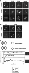
Fig. 1. Cell cycle-dependent localization of Cdh1p, Cdc20p and Cdc23p. (A) Wild-type cells (EY957) expressing the indicated GFP fusion proteins from the GAL promoter were grown at 30°C until mid-log phase, and analysed by GFP microscopy. Cells at different stages of the cell cycle are shown. The arrowheads point to Cdh1p–GFP localized to the mother bud neck. Note that Cdh1p is nuclear during G1, but predominantly cytoplasmic during S phase, G2 and mitosis. (B) Indirect immunofluorescence of HA3-Cdh1p expressed from the GAL promoter using 11HA antibodies. The arrowhead points to HA3-Cdh1p localized to the mother bud neck. (C) Nuclear recovery of Cdh1p–GFP was measured by FRAP as schematically represented in the upper panel. Nuclear recovery was measured in G1 cells (squares), or in late mitosis (late M) during relocalization of Cdh1p (diamonds). The nuclear/cytoplasmic exchange rate was quantified as described in Materials and methods, and is shown as t1/2 with SD. An unbleached nucleus was included as control (triangles).
None
Fig. 2. Phosphorylation of Cdh1p by Cdc28p regulates its subcellular localization. (A) The localization of Cdh1p–GFP was analysed by GFP microscopy in wild-type cells arrested in G1 with α-factor, in S phase with HU or in mitosis with nocodazole (NOCO). The phosphorylation state of HA3-Cdh1p was analysed by immunoblotting (lower panel). The arrow points to the position of unphosphorylated Cdh1p-HA. Immunoblotting with antibodies against actin confirms equal loading of the gel (lower blot). (Bcln1,2,3 pMETCLN2 (EY569) cells expressing Cdh1p–GFP from the ADH1 promoter (upper panel) or HA3-Cdh1p from the GAL1 promoter (lower panel) were arrested in G1 by depletion of Cln2p (time = 0), and released by expression of Cln2p. Aliquots were analysed after the times indicated by GFP microscopy (upper images) or immunoblotting (lower blot). Cells expressing non-phosphorylatable HA3-Cdh1p-m11 were included as a control. (Ccdc16-1 (YMP190), cdc14-3 (YMP809) or wild-type (K699) cells arrested with NOCO expressing either wild-type Cdh1p or non-phosphorylatable Cdh1p-m11 fused to GFP (upper panels) were arrested at the metaphase–anaphase transition or after anaphase, respectively, by shifting the temperature to 37°C for 2 h, and analysed by GFP microscopy. Numbers indicate the percentage of cells with nuclear accumulation of Cdh1p–GFP; at least 200 cells were included in the analysis. For control, the phosphorylation state of HA3-Cdh1p or HA3-Cdh1p-m11 was analysed in wild-type or cdc16-1 cells by immunoblotting (upper blot). Immunoblotting with antibodies against actin controls for the loading of the gel (lower blot). (D) Inhibition of Cdc28 kinase activity is sufficient for nuclear accumulation of Cdh1p–GFP. cdc16-1 cdc28-as1 cells (YMJ1055) expressing Cdh1p–GFP were arrested in mitosis by shifting to 37°C. After 90 min, the culture was divided: one half was incubated with the drug Na-PP1 specifically to inhibit Cdc28p-as1 (+Na-PP1), while only the solvent DMSO was added to the other half (–Na-PP1). The localization of Cdh1p–GFP was analysed by GFP microscopy after 90 min. Numbers indicate the percentage of cells with nuclear accumulation of Cdh1p–GFP; at least 200 cells were included in the analysis. For control, the phosphorylation state of HA3-Cdh1p was examined in cdc16-1 cdc28-as1 cells by immunoblotting (lower panel). An immunoblot against actin (arrow) controls for equal loading (lower blot).
None
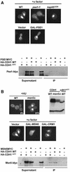
Fig. 3. The nuclear localization of Cdh1p is controlled by the importin Pse1p and the exportin Msn5p. (A) Wild-type (K699) cells or temperature-sensitive mutants in the import receptors Pse1p (pse1-1, YBL1) or Kap95p (kap95L63A, YBL14) were arrested in G1 by α-factor, and the localization of Cdh1p–GFP was analysed by GFP microscopy (upper panel). Note that nuclear accumulation of Cdh1p–GFP requires functional Pse1p. Overexpression of Pse1p from the inducible GAL promoter was sufficient for nuclear accumulation of Cdh1p–GFP (middle panels) at cell cycle stages where Cdh1p is predominantly cytoplasmic (vector). Pse1p-myc was able to co-immunoprecipitate (IP) with both wild-type HA3-Cdh1p and unphosphorylatable Cdh1p-m11-HA (lower panel). For co-immunoprecipitation, extracts prepared from cells expressing Pse1p-myc and, as indicated, either no protein (vector), wild-type HA3-Cdh1p or non-phosphorylatable HA3-Cdh1p-m11 were incubated with HA11 antibodies. The immunoprecipitates (IP) were analysed by immunoblot analysis with anti-Myc antibodies (9E10). For control, an aliquot of the extracts before immunoprecipitation (supernatant) was included. (B) The localization of Cdh1p–GFP was examined in wild-type (K699) and msn5Δ (YMJ1171) cells arrested with HU. Numbers indicate the percentage of cells with nuclear accumulation of Cdh1p–GFP; at least 200 cells were included in the analysis. The phosphorylation state of HA-Cdh1p and HA3-Cdh1p-m11 was analysed by immunoblotting in wild-type and msn5Δ cells as indicated (right blot). Overexpression of the exportin Msn5p but not Crm1p from the inducible GAL promoter in wild-type cells arrested with α-factor was sufficient to promote nuclear export of Cdh1p–GFP (middle panel). Co-immunoprecipitation experiments between Msn5p-myc and Cdh1p (lower panel) were performed as described in (A).
None
Fig. 4. Characterization of functional domains of Cdh1p for Msn5p binding and subcellular localization. (A) The interaction of Msn5p with full-length Cdh1p or the indicated deletion mutants was analysed by two-hybrid analysis. The numbers indicate Miller units ± SD; the background of the vector controls has already been subtracted. A schematic representation of the various constructs is shown in the left panel. Stippled box: C-box, required for the interaction with the APC/C; hatched bars: WD repeats involved in substrate interaction. The localization of the indicated proteins fused to GFP was analysed by GFP microscopy in wild-type cells at the G1 and G2/M phase of the cell cycle. Note that the 90 N-terminal amino acids are important for binding to Msn5p, but also contain a functional NLS. (B) The localization of various Cdh1p mutant proteins as indicated schematically on the left was analysed by GFP microscopy. Note that an intact substrate interaction site is required for localization of Cdh1p to the mother bud neck (arrowhead). (C) The ability of the indicated proteins to complement the defect of cdh1Δ cells (YMJ1075) to degrade endogenous Clb2p (arrow) in cells arrested in G1 by α-factor was analysed by immunoblotting with Clb2p antibodies.
None
Fig. 5. Nuclear Cdh1p has increased activity, but accumulation of endogenous Cdh1p is not sufficient to induce degradation of Clb2p. (A) The accumulation of endogenous Clb2p after bud emergence was examined by immunoblotting in wild-type and msn5Δ cells released from an α-factor block. For control, an extract of exponentially growing cells was included (expo). Note that nuclear Cdh1p is not sufficient to delay accumulation of Clb2p (arrow). The asterisk marks the position of an unknown protein, which is recognized by the Clb2p antibodies. The images on the right show the localization of Cdh1p–GFP in the same strains. (B) The half-life of Clb2p was compared in cdc15-1 (YMP690) or cdc15-1 msn5Δ (YMJ1185) cells after addition of cycloheximide (CX, time = 0). The levels of endogenous Clb2p were examined by immunobloting at the times indicated after addition of CX (in minutes). Note that nuclear Cdh1p is not sufficient to induce degradation of Clb2p (arrow). The images on the right show the localization of Cdh1p–GFP in the same strains. (C) Wild-type (CDH1) or the indicated Cdh1p mutants fused to GFP were overexpressed from the inducible GAL promoter in either wild-type (K699; WT) or msn5Δ (YMJ1171) cells. The plates show 5-fold serial dilutions of the cells spotted in plates containing either glucose (SD; GAL promoter off) or galactose (GAL; GAL promoter on) incubated for 3 days at 30°C. The accumulation of Clb2p (arrow) in wild-type (upper blot) or msn5Δ cells overexpressing Cdh1p from the GAL promoter was examined by immunoblotting after an α-factor release, as described in (A). The phase-contrast images on the right show the morphology of cells at the 120 min time point.
None
Fig. 6. Nuclear Cdh1p is required for degradation of Clb2p. (A) The NLS of Far1p (NLS) or, for control, a non-functional mutant form (nls) was fused to the N-terminus of Cdh1p-Δ50–GFP as schematically indicated on the left. The proteins were expressed as GFP fusions in either wild-type (K699; left panels) or cdc16-1 cells (YMP190; right panels) and analysed by GFP microscopy. Where indicated, wild-type cells were arrested in G1 by addition of α-factor for 2 h. Note that NLS–Cdh1p-Δ50 (upper panels) but not nls–Cdh1p-Δ50 (lower panels) is nuclear at all stages of the cell cycle. (B and C) Wild-type (K699) cells overexpressing either no protein (vector), untagged (B) or HA3-tagged (C) wild-type Cdh1p or the indicated Cdh1p mutants from the inducible GAL promoter were analysed for their ability to form colonies by serial dilution on plates containing glucose (SD; GAL promoter off) or galactose (GAL; GAL promoter on). The plasmids used in (C) were integrated at the URA3 locus of K699 cells. The morphology of the cells was analysed by differential interference contrast microscopy (DIC, right panels). Numbers indicate the percentage of cells with elongated buds. The accumulation of endogenous Clb2p (arrow) was analysed by immunoblotting with specific antibodies (blot). The asterisk marks the position of an unknown protein, which is recognized by the Clb2p antibodies and serves as a loading control.
None
Fig. 7. Nuclear export of Cdh1p may contribute to the inactivation of APC/CCdh1. Model for the inactivation of APC/CCdh1 after bud emergence. Phosphorylation of Cdh1p by Cdk1 (predominantly Cdc28p–Clb5p) prevents its interaction with the APC/C core complex (Zachariae et al., 1998), and promotes nuclear export by Msn5p. Cytoplasmic sequestration of Cdh1p prevents the assembly of functional APC/CCdh1 complexes during cell cycle stages other than G1, thereby contributing to the inactivation of APC/CCdh1.

Similar articles

Cited by

References

    1. Akada R., Kallal,L., Johnson,D.I. and Kurjan,J. (1996) Genetic relationships between the G protein by complex, Ste5p, Ste20p and Cdc42p: investigation of effector roles in the yeast pheromone response pathway. Genetics, 143, 103–117. - PMC - PubMed
    1. Amon A., Irniger,S. and Nasmyth,K. (1994) Closing the cell cycle circle in yeast: G2 cyclin proteolysis initiated at mitosis persists until the activation of G1 cyclins in the next cycle. Cell, 77, 1037–1050. - PubMed
    1. Ausubel F.M., Brent,R., Kingston,R.E., Moore,D.D., Seidman,J.G., Smith,J.A. and Struhl,K. (1991) Current Protocols in Molecular Biology. Greene Publishing Associates and Wiley-Interscience, New York, NY.
    1. Barral Y., Parra,M., Bidlingmaier,S. and Snyder,M. (1999) Nim1-related kinases coordinate cell cycle progression with the organization of the peripheral cytoskeleton in yeast. Genes Dev., 13, 176–187. - PMC - PubMed
    1. Baumer M., Kunzler,M., Steigemann,P., Braus,G.H. and Irniger,S. (2000) Yeast Ran-binding protein Yrb1p is required for efficient proteolysis of cell cycle regulatory proteins Pds1p and Sic1p. J. Biol. Chem., 275, 38929–38937. - PubMed

Publication types

MeSH terms