

Review
doi: 10.1085/jgp.20028708.
Proton conduction through gp91phox
Affiliations
- PMID: 12451045
- PMCID: PMC2229565
- DOI: 10.1085/jgp.20028708
Item in Clipboard
Review
Proton conduction through gp91phox
J Gen Physiol.
2002 Dec.
No abstract available
Figures

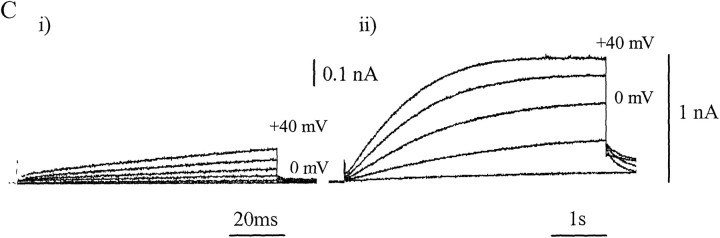
Expression of NADPH Oxidase (Nox) protein in PLB and PLBKO cells. (A) Nox protein immunodetected in undifferentiated (i) and differentiated (ii) PLB-298 cells after incubation with an anti–COOH-terminal gp91phox polyclonal antibody and FITC-labeled anti–rabbit antibody. The polyclonal anti-gp91phox was raised to the COOH-terminal 14-amino acid peptide coupled to keyhole limpet hemocyanin. It was verified by Western blot against detergent-solubilized membrane proteins from human neutrophils (Henderson et al., 1995). The images, which show an optical slice close to the cell center, were obtained using a BioRad MRC 600 inverted confocal microscope. A pseudocolor scale in which the highest intensity of emitted fluorescence is denoted by red through orange, yellow and green to blue, low fluorescence, indicates the presence of a Nox protein. Differentiated PLB-985 cells treated in the absence of the anti-gp91phox antibody are shown in iii. (B) Immunodetection of Nox protein in PLBKO cells using the same technique and antibody as described for A. As before, undifferentiated (i), differentiated (ii), and control cells in the absence of anti-gp91phox antibody (iii) are shown. (C) Whole-cell currents recorded from a single differentiated PLB-985 cell under conditions designed to maximize the amplitude of H+ current (see Henderson and Meech, 1999). The patch pipette–filling solution, which was adjusted to pH 6.5, contained 119 mM TMA hydroxide, to block potassium currents, a small quantity of calcium buffer (3.7 mM EGTA, 0.74 mM CaCl2) and ∼120 mM MES pH buffer. The bathing solution contained 110 mM TMA methane-sulphonate, 2 mM Ca(OH)2, 2 mM Mg(OH)2, 5 mM glucose, and 100 mM EPPS buffer adjusted to pH 8.0. Low amplitude prepulses in the linear current–voltage range (−60 to −100 mV) were scaled by PCLAMP 6 software (Axon Instruments, Inc.) and used for online subtraction of linear capacitive and leakage currents (Armstrong and Bezanilla, 1974). The superimposed currents shown were recorded in response to a standard stepped-voltage protocol; commands were in the range -40 to 40 mV (20-mV increments). The command duration was 100 ms (i) or 5 s (ii). The immunostaining of PLB-985 and PLBKO cells and recording of whole cell currents are the work of X. Wen Hu. Mary C. Dinauer provided the PLBKO cells. EPPS is N-[2-hydroxyethyl]-piperazine-N'-[3-propane-sulphonic acid]; TMA, tetramethylammonium.
Expression of NADPH Oxidase (Nox) protein in PLB and PLBKO cells. (A) Nox protein immunodetected in undifferentiated (i) and differentiated (ii) PLB-298 cells after incubation with an anti–COOH-terminal gp91phox polyclonal antibody and FITC-labeled anti–rabbit antibody. The polyclonal anti-gp91phox was raised to the COOH-terminal 14-amino acid peptide coupled to keyhole limpet hemocyanin. It was verified by Western blot against detergent-solubilized membrane proteins from human neutrophils (Henderson et al., 1995). The images, which show an optical slice close to the cell center, were obtained using a BioRad MRC 600 inverted confocal microscope. A pseudocolor scale in which the highest intensity of emitted fluorescence is denoted by red through orange, yellow and green to blue, low fluorescence, indicates the presence of a Nox protein. Differentiated PLB-985 cells treated in the absence of the anti-gp91phox antibody are shown in iii. (B) Immunodetection of Nox protein in PLBKO cells using the same technique and antibody as described for A. As before, undifferentiated (i), differentiated (ii), and control cells in the absence of anti-gp91phox antibody (iii) are shown. (C) Whole-cell currents recorded from a single differentiated PLB-985 cell under conditions designed to maximize the amplitude of H+ current (see Henderson and Meech, 1999). The patch pipette–filling solution, which was adjusted to pH 6.5, contained 119 mM TMA hydroxide, to block potassium currents, a small quantity of calcium buffer (3.7 mM EGTA, 0.74 mM CaCl2) and ∼120 mM MES pH buffer. The bathing solution contained 110 mM TMA methane-sulphonate, 2 mM Ca(OH)2, 2 mM Mg(OH)2, 5 mM glucose, and 100 mM EPPS buffer adjusted to pH 8.0. Low amplitude prepulses in the linear current–voltage range (−60 to −100 mV) were scaled by PCLAMP 6 software (Axon Instruments, Inc.) and used for online subtraction of linear capacitive and leakage currents (Armstrong and Bezanilla, 1974). The superimposed currents shown were recorded in response to a standard stepped-voltage protocol; commands were in the range -40 to 40 mV (20-mV increments). The command duration was 100 ms (i) or 5 s (ii). The immunostaining of PLB-985 and PLBKO cells and recording of whole cell currents are the work of X. Wen Hu. Mary C. Dinauer provided the PLBKO cells. EPPS is N-[2-hydroxyethyl]-piperazine-N'-[3-propane-sulphonic acid]; TMA, tetramethylammonium.
Similar articles
-
The gp91phox component of NADPH oxidase is not a voltage-gated proton channel.J Gen Physiol. 2002 Dec;120(6):773-9. doi: 10.1085/jgp.20028704. J Gen Physiol. 2002. PMID: 12451047 Free PMC article. Review. No abstract available.
-
NADPH oxidase of chondrocytes contains an isoform of the gp91phox subunit.Biochem J. 1998 Feb 1;329 ( Pt 3)(Pt 3):449-51. doi: 10.1042/bj3290449. Biochem J. 1998. PMID: 9445369 Free PMC article.
-
NADPH oxidase subunit gp91phox: a proton pathway.Protoplasma. 2001;217(1-3):37-42. doi: 10.1007/BF01289411. Protoplasma. 2001. PMID: 11732336 Review.
-
NOX family NADPH oxidases: do they have built-in proton channels?J Gen Physiol. 2002 Dec;120(6):781-6. doi: 10.1085/jgp.20028713. J Gen Physiol. 2002. PMID: 12451048 Free PMC article. Review. No abstract available.
-
Voltage-gated proton "channels": a spectator's viewpoint.J Gen Physiol. 2002 Dec;120(6):767-71. doi: 10.1085/jgp.20028706. J Gen Physiol. 2002. PMID: 12451046 Free PMC article. Review. No abstract available.
Cited by
-
Deficiency of gp91phox inhibits allergic airway inflammation.Am J Respir Cell Mol Biol. 2013 Sep;49(3):396-402. doi: 10.1165/rcmb.2012-0442OC. Am J Respir Cell Mol Biol. 2013. PMID: 23590311 Free PMC article.
-
Interactions between electron and proton currents in excised patches from human eosinophils.J Gen Physiol. 2003 Dec;122(6):713-26. doi: 10.1085/jgp.200308891. J Gen Physiol. 2003. PMID: 14638931 Free PMC article.
-
Voltage-gated proton channels: what's next?J Physiol. 2008 Nov 15;586(22):5305-24. doi: 10.1113/jphysiol.2008.161703. Epub 2008 Sep 18. J Physiol. 2008. PMID: 18801839 Free PMC article. Review.
-
Insights into the structure and function of HV1 from a meta-analysis of mutation studies.J Gen Physiol. 2016 Aug;148(2):97-118. doi: 10.1085/jgp.201611619. J Gen Physiol. 2016. PMID: 27481712 Free PMC article. Review.
-
Expression of gp91phox/Nox2 in COS-7 cells: cellular localization of the protein and the detection of outward proton currents.Biochem J. 2005 Feb 1;385(Pt 3):649-57. doi: 10.1042/BJ20040829. Biochem J. 2005. PMID: 15377283 Free PMC article.
References
-
- Banfi, B., A. Maturana, S. Jaconi, S. Arnaudeau, T. Laforge, B. Sinha, E. Ligeti, N. Demaurex, and K.H. Krause. 2000. A mammalian H+ channel generated through alternative splicing of the NADPH oxidase homolog NOH-1. Science. 287:138–142. - PubMed
-
- Banfi, B., G. Molnar, A. Maturana, K. Steger, B. Hegedus, N. Demaurex, and K.H. Krause. 2001. A Ca2+-activated NADPH oxidase in testis, spleen, and lymph nodes. J. Biol. Chem. 276:37594–37601. - PubMed
Publication types
MeSH terms
Substances
LinkOut - more resources
Full Text Sources
Miscellaneous
