. 2003 Feb 14;278(7):5009-20.
doi: 10.1074/jbc.M210436200. Epub 2002 Nov 20.

Vps51 is part of the yeast Vps fifty-three tethering complex essential for retrograde traffic from the early endosome and Cvt vesicle completion

Affiliations

Vps51 is part of the yeast Vps fifty-three tethering complex essential for retrograde traffic from the early endosome and Cvt vesicle completion

Fulvio Reggiori et al. J Biol Chem. .

Abstract

Autophagy, pexophagy, and the Cvt pathway are processes that deliver hydrolytic enzymes and substrates to the yeast vacuole/lysosome via double-membrane cytosolic vesicles. Whereas these pathways operate under different nutritional conditions, they all employ common machinery with only a few specific factors assisting in the choice of the delivery program and the membrane source for the sequestering vesicle. We found that the YKR020w gene product is essential for Cvt vesicle formation but not for pexophagy or induction of autophagy. Autophagosomes in the ykr020wdelta mutant, however, have a reduced size. We demonstrate that Ykr020 is a subunit of the Vps fifty-three tethering complex, composed of Vps52, Vps53, and Vps54, which is required for retrograde traffic from the early endosome back to the late Golgi, and for this reason we named it Vps51. This complex participates in a fusion event together with Tlg1 and Tlg2, two SNAREs also shown to be necessary for Cvt vesicle assembly. In particular, those factors are essential to correctly target the prApe1-Cvt19-Cvt9 complex to the preautophagosomal structure, the site of Cvt vesicle formation.


Figures
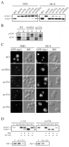
Fig 1
Fig 1
The ykr020wΔ, vps52Δ, vps53Δ, and vps54Δ cells have a defect in Cvt vesicle completion. A, The ykr020wΔ, vps52Δ, vps53Δ, and vps54Δ strains have a reversible inhibition of prApe1 processing similar to that of vac8Δ cells. Wild type (WT), ykr020wΔ, vps52Δ, vps53Δ (FRY107), vps54Δ, apg9Δ, and vac8Δ cells in the BY4742 background grown either in YPD or nitrogen starved in SD-N medium for 4 h were trichloroacetic acid precipitated. Acetone-washed proteins were then resolved by SDS-PAGE and prApe1 maturation analyzed by immunoblot. B, Prc1 is mislocalized to the periplasmic space in the ykr020wΔ mutant similarly to vps52Δ cells. Cells from wild type, ykr020wΔ, and vps52Δ strains in the BY4742 background were pulse-labeled for 10 min and chased for 30 min. Internal (I) and external (E) Prc1 was immunoprecipitated and then resolved by SDS-PAGE. C, The ykr020wΔ, vps52Δ, vps53Δ, and vps54Δ strains have a reversible accumulation of GFP-prApe1 in a cytosolic punctate structure. WT, ykr020wΔ, vps52Δ, vps53Δ (FRY107), and vps54Δ strains in the BY4742 background were transformed with a plasmid expressing the N-terminal GFP-tagged prApe1 (pTS466). Transformed cells were grown either in SMD or nitrogen starved for 3 h in SD-N medium and examined with a fluorescence microscope. DIC, differential interference contrast. D, precursor Ape1 is protease-sensitive in the vps52Δ mutant. The vps52Δ, vps54Δ, ykr020wΔ, and ccz1Δ strains in the BY4742 background were grown in YPD until early log phase. The cells were converted to spheroplasts and lysed. The total cells lysates (T) were centrifuged at 13,000 rpm for 5 min to separate the pellet (P13) and the supernatant (S13) fractions. The P13 fraction was resuspended in lysis buffer (27) and then mixed with equal volumes of lysis buffer, 40 μg/ml proteinase K, 0.4% Triton X-100, or proteinase K plus 0.4% Triton X-100, and incubated on ice for 30 min. The reactions were stopped by adding trichloroacetic acid. Samples were resolved by SDS-PAGE and examined by immunoblot with serum to Ape1. The ccz1Δ control cells accumulate completed Cvt vesicles and prApe1 is accessible to proteinase K only in the presence of Triton X-100. Even in the absence of detergent, prApe1 in the vps52Δ strain was digested to its mature form by the same protease indicating that Cvt vesicles were not completely assembled. Essentially identical results were obtained for the vps52Δ and ykr020wΔ strains. The T, S13, and P13 fractions were also probed for the cytosolic marker Pgk1. Recovery of Pgk1 in the S13 fractions indicates efficient lysis of spheroplasts.
Fig. 2
Fig. 2
The ykr020wΔ, vps52Δ, vps53Δ, and vps54Δ mutants are specifically defective in the Cvt pathway. A, precursor Ape1 is not imported to the vacuole by the Cvt pathway under starvation conditions in the vps52Δ strain. Wild type (WT), vps52Δ, vac8Δ, and vps52Δ vac8Δ (FRY119) cells in the BY4742 background were analyzed as in Fig. 1A. The vps52Δ vac8Δ double mutant was able to import and mature prApe1. Y, YPD; -N, SD-N. B, the ykr020wΔ and VFT mutant strains are sensitive to nitrogen starvation. Wild type, ykr020wΔ, vps52Δ, vps53Δ (FRY107), vps54Δ, cvt9Δ, and apg1Δ strains in the BY4742 background were grown in YPD medium until early log phase and then shifted to SD-N. At the indicated time points, equal volumes of culture were withdrawn and plated on YPD plates. After 3 days the number of colonies representing the viable cells was counted, and the percentage of survival calculated and plotted against time in starvation medium. C, autophagy is functional in the vps52Δ and ykr020wΔ mutants. Wild type (TN124), ykr020wΔ (FRY122), vps52Δ (FRY122), and apg13Δ (D3Y103) cells expressing Pho8Δ60 were shifted from YPD medium (black bars) to SD-N medium (white bars) for 4 h. Autophagy induction was determined by a Pho8 activity assay. Results were expressed as percentage of the activity measured for the wild type strain starved for nitrogen. Error bars represent the standard deviation from three separate experiments. D, viability of vps mutants is sensitive to nitrogen starvation. Wild type (WT), vps4Δ, vps5Δ, vps27Δ, and vps29Δ strains in the BY4742 background were treated as in panel D. E, the ykr020wΔ, vps52Δ, and vps53Δ strains are not defective in pexophagy. Cells from wild type, apg1Δ, ykr020wΔ, vps52Δ, and vps53Δ (FRY107) strains in the BY4742 background were grown in conditions that induce peroxisomes, washed, and resuspended in SD-N medium for the times indicated. The presence of the peroxisomal thiolase enzyme, Fox3, was detected by immunoblotting.
Fig. 3
Fig. 3
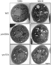
The ykr020wΔ and vps52Δ strains have smaller auto-phagosomes. Wild type (WT, TDY2), ykr020wΔ (FRY124), and vps52Δ (FRY125) cells were grown in YPD medium at 26°C to early log phase. Cultures were split in half and centrifuged. One sample was resus-pended again in YPD medium whereas the other was resuspended in SD-N medium. Cells were then grown at 37°C for 3 h. Permanganate fixation, dehydration, and embedding were carried out as described by Kaiser and Schekman (78). Uranyl acetate-stained sections were observed using a Philips CM10 transmission electron microscope. Examples of autophagosomes are indicated with an arrow. The bar is 1 μm.
Fig. 4
Fig. 4
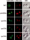
The ykr020wΔ, vps52Δ, vps53Δ, and vps54Δ cells have a defect in Snc1 recycling and display abnormal vacuole morphology. Wild type (WT), ykr020wΔ, vps52Δ, vps53Δ (FRY107), and vps54Δ cells in the BY4742 background were transformed with a plasmid expressing the GFP-Snc1 chimera (pGS416). Transformed cells were grown to a mid-log stage in SMD medium and FM 4-64 was added to the culture medium for 15 min. Cells were then collected, incubated in the same medium without FM 4-64 for an additional 30 min to chase the dye, and finally imaged with a fluorescence microscope. In wild type cells, Snc1 was concentrated on the plasma membrane while it was redirected to the vacuole lumen in the mutants. Pictures represent cells with an average level of GFP-Snc1 expression. In cells where those levels were lower, the staining pattern was primarily cytosolic punctate dots. FM 4-64 staining showed that deletion strains have a tubular, fragmented morphology of the vacuole. DIC, differential interference contrast.
Fig. 5
Fig. 5
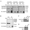
The YKR020w gene product is a component of the VFT complex. A, Ykr020 binds Vps52, Vps53, and Vps54. Strains expressing Vps52-HA (PSY118), Vps53-HA (PSY119), or Vps54-HA (PSY120) and carrying a plasmid expressing either the PA-Ykr020 construct (pCuPAYKR020(416)) or PA alone (pRS416-CuProtA) were used to prepare detergent-solubilized extracts (Ext) as described under “Experimental Procedures.” IgG-Sepharose beads were used to affinity purify the PA fusions together with the associated proteins (IP). Eluted polypeptides were separated with SDS-PAGE and then visualized by immunoblotting with antiserum to HA. For each experiment 0.2% of the total lysate or 20% of the total eluate were loaded per gel lane. B, there is only one Ykr020 molecule per VFT complex. The experiment performed in panel A was repeated with a strain (PSY116) expressing Ykr020-Myc. C, Ykr020 is not required for the stability of Vps53. Wild type (WT, PSY119), vps52Δ (FRY116), and ykr020wΔ (FRY117) strains expressing Vps53-HA were grown to early log phase and proteins were precipitated with trichloroacetic acid. Proteins were separated by SDS-PAGE and analyzed by Western blot with anti-HA antibodies. Pgk1 was used to verify that the same amount of material was loaded on each gel lane. D, Vps52 is not necessary to maintain normal cellular levels of Ykr020. Wild type (WT, PSY116) and vps52Δ (FRY118) cells expressing Ykr020-Myc were analyzed as in panel C using anti-Myc antibodies.
Fig. 6
Fig. 6
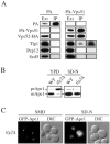
The Vps51-containg VFT complex interacts with Tlg1. A, Tlg1 interacts with the Vps51-containing VFT complex. Spheroplasts from wild type (PSY118) cells expressing Vps52-HA and transformed with either the plasmid expressing PA-Vps51 (pCuPAYKR020(416)) or PA alone (pRS416-CuProtA) were treated with 1.5 mm of the cross-linker (dithiobis(succinimidyl propionate) (DSP), detergent solubilized, and the PA fusions were affinity purified on IgG-Sepharose as described in the legend to Fig. 5A. Samples from the extracts (Ext) or purified fractions (IP) were analyzed by Western blot using anti-HA, Tlg1, Sed5, and Pep12 antibodies or antiserum. A longer film exposure is also shown to demonstrate the total absence of cross-linking between the VFT complex and the control tSNAREs. B, the tlg2Δ strain accumulates prApe1 in YPD medium and this defect is bypassed in nitrogen starvation conditions. Wild type (WT) and tlg2Δ cells in the BY4742 background were essentially treated as in Fig. 1A. C, the tlg2Δ strain has a reversible accumulation of GFP-prApe1 in a cytosolic punctate structure. The same strains used in panel B were transformed with the plasmid carrying the GFP-prApe1 construct (pTS466) and analyzed as described in the legend to Fig. 1C. DIC, differential interference contrast.
Fig. 7
Fig. 7
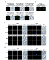
In the absence of the VFT complex, the prApe1-Cvt19-Cvt9 complex is not properly targeted to the PAS. A, Cvt9 is mislocalized in the absence of Vps51. Wild type (WT, SEY6210) and vps51Δ (FRY126) cells were co-transformed with the following pairs of plasmids: pRS414 (empty vector) and pTS495 (promGFP-CVT9); pCuPAYKR020 (414) (pPA-VPS51) and pTS495; pRS414 and pCuGFPCVT9(416) (pCuGFP-CVT9); pCuPAYKR020(414) and pCuGFPCVT9(416). The transformed cells were grown in SMD medium to early log phase and visualized with a fluorescence microscope. It should be noted that there are additional very faint dots in the vps51Δ cells expressing GFP-Cvt9 under the control of its native promoter (promGFP-CVT9) that are easily detected when the strong CUP1 promoter drives expression of the same chimera. B, Cvt9 mislocalization in vps51Δ cells is reversed by nitrogen starvation conditions. The vps51Δ strain in the BY4742 background was transformed with pCuGFPCVT9(416) (pCuGFP-CVT9). Transformed mutants were either grown in SMD medium to early log phase or starved for nitrogen in SD-N medium for 3 h and then analyzed with a fluorescence microscope. Identical results were obtained with tlg2Δ, vps52Δ, vps53Δ, and vps54Δ strains in the BY4742 background (data not shown). C, the prApe1-Cvt19 complex is not correctly targeted to the PAS in the absence of the VFT complex. Wild type (SEY6210) and vps52Δ (PSY113) strains were transformed with the following two pairs of plasmid: pTS470 (CFP-Ape1) and pRS414EYFP-Aut7 (YFP-Aut7); pCVT19CFP(414) (Cvt19-CFP) and pRS414EYFP-Aut7. Transformed cells were either grown in SMD medium to early log phase or starved for nitrogen in SD-N medium for 3 h and then visualized with a fluorescence microscope. The CFP-Ape1 and Cvt19-CFP did not co-localize with the PAS (YFP-Aut7) when cells were grown in SMD medium. Nitrogen starvation conditions induced the correct targeting of CFP-Ape1 and Cvt19-CFP to the PAS because those two chimeras were in the same punctate structure as YFP-Aut7 in all cells. These observations were confirmed by repeating the same experiment in vps51Δ and vps53Δ cells (data not shown). D, Cvt9 is associated with the prApe1-Cvt19 complex away from the PAS in the vps52Δ mutant. Wild type (SEY6210) and vps52Δ (PSY113) strains were transformed with the following two pairs of plasmid: pPS98 (CFP-Cvt9) and pRS414EYFP-Aut7 (YFP-Aut7); pPS97 (YFP-Cvt9) and pCVT19CFP(414) (Cvt19-CFP). Transformed cells were grown in SMD medium to early log stage and analyzed with a fluorescence microscope. The CFP-Cvt9 fluorescent dots were separated from the PAS (YFP-Aut7), whereas co-localization between Cvt19 and Cvt9 was observed in all the images taken. DIC, differential interference contrast.
Fig. 8
Fig. 8
Model for VFT complex function in the Cvt pathway. The VFT complex, including Vps51, functions as a tethering factor that is required for retrieval (retrograde traffic) from the early endosome to the Golgi complex (33, 45, 46). In the Cvt pathway, mutations in the VFT proteins result in mislocalization of the PAS component Cvt9, suggesting a tethering role in anterograde transport from the Golgi complex to the PAS. By analogy with its role in retrieval from the endosome, it is also possible that the VFT complex is needed for retrograde recovery of certain components from the PAS. Most of the Apg/Cvt proteins that localize to the PAS are not found in completed Cvt vesicles suggesting that they are excluded from the vesicle and retained at the PAS or that they are recycled back to another compartment such as the Golgi complex. See text for details.

Similar articles

Cited by

References

    1. Reggiori F, Klionsky DJ. Euk. Cell. 2002;1:11–21. - PMC - PubMed
    1. Klionsky DJ, Emr SD. Science. 2000;290:1717–1721. - PMC - PubMed
    1. Harding TM, Hefner-Gravink A, Thumm M, Klionsky DJ. J. Biol. Chem. 1996;271:17621–17624. - PubMed
    1. Scott SV, Hefner-Gravink A, Morano KA, Noda T, Ohsumi Y, Klionsky DJ. Proc. Natl. Acad. Sci. U. S. A. 1996;93:12304–12308. - PMC - PubMed
    1. Scott SV, Baba M, Ohsumi Y, Klionsky DJ. J. Cell Biol. 1997;138:37–44. - PMC - PubMed

Publication types

MeSH terms