

TGFbeta-Smad signalling in postoperative human lens epithelial cells
- PMID: 12446380
- PMCID: PMC1771405
- DOI: 10.1136/bjo.86.12.1428
TGFbeta-Smad signalling in postoperative human lens epithelial cells
Abstract
Aims: To localise Smads3/4 proteins in lens epithelial cells (LECs) of fresh and postoperative human specimens. Smads3/4 are involved in signal transduction between transforming growth factor beta (TGFbeta) cell surface receptors and gene promoters. Nuclear localisation of Smads indicates achievement of endogenous TGFbeta signalling in cells.
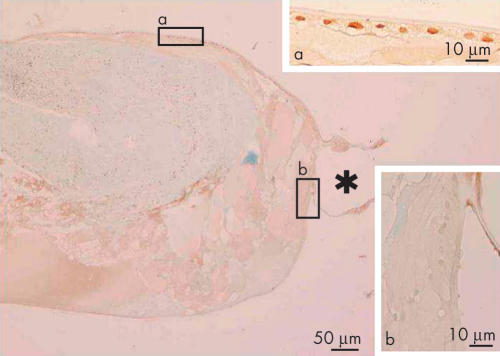
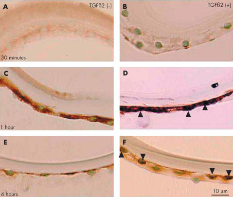
Methods: Three circular sections of the anterior capsule, one lens, and 17 capsules undergoing postoperative healing were studied. Immunohistochemistry was performed for Smads3/4 in paraffin sections of the specimens. The effect of exogenous TGFbeta2 on Smad3 subcellular localisation was examined in explant cultures of extracted human anterior lens epithelium.
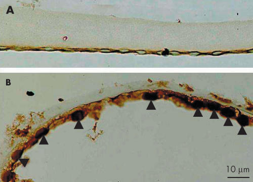
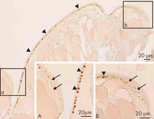
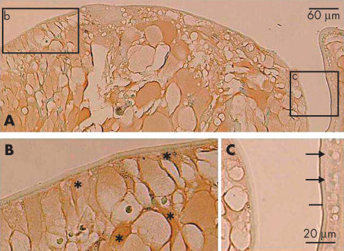
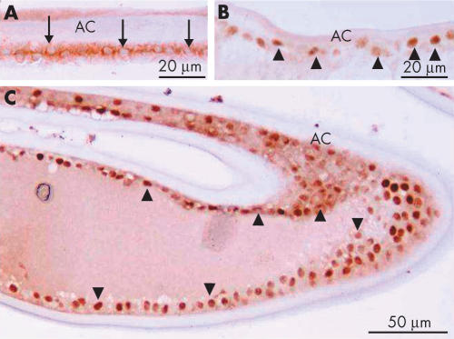
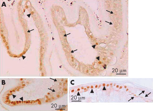
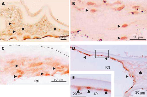
Results: The cytoplasm, but not the nuclei, of LECs of uninjured lenses was immunoreactive for Smads3/4. In contrast, nuclear immunoreactivity for Smads3/4 was detected in LECs during capsular healing. Nuclei positive for Smads3/4 were observed in monolayered LECs adjacent to the regenerated lens fibres of Sommerring's ring. Interestingly, the nuclei of LECs that were somewhat elongated, and appeared to be differentiating into fibre-like cells, were negative for Smads3/4. Fibroblast-like, spindle-shaped lens cells with nuclear immunoreactivity for nuclear Smads3/4 were occasionally observed in the extracellular matrix accumulated in capsular opacification. Exogenous TGFbeta induced nuclear translocation of Smad3 in LECs of anterior capsule specimens in explant culture.
Conclusions: This is consistent with TGFbeta induced Smad signalling being involved in regulating the behaviour of LECs during wound healing after cataract surgery.
Figures

Similar articles
-
Smad translocation and growth suppression in lens epithelial cells by endogenous TGFbeta2 during wound repair.Exp Eye Res. 2001 Jun;72(6):679-86. doi: 10.1006/exer.2001.1002. Exp Eye Res. 2001. PMID: 11384156
-
Collagens XII and XIV (FACITs) in capsular opacification and in cultured lens epithelial cells.Curr Eye Res. 2001 Dec;23(6):463-8. doi: 10.1076/ceyr.23.6.463.6971. Curr Eye Res. 2001. PMID: 12045897
-
Enhanced EGF receptor-signaling potentiates TGFβ-induced lens epithelial-mesenchymal transition.Exp Eye Res. 2019 Aug;185:107693. doi: 10.1016/j.exer.2019.107693. Epub 2019 Jun 12. Exp Eye Res. 2019. PMID: 31201806
-
Transforming growth factor-beta-induced epithelial-mesenchymal transition in the lens: a model for cataract formation.Cells Tissues Organs. 2005;179(1-2):43-55. doi: 10.1159/000084508. Cells Tissues Organs. 2005. PMID: 15942192 Review.
-
Fibrosis in the lens. Sprouty regulation of TGFβ-signaling prevents lens EMT leading to cataract.Exp Eye Res. 2016 Jan;142:92-101. doi: 10.1016/j.exer.2015.02.004. Epub 2015 May 21. Exp Eye Res. 2016. PMID: 26003864 Free PMC article. Review.
Cited by
-
Lanosterol Synthase Prevents EMT During Lens Epithelial Fibrosis Via Regulating SREBP1.Invest Ophthalmol Vis Sci. 2023 Dec 1;64(15):12. doi: 10.1167/iovs.64.15.12. Invest Ophthalmol Vis Sci. 2023. PMID: 38079167 Free PMC article.
-
Lens Fibrosis: Understanding the Dynamics of Cell Adhesion Signaling in Lens Epithelial-Mesenchymal Transition.Front Cell Dev Biol. 2022 May 17;10:886053. doi: 10.3389/fcell.2022.886053. eCollection 2022. Front Cell Dev Biol. 2022. PMID: 35656546 Free PMC article. Review.
-
Role of Decorin in the Lens and Ocular Diseases.Cells. 2022 Dec 24;12(1):74. doi: 10.3390/cells12010074. Cells. 2022. PMID: 36611867 Free PMC article. Review.
-
Fibronectin regulates growth factor signaling and cell differentiation in primary lens cells.J Cell Sci. 2018 Nov 20;131(22):jcs217240. doi: 10.1242/jcs.217240. J Cell Sci. 2018. PMID: 30404825 Free PMC article.
-
HPV16-E6 Oncoprotein Activates TGF-β and Wnt/β-Catenin Pathways in the Epithelium-Mesenchymal Transition of Cataracts in a Transgenic Mouse Model.Biomed Res Int. 2018 May 16;2018:2847873. doi: 10.1155/2018/2847873. eCollection 2018. Biomed Res Int. 2018. PMID: 29888254 Free PMC article.
References
-
- McDonnell PJ, Zarbin MA, Green WR. Posterior capsule opacification in pseudophakic eyes. Ophthalmology 1983;90:1548–53. - PubMed
-
- Kappelhof JP, Vrensen GFJM, de Jong PTVM, et al. An ultrastructural study of Elschnig’s pearls in the pseudophakic eye. Am J Ophthalmol 1986;101:58–69. - PubMed
-
- Apple DJ, Solomon KD, Tetz MR, et al. Posterior capsule opacification. Surv Ophthalmol 1992;37:73–116. - PubMed
-
- Saika S, Ohmi S, Tanaka S, et al. Outgrowth of lens epithelial cells and matrix formation on intraocular lenses in rabbit eyes. J Cataract Refract Surg 1996;22:835–40. - PubMed
MeSH terms
Substances
LinkOut - more resources
Full Text Sources
Molecular Biology Databases
Miscellaneous