. 2002 Dec;76(24):12877-89.
doi: 10.1128/jvi.76.24.12877-12889.2002.

ICP27 interacts with the RNA export factor Aly/REF to direct herpes simplex virus type 1 intronless mRNAs to the TAP export pathway

Affiliations

ICP27 interacts with the RNA export factor Aly/REF to direct herpes simplex virus type 1 intronless mRNAs to the TAP export pathway

I-Hsiung Brandon Chen et al. J Virol. 2002 Dec.

Abstract

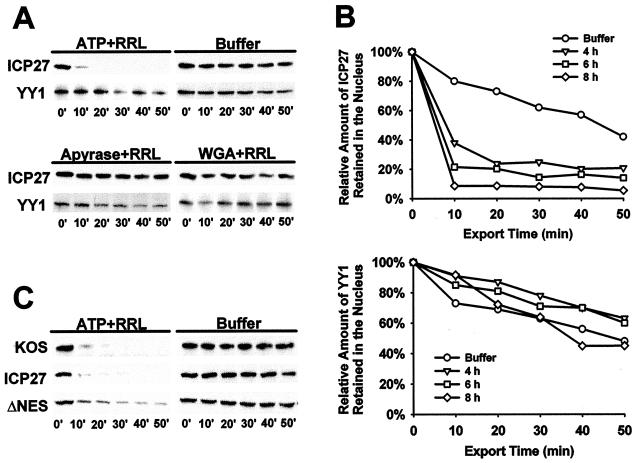
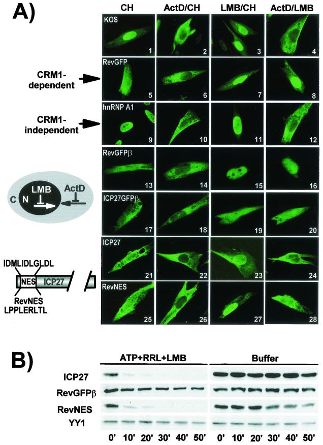
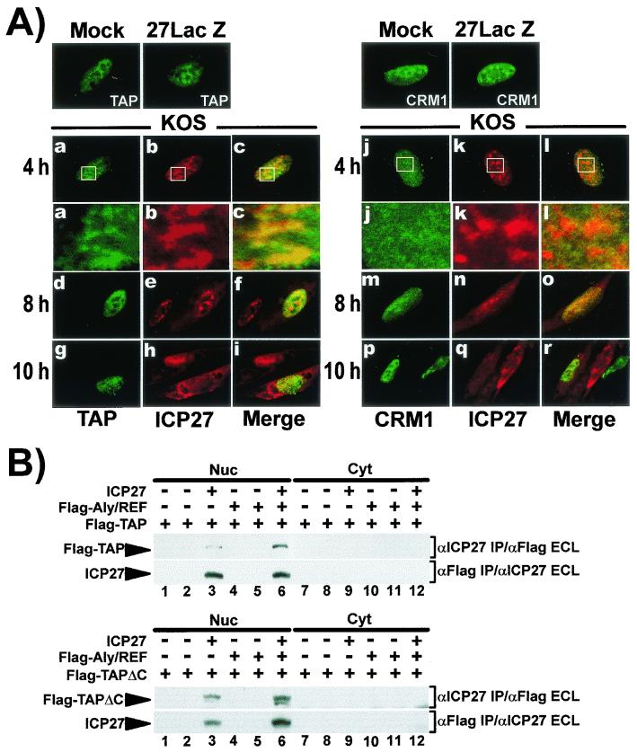
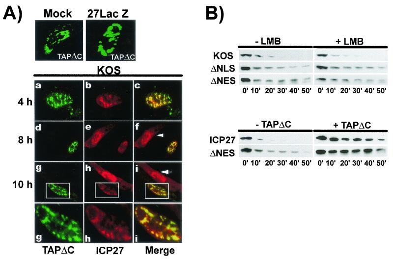
Herpes simplex virus type 1 (HSV-1) protein ICP27 facilitates the export of viral intronless mRNAs. ICP27 shuttles between the nucleus and cytoplasm, which has been shown to require a leucine-rich nuclear export sequence (NES). ICP27 export was reported to be sensitive to the CRM1 inhibitor leptomycin B (LMB) in HSV-1-infected cells but not in Xenopus oocytes, where ICP27 interacts with the export factor Aly/REF to access the TAP export pathway. Here, we show that ICP27 interacts with Aly/REF in HSV-1-infected mammalian cells and that Aly/REF stimulates export of viral intronless RNAs but does not cross-link to these RNAs. During infection, Aly/REF was no longer associated with splicing factor SC35 but moved into structures that colocalized with ICP27, suggesting that ICP27 recruits Aly/REF from spliceosomes to viral intronless RNAs. Further, ICP27 was found to interact in vivo with TAP but not with CRM1. In vitro export assays showed that ICP27 export was not sensitive to LMB but was blocked by a dominant-negative TAP deletion mutant lacking the nucleoporin interaction domain. These data suggest that ICP27 uses the TAP pathway to export viral RNAs. Interestingly, the leucine-rich N-terminal sequence was required for efficient export, even though ICP27 export was LMB insensitive. Thus, this region is required for efficient ICP27 export but does not function as a CRM1-dependent NES.


Figures

FIG. 1.
FIG. 1.
ICP27 interacts with Aly/REF. (A) The coding region of ICP27 is shown schematically, including the leucine-rich putative NES, NLS, two arginine-rich regions (R1 or R2), three putative KH domains, and a zinc finger-like motif (CHCC). ICP27 mutations fused to the Gal4 DNA-binding domain were tested for interaction with Aly/REF fused to the Gal4 activation domain as measured by β-galactosidase production. (B) 293 cells were transfected with pFlag-Aly/REF and then mock- or HSV-1 KOS-infected 24 h later. Cells were labeled with [35S]methionine for 5 h beginning 1 h after infection. Extracts were immunoprecipitated with anti-Flag or anti-ICP27 antibodies in the presence or absence of RNase as indicated. (C) 293 cells were cotransfected with pFlag-Aly/REF and plasmids expressing mutants D2ΔS5, ΔNLS, R1, S5, or H17 and later infected with 27-LacZ or the ICP27 viral mutant, tsLG4 at 39.5°C. Labeling with [35S]methionine and immunoprecipitations with anti-ICP27 and anti-Flag antibodies were performed as described above. The positions of the mutants tested for interaction by immunoprecipitation, and the results are shown schematically in the right panel.
FIG. 2.
FIG. 2.
Aly/REF colocalizes with ICP27. RSF cells were transfected with pEGFP-Aly/REF and were infected with HSV-1 KOS 24 h later. Cells were fixed at the times indicated. Immunofluorescence staining was performed with anti-ICP27 antibody H1119. EGFP expression was detected by direct fluorescence.
FIG. 3.
FIG. 3.
Aly/REF colocalization with splicing factor SC35 is altered during HSV infection. RSF cells were transfected with pEGFP-Aly/REF and were later mock infected (panels a to c) or infected with the ICP27 null mutant, 27-LacZ (panels d to i) or wild-type KOS (panels j to u). At the times indicated, cells were fixed and stained with anti-SC35 monoclonal antibody. EGFP fluorescence was detected directly. The merged images show areas of colocalization in yellow. Arrows in panels q, r, t, and u show examples of SC35 structures that do not contain Aly/REF.
FIG. 4.
FIG. 4.
Overexpression of Aly/REF facilitates export of viral intronless mRNAs. (A) RSF cells were transfected with pFlag-Aly/REF or a control vector plasmid and were infected with HSV-1 KOS. Nuclear and cytoplasmic RNA fractions were prepared and analyzed by RNase protection with probes specific for gB or ICP27 mRNA (left panels). Portions of the 8-h nuclear and cytoplasmic fractions were immunoprecipitated with anti-ICP27 or anti-Flag antibody as indicated (right panels). Blots were subsequently probed first with the antibody used in the immunoprecipitation and detected by ECL. The signals were allowed to decay and the blots were then probed with the other antibody as indicated and again detected by ECL. Asterisks mark heavy-chain immunoglobulin G from the immunoprecipitation that reacted with the secondary antibody used in the immunoblot analysis. (B) Transfected RSF cells expressing Flag-Aly/REF were infected with KOS for 6 h, and nuclear and cytoplasmic RNA fractions were analyzed by RNase protection with probes for UL49, gB, gC, gD, Aly/REF, and ICP27 mRNAs. (C) Transfected RSF cells were infected with KOS, and UV cross-linking was performed 6 h later as described previously (41, 42). RNA-protein complexes were immunoprecipitated with anti-ICP27 or anti-Flag antibodies and RNase protection analysis was performed with probes for gB, gC, gD, and Aly/REF mRNAs as described previously (41). (D) RSF cells were transfected with pFlag-Aly/REF and pCMV-ICP27 or mutants pCMV-R1 or pCMV-L/R and were infected with 27-LacZ virus 24 h after transfection. Nuclear and cytoplasmic RNA fractions prepared 6 h after infection were analyzed by RNase protection.
FIG. 5.
FIG. 5.
An in vitro nuclear export assay to quantify ICP27 export. (A) RSF cells infected with KOS for 6 h were permeabilized by adding digitonin. After depeltion of the cytoplasmic factors, 50% RRL and an ATP regeneration system (ATP) (upper left panel), transport buffer (buffer) alone (upper right panel), apyrase (20 U/ml) and RRL (lower left panel), or wheat germ agglutinin (WGA; 200 μg/ml) and RRL were added. At the times indicated, permeabilized cells were washed in ice-cold transport buffer, and proteins that remained in the nucleus were harvested by adding 2× SDS-PAGE loading buffer. Western blot analysis was performed with anti-ICP27 or anti-YY1 monoclonal antibodies, and the bands were visualized by ECL. (B) Nuclear export assays were performed at 4, 6, and 8 h after viral infection in the presence of 50% RRL and ATP. The results were quantified from scanned X-ray films by using SigmaScan (Jandel Scientific Software). (C) RSF cells were either infected with HSV-1 KOS or were transfected with pCMV-ICP27 (ICP27) or pCMV-DNES, followed by infection with 27-LacZ virus. In vitro export assays were performed 6 h after infection.
FIG. 6.
FIG. 6.
Export of ICP27 is not sensitive to LMB. (A) RSF cells were infected with HSV-1 KOS for 6 h (panels 1 to 4 and 9 to 12) or were transfected with pRevGFP (panels 5 to 8), pRevGFPβ (panels 13 to 16), pICP27GFPβ (panels 17 to 20), pCMV-ICP27 (panels 21 to 24), or pRevNES (panels 25 to 28). Transfected cells were infected with 27-lacZ virus. Cells were treated with CH (100 μg/ml), ActD (10 μg/ml), and LMB (25 ng/ml) as indicated beginning 5 h after infection for 4 h. Cells were fixed in 3.7% formaldehyde. Immunofluorescent staining was performed with anti-ICP27 (panels 1 to 4 and 21 to 28) and anti-hnRNP A1 antibody (panels 9 to 12). GFP fluorescence was visualized directly. (B) RSF cells were transfected with pCMV-ICP27, pRevGFPβ, or pRevNES for 24 h and then were infected with 27-LacZ. LMB (25 ng/ml) was added 5 h after infection and was present throughout the export assays.
FIG. 7.
FIG. 7.
ICP27 interacts with TAP but not with CRM1. (A) RSF cells were transfected with pEGFP-TAP or pEGFP-CRM1 for 24 h and then were mock infected, infected with 27-LacZ, or infected with KOS. Cells were fixed at 8 h for mock and 27-LacZ samples and at the times indicated for KOS-infected cells. EGFP was detected by direct fluorescence. Enlargements of the areas marked by rectangles in panels a to c and j to l are shown beneath. (B) RSF cells were transfected with pFlag-TAP (upper panels) or pFlag-TAPΔC, which has a deletion of C-terminal residues 518 to 619 (lower panels). pFlag-Aly/REF was cotransfected where indicated. Transfected cells were mock infected (lanes 1, 4, 7, and 10), infected with 27-LacZ (lanes 2, 5, 8, and 11), or KOS (lanes 3, 6, 9, and 12) for 6 h. Nuclear and cytoplasmic fractions were immunoprecipitated with anti-ICP27 or anti-Flag antibodies as indicated. Immunoblot analysis was performed with the antibodies indicated.
FIG. 8.
FIG. 8.
Dominant-negative TAPΔC blocks export of ICP27. (A) RSF cells were transfected with pEGFP-TAPΔC and then were mock, 27-LacZ, or KOS infected. Cells were fixed at the times indicated. Enlargements of the areas marked by rectangles in panels g to i are shown below. Arrows mark cells where ICP27 is expressed but EGFP-TAPΔC is not. (B) In vitro export assays were performed on cells infected with KOS or transfected with pCMV-ICP27, ΔNLS, or ΔNES, followed by infection with 27-LacZ virus. In the upper panels, LMB or buffer alone (−LMB) was added 5 h after infection for 2 h, at which time the in vitro export assay was performed. In the lower panels, cells were cotransfected with pFlag-TAPΔC or with a control plasmid (−TAPΔC) and ICP27 or ΔNES and then infected with 27-LacZ virus. The export assay was performed 6 h after infection.

Similar articles

Cited by

References

    1. Bachi, A., I. C. Braun, J. P. Rodrigues, N. Pante, K. Ribbeck, C. von Kobbe, U. Kutay, M. Wilm, D. Gorlich, M. Carmo-Fonseca, and E. Izaurralde. 2000. The C-terminal domain of TAP interacts with the nuclear pore complex and promotes export of specific CTE-bearing RNA substrates. RNA 6:136-158. - PMC - PubMed
    1. Bear, J., W. Tan, A. S. Zolotukhin, C. Tabernero, E. A. Hudson, and B. K. Felber. 1999. Identification of novel import and export signals of human TAP, the protein that binds to the Constitutive Transport Element of the type D retrovirus mRNAs. Mol. Cell. Biol. 19:6306-6317. - PMC - PubMed
    1. Braun, I. C., A. Herold, M. Rode, E. Conti, and E. Izaurralde. 2001. Overexpression of TAP/p15 heterodimers bypasses nuclear retention and stimulates nuclear mRNA export. J. Biol. Chem. 276:20536-20543. - PubMed
    1. Bryant, H. E., S. Wadd, A. I. Lamond, S. J. Silverstein, and J. B. Clements. 2001. Herpes simplex virus IE63 (ICP27) protein interacts with spliceosome-associated protein 145 and inhibits splicing prior to the first catalytic step. J. Virol. 75:4376-4385. - PMC - PubMed
    1. Cullen, B. R. 2000. Connections between the processing and nuclear export of mRNA: evidence for an export license? Proc. Natl. Acad. Sci. USA 97:4-6. - PMC - PubMed

Publication types

LinkOut - more resources