. 2002 Nov 15;22(22):9733-41.
doi: 10.1523/JNEUROSCI.22-22-09733.2002.

Tau-mediated cytotoxicity in a pseudohyperphosphorylation model of Alzheimer's disease

Affiliations

Tau-mediated cytotoxicity in a pseudohyperphosphorylation model of Alzheimer's disease

Thomas Fath et al. J Neurosci. .

Abstract

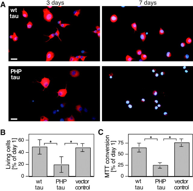
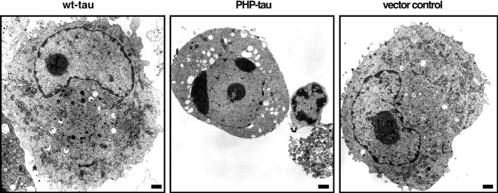
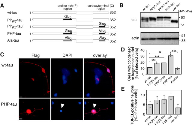
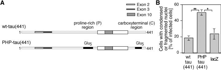
Aggregation and increased phosphorylation of tau at selected sites ("hyperphosphorylation") are histopathological hallmarks of Alzheimer's disease (AD). However, it is not known whether the tau pathology has a primary role during neuronal degeneration. To determine the role of tau hyperphosphorylation in AD, pseudohyperphosphorylated tau (PHP-tau) that simulates disease-like permanent, high stoichiometric tau phosphorylation and mimics structural and functional aspects of hyperphosphorylated tau was expressed in neural cells. In differentiated PC12 cells, PHP-tau exhibited reduced microtubule interaction and failed to stabilize the microtubule network compared with exogenously expressed wild-type tau (wt-tau). During longer culture, PHP-tau exerted a cytotoxic effect, whereas wt-tau was neutral. PHP-tau-mediated cytotoxicity was associated with an induction of apoptotic cell death as characterized by chromatin condensation, DNA fragmentation, and caspase-3 activation in the absence of detectable protein aggregates. Furthermore, PHP-tau expression specifically sensitized the cells for other apoptotic stimuli (colchicine and staurosporine). Herpes simplex virus-mediated overexpression of PHP-tau induced degeneration associated with an induction of apoptotic mechanisms also in terminally differentiated human CNS model neurons. Partially pseudophosphorylated constructs caused an intermediate toxicity. The data provide evidence for a neurotoxic "gain of function" of soluble tau during AD as a result of structural changes that are induced by a cumulative, high stoichiometric tau phosphorylation. PHP-tau-expressing cells and organisms could provide a useful system to identify mechanisms that contribute to tau-mediated toxicity.


Figures

Fig. 1.
Fig. 1.
Simulation of a disease-like tau hyperphosphorylation in cells. A, Residues that constitute sites with an increased phosphorylation in PHF-tau (Morishima-Kawashima et al., 1995) and have been substituted for glutamate to simulate a disease-like hyperphosphorylation (PHP-tau) are indicated by asterisks. The microtubule-binding repeats of tau are indicated by the thick black box. Adult-specific exons are shaded. B, Fluorescence staining for exogenous tau expression in undifferentiated PC12 cells stably transfected with wt-tau, PHP-tau, or a vector control (vec). C, Immunoblots of cellular lysates from undifferentiated PC12 cells stably transfected with wt-tau or PHP-tau. Lysates remained untreated (left) or were preincubated with alkaline phosphatase (AP) to remove phosphate residues (middle). For comparison, recombinant wt-tau and PHP-tau are shown on the right. B, Cells were plated at 2 × 10⁴ cells per squared centimeter on collagen-coated coverslips, fixed on the next day with 4% paraformaldehyde, and processed for immunofluorescence, as described in Materials and Methods, using a mouse monoclonal antibody against the FLAG-epitope (M5, top) and a rat monoclonal antibody against tubulin (YL1/2,bottom). C, Lysates corresponding to 2.5 × 10⁵ cells were loaded, separated by SDS-PAGE on 10% acrylamide, and detected with anti-tau (Tau5) antibody. To remove covalently bound phosphates, lysates were treated with 5 U of alkaline phosphatase overnight at 37°C. Scale bars, 10 μm.
Fig. 2.
Fig. 2.
Expression and microtubule association of wt-tau and PHP-tau in differentiated PC12 cells. A, Fluorescence staining for exogenous tau expression in differentiated PC12 cells stably transfected with wt-tau and PHP-tau.B, Fluorescence staining for exogenous tau and cellular microtubules of differentiated wt-tau- and PHP-tau-expressing PC12 cells after a combined extraction–fixation protocol to demonstrate cytoskeletal association. Note the colocalization of tau and microtubules that is evident in growth cone-like structures (arrowheads). C, Immunoblot of cytoskeletally associated pellet (P) and nonassociated supernatant (S) fraction after fractionation of lysates from differentiated PC12 cells. Populations of tau phosphoisoforms with fast and slow electrophoretic mobilities are indicated. Fractionations were also performed in the presence of 10 μm taxol to completely polymerize the microtubule network. D, Ratio of acetylated/total tubulin as determined by immunoblot analysis of cellular lysates from differentiated PC12 cells. Numbers are normalized to the ratio of acetylated/total tubulin in control cells (set as 1). For immunofluorescence analysis, cells were differentiated for 4 d with NGF, fixed using 4% paraformaldehyde (A) or a combined NP-40 extraction–fixation protocol (B), and processed for immunofluorescence using anti-FLAG (M5) and anti-tubulin (YL1/2) antibody as described in Materials and Methods. Scale bars, 10 μm. For immunoblots, lysates corresponding to 0.5–1.5 × 10⁶ cells were loaded and detected with anti-tau (Tau5), anti-tubulin (DM1A), or anti-acetylated tubulin (6-11B-1) antibody. Quantifications were from four experiments using two independent clonal lines per construct. Mean and SE are shown.
Fig. 3.
Fig. 3.
Reduced survival of differentiated PC12 cells stably transfected with PHP-tau. A, Fluorescence staining for exogenous tau (red) and nuclei (blue) of wt-tau-expressing (top) and PHP-tau-expressing (bottom) PC12 cells 3 and 7 d after induction of differentiation. B, C, Number (B) and MTT conversion (C) of differentiated PC12 cells. Survival was significantly reduced in PHP-tau-expressing cells. Cultures were for 1 week (B, C) or indicated times (A) after the induction of differentiation with NGF. Cells were fixed with 4% paraformaldehyde and stained against the FLAG-epitope tag (M5) and with DAPI. Quantifications were from six experiments using two independent clonal lines per construct. Mean and SE are shown. Scale bars, 20 μm.
Fig. 4.
Fig. 4.
Ultrastructural changes in PC12 cells expressing PHP-tau. Compared with wt-tau-expressing and vector control cells, PHP-tau-expressing cells showed chromatin condensation. No evidence for abnormal protein aggregates was observed in any of the lines analyzed. Cells were cultured in serum-free medium for 1 week after the induction of differentiation with NGF and processed for electron microscopy as described in Materials and Methods. Scale bars, 1 μm.
Fig. 5.
Fig. 5.
Induction of apoptosis in PHP-tau-expressing PC12 cells. A, B, Quantification of TUNEL-positive cells (A) and caspase-3 activity (B) in cell lines expressing wt-tau, PHP-tau, or a control line. The ratio of TUNEL-positive cells/level of caspase-3 activity is increased in PHP-tau-expressing cells. C, Effect of colchicine (left) and staurosporine (right) on the relative survival of stably transfected PC12 lines as analyzed by MTT conversion. PHP-tau-expressing cells show a reduced relative survival in the presence of both drugs compared with wt-tau-expressing cells or control lines. Cells were cultured in serum-free medium for 5 d (A, B) or 1 week (C) after the induction of differentiation with NGF. A, The ratio of TUNEL-positive cells/total cells was determined by visual inspection from 10 microscopic frames. An independent experiment gave very similar results. B,C, Four experiments with two independent clonal lines were evaluated. Mean and SE are shown. C, Relative survival of cells treated with colchicine or staurosprine (0.1 μg/ml and 100 nm, respectively; added twice, after 4 and 6 d) was measured against the survival of cultures in the absence of drugs.
Fig. 6.
Fig. 6.
Virus-mediated overexpression of fetal PHP-tau and partially pseudophosphorylated constructs (PP(P)-tau and PP(C)-tau) in human model neurons induce neuronal degeneration. A, Schematic of the different tau constructs generated by site-directed mutagenesis. Clusters of five serine/threonine residues in the proline-rich (P) region or the C-terminal (C) region were changed to glutamate or alanine as indicated by Glu5 orAla5, respectively. B, Immunoblot of lysates from infected model neurons expressing the indicated constructs. All constructs are expressed to a comparable level in the cell. As a control for equal loading, an actin blot is shown. Some of the constructs are present as different phosphoisoforms, as indicated by the presence of multiple bands with different electrophoretic mobilities. C, Fluorescence staining for exogenous tau (red) and nuclei (blue) of NT2-N neurons infected with a HSV[wt-tau] (top) or HSV[PHP-tau] (bottom) construct. Condensed nuclei in infected NT2-N neurons are indicated by arrowheads. D, E, Quantification of the percentage of condensed or fragmented nuclei (D) and TUNEL-positive cells (E) after infection with the indicated constructs. Cells infected with PHP-tau show an increased fraction of degenerated and TUNEL-positive neurons compared with wt-tau- or Ala-tau-expressing cells. The partially pseudophosphorylated constructs induce intermediate toxicity. For immunoblot analysis, 15,000 cells per well were plated on Matrigel-coated four-well plates, infected 3 d later, and lysed 1 d after infection. Lysates corresponding to 750 cells were separated by SDS-PAGE on 10% acrylamide and detected with anti-tau (Tau5) and anti-actin antibody. For immunocytochemistry, cells were plated at 2500–5000 cells per squared centimeter on Matrigel-coated coverslips, infected 3 d later with the respective HSV-1 construct, fixed with 4% paraformadehyde 3–4 d later, and stained against the Flag-epitope tag (M5) and with DAPI as described in Materials and Methods.D, The percentage of infected cells with condensed or fragmented nuclei was determined by visual inspection. Between 476 and 3478 infected neurons from three sets of experiments were evaluated per construct. Experiments with independently prepared virus stocks gave very similar results. E, The ratio of TUNEL-positive/total cells was determined by visual inspection of infected cells on four coverslips from two independent experiments. Mean and SE are shown. Scale bars, 10 μm.
Fig. 7.
Fig. 7.
Virus-mediated expression of adult PHP-tau in human model neurons induces neuronal degeneration similar to the fetal constructs. A, Schematic of adult wt-tau and PHP-tau constructs derived from the longest low-molecular weight tau isoform. The microtubule-binding repeats of tau are indicated by thethick white box. Adult-specific exons areshaded. B, Quantification of the percentage of condensed or fragmented nuclei from neurons infected with wt-tau, PHP-tau, or, as a control, lacZ. Similar to the fetal isoforms, the longest low-molecular weight adult PHP-tau also induces neuronal degeneration. Cells were plated, infected, fixed 4 d after infection, and processed for immunofluorescence as described in the legend of Figure 6. The percentage of infected cells with condensed or fragmented nuclei was determined by visual inspection. Between 86 and 203 infected neurons from four sets of experiments were evaluated per construct. Experiments with independently prepared virus stocks for wt-tau and PHP-tau gave very similar results.

Similar articles

Cited by

References

    1. Alonso AC, Zaidi T, Grundke-Iqbal I, Iqbal K. Role of abnormally phosphorylated tau in the breakdown of microtubules in Alzheimer disease. Proc Natl Acad Sci USA. 1994;91:5562–5566. - PMC - PubMed
    1. Alves da Costa C, Paitel E, Mattson MP, Amson R, Telerman A, Ancolio K, Checler F. Wild-type and mutated presenilins 2 trigger p53-dependent apoptosis and down-regulate presenilin 1 expression in HEK293 human cells and in murine neurons. Proc Natl Acad Sci USA. 2002;99:4043–4048. - PMC - PubMed
    1. Biernat J, Gustke N, Drewes G, Mandelkow E-M, Mandelkow E. Phosphorylation of Ser262 strongly reduces binding of tau to microtubules: distinction between PHF-like immunoreactivity and microtubule binding. Neuron. 1993;11:153–163. - PubMed
    1. Braak E, Braak H, Mandelkow EM. A sequence of cytoskeleton changes related to the formation of neurofibrillary tangles and neuropil threads. Acta Neuropathol Berl. 1994;87:554–567. - PubMed
    1. Brandt R, Lee G, Teplow DB, Shalloway D, Abdel-Ghany M. Differential effect of phosphorylation and substrate modulation on tau's ability to promote microtubule growth and nucleation. J Biol Chem. 1994;269:11776–11782. - PubMed

Publication types

MeSH terms