. 2002 Nov 15;21(22):6236-45.
doi: 10.1093/emboj/cdf616.

MDM2-HDAC1-mediated deacetylation of p53 is required for its degradation

Affiliations

MDM2-HDAC1-mediated deacetylation of p53 is required for its degradation

Akihiro Ito et al. EMBO J. .

Abstract

The tumor suppressor p53 is stabilized and activated in response to cellular stress through post-translational modifications including acetylation. p300/CBP-mediated acetylation of p53 is negatively regulated by MDM2. Here we show that MDM2 can promote p53 deacetylation by recruiting a complex containing HDAC1. The HDAC1 complex binds MDM2 in a p53-independent manner and deacetylates p53 at all known acetylated lysines in vivo. Ectopic expression of a dominant-negative HDAC1 mutant restores p53 acetylation in the presence of MDM2, whereas wild-type HDAC1 and MDM2 deacetylate p53 synergistically. Fibroblasts overexpressing a dominant negative HDAC1 mutant display enhanced DNA damage-induced p53 acetylation, increased levels of p53 and a more pronounced induction of p21 and MDM2. These results indicate that acetylation promotes p53 stability and function. As the acetylated p53 lysine residues overlap with those that are ubiquitylated, our results suggest that one major function of p53 acetylation is to promote p53 stability by preventing MDM2-dependent ubiquitylation, while recruitment of HDAC1 by MDM2 promotes p53 degradation by removing these acetyl groups.


Figures
None
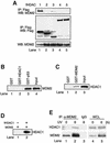
Fig. 1. Interaction between MDM2 and HDAC1. (A) 293T cells were cotransfected with 10 µg of MDM2 and 5–10 µg of either Flag-tagged HDAC1 (lane 1), HDAC2 (lane 2), HDAC3 (lane 3), HDAC4 (lane 4) or HDAC5 (lane 5). Cellular extracts were immunoprecipitated with anti-Flag antibody followed by immunoblotting with anti-MDM2 antibody (top panel) or anti-Flag antibody (middle panel). Total MDM2 protein was detected with anti-MDM2 antibody (bottom panel). (B) Either GST (lane 1), GST–HDAC1 (lane 2) or GST–p53 (lane 3) were incubated with recombinant MDM2 protein followed by immunoblotting with anti-MDM2 antibody. (C) H1299 cells were transfected with Flag-tagged HDAC1 and the cellular extracts were incubated with either GST (lane 1) or GST–MDM2 (lane 2) followed by immunoblotting with anti-Flag antibody. (D) H1299 cells were transfected with MDM2 alone (lane 1) or cotransfected with Flag-tagged HDAC1 (lane 2) and cellular extracts were immunoprecipitated with anti-MDM2 antibody followed by immunoblotting with anti-Flag antibody. (E) A549 cells were exposed to UV-B irradiation (75 J/m2) for 6 h and cellular extracts were immunoprecipitated with either anti-MDM2 antibody (lanes 1 and 2) or mouse IgG as a control (lane 3) followed by immunoblotting with anti-HDAC1 antibody (top panel) or anti-MDM2 antibody (bottom panel). Total HDAC1 and MDM2 protein were detected with either anti-HDAC1 antibody (top panel, lanes 4 and 5) or anti-MDM2 antibody (bottom panel, lanes 4 and 5).
None
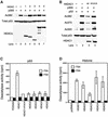
Fig. 2. Deacetylation of p53 by HDAC1. (A) 293T cells were transfected with an empty vector (lane 1) or cotransfected with either 2 µg of p300 alone (lane 2) or cotransfected with 2 µg of Flag-tagged HDAC1 (lane 3), 2 µg of HDAC2 (lane 4), 2 µg of HDAC3 (lane 5), 4 µg of HDAC4 (lane 6) or 4 µg of HDAC5 (lane 7). The levels of endogenous acetylated p53, total p53, and each HDAC were detected by immunoblotting with anti-acetyl ated p53 (Lys382) (top panel), anti-p53 antibody (middle panel) and anti-Flag antibody (bottom panel). (B) H1299 cells (p53–/–) were transfected with 0.2 µg of p53 alone (lane 1) or cotransfected with either 2 µg of p300 alone (lane 2), with 2 µg of HDAC1 wild-type (lane 3) or 2 µg of enzyme-dead H141A mutant (lane 4). The level of acetylated p53 was assessed using either antibody specific for acetylated Lys382 (Ac382), acetylated Lys373 (Ac373) or acetylated Lys320 (Ac320). The levels of total p53 and Flag-tagged HDAC1 were detected as described in (A). (C and D) 293T cells were transfected with empty vector or Flag-tagged HDACs and cellular extracts were prepared as described in Materials and methods. Deacetylase activity was measured against acetylated GST–p53 (C) or histone H4 peptide (D) in the presence or absence of TSA. Results are representative of three independent experiments. Note that all HDAC family members possess deacetylase activity towards histones, but only HDAC1 can efficiently deacetylate p53.
None
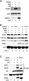
Fig. 3. HDAC1 and MDM2 work cooperatively to deacetylate p53. (A) MEF (p53–/–;MDM2–/–) cells were transfected with 0.1 µg of p53 (lane 1), or cotransfected either with 2 µg of p300 alone (lane 2) or with p300 and 2 µg of Flag-tagged HDAC1 (lane 3). The level of total p53 (middle panel) and acetylated p53 (top panel) were detected as described in Figure 2. (B) MEF (p53–/–;MDM2–/–) cells were transfected with either 0.1 µg of p53, 2 µg of p300 and 0.5 µg of internal control GFP (lane 1), or cotransfected either with 12.5 ng of Flag-tagged HDAC1 wild-type (lane 2), with 12.5 ng of Flag-tagged HDAC1 H141A mutant (lane 3), with 0.5 µg of MDM2 (lane 4), with MDM2 and 12.5 ng of Flag-tagged HDAC1 wild-type (lane 5) or 0.5 µg of MDM2 and 12.5 ng of Flag-tagged HDAC1 H141A mutant (lane 6). The levels of indicated proteins were determined by immunoblotting. Of note, we used 160× the amount of Flag-tagged HDAC1 expression vectors in (A) when compared with (B). (C) MEF (p53–/–;MDM2–/–) cells were transfected with either 1 µg of Flag-tagged HDAC1 wild-type alone, or cotransfected either with 0.3 µg of p53, with 0.3 µg of p53 and 4µg of MDM2 wild-type, or 0.3 µg of p53 and 4µg of p53-binding-deficient MDM2 mutant (Δ58–92). Cells were also treated 24 h post-transfection with the protease inhibitor LLnV (10 µM) for 4 h to inhibit MDM2-mediated p53 degradation. Cellular extracts were immunoprecipitated with anti-goat p53 antibody followed by immunoblotting with Flag antibody (top panel), anti-MDM2 antibody (second panel) or anti-p53 antibody (third panel). Total HDAC1 and MDM2 protein were detected with either anti-Flag antibody (fourth panel) or anti-MDM2 antibody (bottom panel). Of note, the interaction between p53 and p53-binding-deficient MDM2 mutant (Δ58–92) is likely mediated by HDAC1 through the ternary complex formation (second panel, lane 4).
None
Fig. 4. MDM2 induces p53 deacetylation through HDAC1. H1299 cells were transfected with 0.2 µg of p53 wild-type and 0.5 µg of GFP (all lanes), and cotransfected with one or more of the following; 2 µg of p300 (lanes 2–7), 2 µg of MDM2 (lanes 3, 6 and 7), 2 µg of Flag-tagged HDAC1 wild-type (lanes 4 and 6), 2 µg of Flag-tagged HDAC1 H141A mutant (lanes 5 and 7). The levels of indicated proteins were determined by immunoblotting.
None
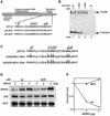
Fig. 5. Mutations of lysine residues of acetylation sites prevent MDM2-mediated p53 degradation. (A) Schematic structure of p53 mutations of C-terminal lysines to alanines or arginines. (B) GST–p53 wild-type (lane 1), GST–p53 3KA mutant with mutated lysine residues 320, 373 and 382 to alanine residues (lane 2), or GST–p53 6KR mutant with mutated lysine residues 320, 370, 372, 373, 381 and 382 to arginine residues (lane 3) were acetylated by recombinant CBP in the presence of the [ 14C]acetyl-CoA and analyzed by SDS–PAGE followed by autoradiograph. Acetylated p53 and CBP are indicated by arrows. As a negative control, wild-type GST–p53 was incubated without recombinant CBP in the presence of the [14C]acetyl-CoA (lane 4). (C) Acetylation sites of p53 overlap with ubiquitylation sites. The lysine residues susceptible to acetylation and ubiquitylation in the C-terminus of p53 are indicated by arrows. (D) H1299 cells were transfected with 0.5 µg of an expression plasmid encoding GFP as an internal control, 0.2 µg of wild-type p53 or p53 6KR mutant, together with empty pcDNA3 vector or the indicated amount of MDM2 vector. Thirty-six hours after transfection, cell extracts were prepared and analyzed by immunoblotting with anti-p53, anti-MDM2 and anti-GFP antibodies. (E) The band intensity of p53 and GFP protein levels was measured with NIH imaging software. p53 (empty circles) and 6KR mutant (filled circles) levels were normalized to GFP levels and were set to 1 in the absence of MDM2.
None
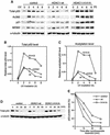
Fig. 6. Effect of HDAC1 on p53 acetylation, stability and activity in response to DNA damage. (A) NIH 3T3 cells infected with mock vector (control), pBabe-HDAC1 wild-type (HDAC1-wt), or pBabe-HDAC1 H141A mutant (HDAC1 H141A) were exposed to UV-B (75 J/m2). Cells were harvested at the indicated times. The levels of total p53 (panel 1), acetylated p53 (panel 2), p21 (panel 3), MDM2 (panel 4) and the internal control α-tubulin (panel 5) were assessed by immunoblotting. (B and C) The band intensity of p53, acetylated p53 and α-tubulin protein levels in all three cell lines were measured with NIH imaging software. The levels of p53 (B) and acetylated p53 (C) were normalized to α-tubulin and the highest intensity levels of p53 or acetylated p53 were set to 1. (B) and (C) are representative results of three (B) and two (C) independent experiments. (D) All three stable cell lines were exposed to UV-B (75 J/m2) and 2 h post-irradiation, cyclohexamide (10 µg/ml) was added to inhibit new p53 protein synthesis (designated 0 h). Cells were harvested at the time points indicated after cyclohexamide treatment. The level of total p53 (upper panel) and α-tubulin (lower panel) was determined. (E) The band intensity of p53 and α-tubulin protein levels were measured by NIH imaging software. p53 levels were normalized to α-tubulin levels and calculated against the amount of p53 present at time point 0, which was set at 100%. Results are representative of three independent experiments.
None
Fig. 7. A model for the regulation and interplay of p53 acetylation and ubiquitylation. Under unstressed conditions, p53 is ubiquitylated on lysine residues by MDM2 and targeted for degradation (A). Upon its activation by various cellular insults, p53 becomes acetylated by p300/CBP at the same set of lysine residues also targeted by MDM2 (B). Acetylation on lysine residues thus prevents MDM2-mediated ubiquityl ation and leads to p53 stabilization (C). The stabilized p53 functions as a tumor suppressor and also induces MDM2 (D). The high level of MDM2, in turn, recruits the p53 deacetylase HDAC1 and triggers p53 deacetylation (E). The deacetylated p53 with unmodified lysine residues is now ready to be ubiquitylated by MDM2 and ultimately degraded.

Similar articles

Cited by

References

    1. Appella E. and Anderson,C.W. (2001) Post-translational modifications and activation of p53 by genotoxic stresses. Eur. J. Biochem., 268, 2764–2772. - PubMed
    1. Ashcroft M., Taya,Y. and Vousden,K.H. (2000) Stress signals utilize multiple pathways to stabilize p53. Mol. Cell. Biol., 20, 3224–3233. - PMC - PubMed
    1. Barlev N.A., Liu,L., Chehab,N.H., Mansfield,K., Harris,K.G., Halazonetis,T.D. and Berger,S.L. (2001) Acetylation of p53 activates transcription through recruitment of coactivators/histone acetyltransferases. Mol. Cell, 8, 1243–1254. - PubMed
    1. Boyd S.D., Tsai,K.Y. and Jacks,T. (2000) An intact HDM2 RING-finger domain is required for nuclear exclusion of p53. Nat. Cell Biol., 2, 563–568. - PubMed
    1. Canman C.E., Lim,D.S., Cimprich,K.A., Taya,Y., Tamai,K., Sakaguchi,K., Appella,E., Kastan,M.B. and Siliciano,J.D. (1998) Activation of the ATM kinase by ionizing radiation and phosphorylation of p53. Science, 281, 1677–1679. - PubMed

Publication types

MeSH terms

Substances