. 2002 Nov 1;111(3):319-30.
doi: 10.1016/s0092-8674(02)01047-4.

The Drosophila Wnt, wingless, provides an essential signal for pre- and postsynaptic differentiation

Affiliations

The Drosophila Wnt, wingless, provides an essential signal for pre- and postsynaptic differentiation

Mary Packard et al. Cell. .

Abstract

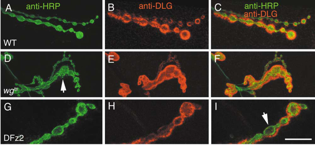
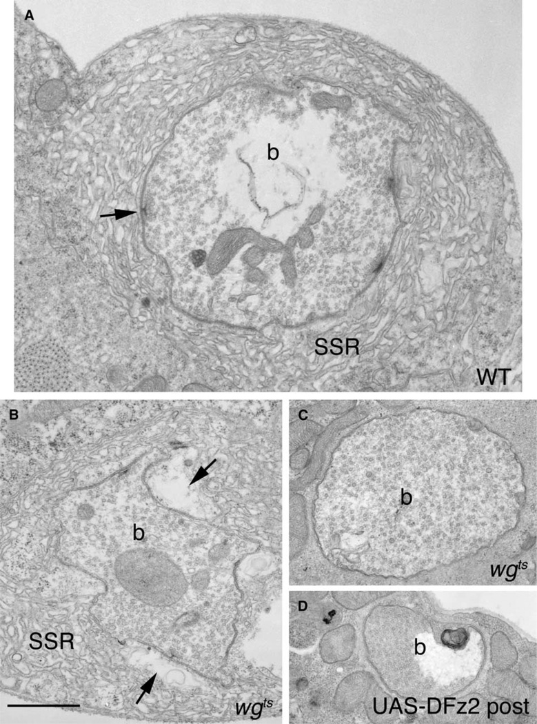
At vertebrate neuromuscular junctions (NMJs), Agrin plays pivotal roles in synapse development, but molecules that activate synapse formation at central synapses are largely unknown. Members of the Wnt family are well established as morphogens, yet recently they have also been implicated in synapse maturation. Here we demonstrate that the Drosophila Wnt, Wingless (Wg), is essential for synapse development. We show that Wg and its receptor are expressed at glutamatergic NMJs, and that Wg is secreted by synaptic boutons. Loss of Wg leads to dramatic reductions in target-dependent synapse formation, and new boutons either fail to develop active zones and postsynaptic specializations or these are strikingly aberrant. We suggest that Wg signals the coordinated development of pre- and postsynaptic compartments.


Figures

Figure 1
Figure 1. Wg and its Receptor, DFz2, Are Localized at the NMJ
(A–C) NMJs at muscles 12 and 13 double-stained with (A) anti-Wg and (B) anti-HRP to stain the presynaptic arbor. (C) Merged Wg and HRP images show that Wg is enriched at type Ib endings. (D–F) NMJs at muscles 12 and 13 double-stained with (D) anti-Wg and (E) anti-DLG, which stains primarily the postsynaptic muscle region of type I boutons. (F) Merged Wg and DLG images show that Wg occupies much of the DLG positive region. Insets: high magnification views of single confocal slices through type Ib boutons. (G–I) NMJs labeled with (G) anti-DFz2, (H) anti-DLG, and (I) both merged to show their colocalization. Insets correspond to high magnification views of a single confocal slice through type Ib boutons. Calibration bar is 50 µm in (A–I) and 12 µm in insets.
Figure 2
Figure 2. Wg Is Secreted by Type Ib Boutons and Endocytosed by Muscles
Images show single confocal slices of (A and G) wild-type NMJs at muscles 6 and 7 double-stained with (A) anti-Wg and anti-HRP, and (G) anti-Wg and anti-CSP. (B and H) NMJs of larvae with presynaptically driven Wg double-stained with (B) anti-Wg and anti-HRP, and (H) anti-Wg and anti-CSP. Note the postsynaptic buildup of Wg. (C and I) NMJs in Wg secretion-defective porc mutants double labeled with (C) anti-Wg and anti-HRP, and (I) anti-Wg and anti-CSP. (D and J) NMJs in temperature-shifted wgts mutants double-stained with (D) anti-Wg and anti-HRP, and (J) anti-Wg and anti-CSP. Note that postsynaptic Wg is dramatically reduced in both porc and wgts mutants. (E and K) NMJs in temperature-shifted shits1 double-stained with (E) anti-Wg and anti-HRP, and (K) anti-Wg and anti-CSP. Note the significant increase in postsynaptic Wg in this mutant. (F) Merged anti-HRP and GFP at an NMJ of a wg-Gal4 larva driving expression of Wg-GFP. (M) NMJ of a larva where UAS-Wg was expressed in muscle, double-stained with anti-Wg and anti-HRP. Note the accumulation of Wg around muscle nucleus (n), whereas levels of Wg at the NMJ are similar to wild-type. (N) Summary of the distribution of Wg (green) at the NMJ of the various mutants shown in diagrammatic form. (L and O) Single confocal slices through the dorsal region of the CNS showing motorneurons in (L) a C380 X UAS-LacZ larva double-stained with anti-Wg and anti-βgal, and (O) a wild-type larva double-stained with anti-Wg and anti-glutamate. Arrow indicates the CNS midline; anterior is left. Calibration bar is 20 µm in (A–K), (M), and 26 µm in (L and O).
Figure 3
Figure 3. Synaptic Bouton Number and the Proportion of Boutons Containing Unbundled Cytoskeleton Are Altered in wgts Mutants
(A) Number of type Ib, Is, and total boutons in temperature-shifted wild-type, wgts, and wgts expressing Wg presynaptically (red) determined in preparations double-stained with Wg and HRP antibodies. N = 18 for CS, 15 for wgts, and 16 for rescue. (B) Bouton number in non-heat pulsed wild-type and larvae overexpressing Wg presynaptically (pre-Wg+). N = 9 for CS and 11 for pre-Wg+. (C) Number of Wg positive boutons, and the percentage of Wg positive boutons containing unbundled (unbund), looped (loop), or both splayed and punctate Futsch. Measurements were made at segments A2 and A3. For Futsch measurements N = 26 for CS A2, 20 for CS A3, 19 for wgts A2, 20 for wgts A3, 9 for rescue A2, and 9 for rescue A3. ** represent p < 0.0001; * represents p < 0.001. (D and F) wild-type NMJs stained with (D) anti-Futsch and (F) double-stained with anti-Futsch and anti-Wg antibodies. (E and G) wgts NMJs stained as above. In (D) and (E), boutons containing unbundled Futsch have been slightly overexposed (green squares) to facilitate visibility of less intense signal. Arrowheads (F and G) point to boutons with unbundled Futsch; t indicates trachea, which also stain with anti-Wg. (H–J) Examples of unbundled Futsch filaments showing (H) a loop, (I) splayed filaments, and (J) diffuse and punctate Futsch. Calibration bar is 30 µm in (D–G), and 5 µm in (H–J).
Figure 4
Figure 4. Synaptic Morphology and DLG Distribution Are Altered in wgts Mutants
(A–C) Wild-type type I boutons double-stained with (A) anti-HRP, (B) anti-DLG, and (C) both merged. (D–F) Boutons in wgts double labeled as above. Note the abnormally shaped type I boutons in wgts mutants (arrow), and that unlike wild-type, DLG surrounds groups of boutons in the mutant. (G–I) Type Ib boutons in a strain overexpressing DFz2 in postsynaptic muscles stained as above. (I), (G), and (H) merged to show abnormal lack of DLG in areas surrounding boutons (arrow). A similar phenotype is observed in wgts mutants (see Figure 5). Calibration bar = 10 µm.
Figure 5
Figure 5. Glutamate Receptor Distribution Is Altered in wgts Mutants
(A–C) Low and (D–F) high magnification of wild-type boutons stained with (A and D) anti-DGluRIIA, (B and E) anti-DLG, and (C and F) both merged. Note the presence of DGluRIIA immunoreactive clusters, some of which have a doughnut-like appearance (arrow in D). (G–I) Low and (J–L) high magnification of boutons in wgts double labeled as above. Note that in wgts mutants, GluRIIA label appears diffuse and both the regularity and shape of immunoreactive clusters are aberrant. Arrow in (L) points to a lack of DGluRIIA in a bouton bud. (M–O) porc mutant NMJ triple labeled with anti-HRP (Blue), anti-DGluRIIA (Green), and anti-DLG (Red). (M) shows DGluRIIA and HRP labels, (N) shows DLG and HRP labels, and (O) shows the triple labeling. Note that both DLG and DGluRIIA are absent from almost an entire side of this string of boutons (arrow), but appear normal at the other side. Calibration bar = 10 µm in (A–C), (G–I), (M–O), and 4 µm in (D–F), (J–L).
Figure 6
Figure 6. Ultrastructure of Synaptic Boutons Is Abnormal in wg Mutants and in Larvae Overexpressing Dfz2 Postsynaptically
Electron micrographs of type Ib boutons in (A) wild-type, (B and C) wgts mutants, and (D) in a strain overexpressing DFz2 in the postsynaptic muscles (UAS-DFz2 post). Note boutons lacking SSR, active zones, and mitochondria. In (B), a wgts bouton with abnormally enlarged pockets in the postsynaptic region apposed to active zones (compare arrows in A and B) is shown. b = bouton, SSR = Subsynaptic Reticulum. Calibration bar is 1.5 µm.
Figure 7
Figure 7. Active Zones and Postsynaptic Region Are Altered in wgts Mutants
(A and D) High and (B, C, E, and F) lower magnification views of active zones in (A–C) wild-type, and a range of aberrant morphologies of the mutant active zones. In (E) is a dramatically enlarged pocket (asterisk) apposed to an active zone in the mutant. (G and H) Two models on the likely operation of Wg during the development of the NMJ. (G) According to this model, the Wg receptor, DFz2, is localized both pre- and postsynaptically and interacts with Wg secreted by the presynaptic bouton. This triggers independent pre- and postsynaptic transduction cascades leading to the proper positioning and morphology of active zones and to proper development of the postsynaptic apparatus. In (H), DFz2 is localized exclusively at the postsynaptic junctional region. The interaction of DFz2 with Wg, results in proper formation and positioning of postsynaptic apparatus and elicits an unidentified retrograde message that signals the proper formation and positioning of active zones. In both models, secreted Wg is removed from the membrane by endocytosis. Calibration bar is 0.2 µm in (A and D) and 0.5 µm in (B, C, E, and F).

Similar articles

Cited by

References

    1. Aberle H, Haghighi AP, Fetter RD, McCabe BD, Magalhaes TR, Goodman CS. Wishful thinking encodes a BMP type II receptor that regulates synaptic growth in Drosophila. Neuron. 2002;33:545–558. - PubMed
    1. Binari RC, Staveley BE, Johnson WA, Godavarti R, Sasisekharan R, Manoukian AS. Genetic evidence that heparin-like glycosaminoglycans are involved in wingless signaling. Development. 1997;124:2623–2632. - PubMed
    1. Budnik V, Koh YH, Guan B, Hartmann B, Hough C, Woods D, Gorczyca M. Regulation of synapse structure and function by the Drosophila tumor suppressor gene dlg. Neuron. 1996;17:627–640. - PMC - PubMed
    1. Burden SJ. Wnts as retrograde signals for axon and growth cone differentiation. Cell. 2000;100:495–497. - PubMed
    1. Cadigan KM, Fish MP, Rulifson EJ, Nusse R. Wingless repression of Drosophila frizzled 2 expression shapes the wingless morphogen gradient in the wing. Cell. 1998;93:767–777. - PubMed

Publication types

LinkOut - more resources