. 2002 Dec;76(23):11853-65.
doi: 10.1128/jvi.76.23.11853-11865.2002.

RNA incorporation is critical for retroviral particle integrity after cell membrane assembly of Gag complexes

Affiliations

RNA incorporation is critical for retroviral particle integrity after cell membrane assembly of Gag complexes

Shainn-Wei Wang et al. J Virol. 2002 Dec.

Abstract

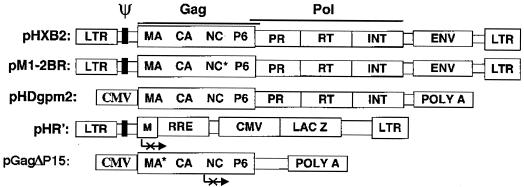
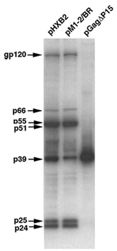
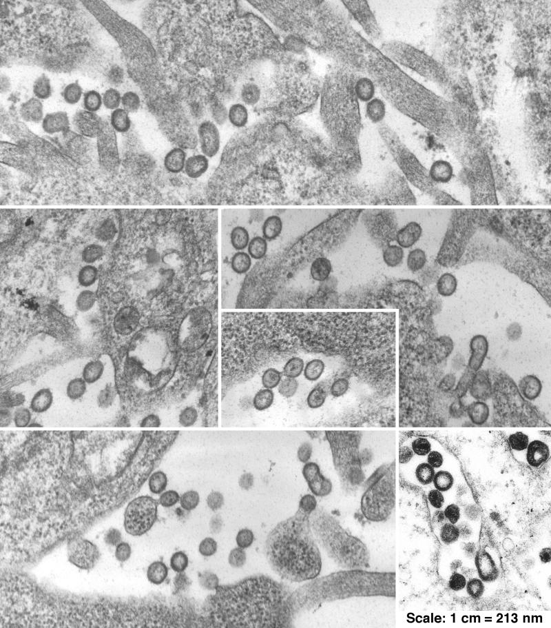
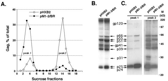
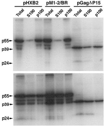
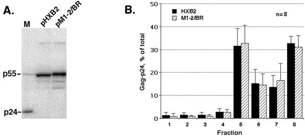
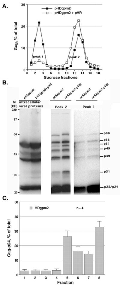
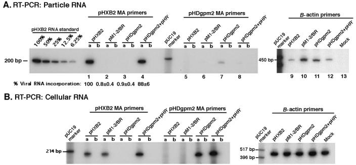
The nucleocapsid (NC) domain of retroviruses plays a critical role in specific viral RNA packaging and virus assembly. RNA is thought to facilitate viral particle assembly, but the results described here with NC mutants indicate that it also plays a critical role in particle integrity. We investigated the assembly and integrity of particles produced by the human immunodeficiency virus type 1 M1-2/BR mutant virus, in which 10 of the 13 positive residues of NC have been replaced with alanines and incorporation of viral genomic RNA is virtually abolished. We found that the mutations in the basic residues of NC did not disrupt Gag assembly at the cell membrane. The mutant Gag protein can assemble efficiently at the cell membrane, and viral proteins are detected outside the cell as efficiently as they are for the wild type. However, only approximately 10% of the Gag molecules present in the supernatant of this mutant sediment at the correct density for a retroviral particle. The reduction of positive charge in the NC basic domain of the M1-2/BR virus adversely affects both the specific and nonspecific RNA binding properties of NC, and thus the assembled Gag polyprotein does not bind significant amounts of viral or cellular RNA. We found a direct correlation between the percentage of Gag associated with sedimented particles and the amount of incorporated RNA. We conclude that RNA binding by Gag, whether the RNA is viral or not, is critical to retroviral particle integrity after cell membrane assembly and is less important for Gag-Gag interactions during particle assembly and release.


Figures

FIG. 1.
FIG. 1.
Schematic representation of the constructs. pHXB2gpt and the pM1-2/BR mutant are, respectively, a wild-type HIV provirus and an HIV provirus with 10 mutations in NC (34, 45). pHDgpm2 expresses HIV Gag and Gag-Pol polyproteins from a sequence whose codons were optimized according to the human codon usage. Although encoding the same protein, this gag sequence is 24% different from the original viral sequence. pHDgpm2 does not contain an HIV-1 packaging sequence, and gag gene expression is driven by the CMV promoter. pHR′ contains the HIV-1 cis-acting sequences necessary for packaging (41). pGagΔ15 expresses an HIV-1 Gag truncated product (stop codon at the end of p2) in a Rev-independent fashion (31). LTR, long terminal repeat.
FIG. 2.
FIG. 2.
Analysis of intracellular viral protein accumulation. Equal amounts of pHXB2, pM1-2/BR, and pGagΔ15 cell protein lysates were analyzed by Western blotting, using an HIV-positive human antiserum. The Gag precursor in the pGagΔ15 mutant is approximately 40 kDa because the NC, p1, and p6 domains are deleted.
FIG. 3.
FIG. 3.
EM of M1-2/BR viral particles. The panels show particles from multiple fields of pM1-2/BR-transfected cells. The last panel at the bottom on the right shows wild-type HIV-1 from pHXB2. Magnification, ×47,000.
FIG. 4.
FIG. 4.
Sucrose gradient and Western blot analyses of HXB2 and M1-2/BR viral particles. (A) Sucrose gradient fractionation of extracellular Gag. Gag protein amounts in each fraction were measured by p24 ELISA and expressed as percentage of the total p24-assocated Gag harvested from the gradient. The densities (in grams per milliliter) of p1 and p2 were 1.03 ± 0.03 and 1.61 ± 0.01 for HXB2 and 1.03 ± 0.01 and 1.15 ± 0.02 for M1-2/BR, respectively. (B) Western blots of pHXB2 and pM1-2/BR cell lysates. HIV-1 proteins were detected with an HIV-positive human serum. (C) Protein profiles of HXB2 and M1-2/BR sucrose fractions corresponding to p1 and p2. M, markers.
FIG. 5.
FIG. 5.
Cellular fractionation of transfected-cell lysates. The S100 and P100 fractions were obtained after ultracentrifugation of the clarified cell lysates. (A) Transfected cells from the individual transfection were lysed in hypotonic buffer in the absence of detergent. (B) Transfected cells were lysed in hypotonic buffer in the presence of Triton X-100.
FIG. 6.
FIG. 6.
Gradient fractionation of intracellular HXB2 and M1-2/BR Gag. (A) Western blotting of total HXB2 and M1-2/BR cell lysates. Lane M, markers. The presence of Gag-related bands smaller than Pr55 could be due to less-than-optimal inhibition of the HIV protease and/or internal initiation at the capsid Met142 (8). (B) Floatation of Triton X-100-extracted cell protein lysates in an eight-fraction Optiprep gradient. The histogram illustrates the results derived from the Western blot and ELISA analyses of the fractions of eight independent gradients. Error bars indicate standard deviations.
FIG. 7.
FIG. 7.
Analysis of mosaic particles composed of HXB2 and M1-2/BR Gag molecules. In each panel lane a corresponds to a sample derived from the transfection of pHXB2 alone; lanes b, c, d, and e correspond to samples derived from the transfection of pHXB2 and pM1-2/BR at ratios of 4:1, 3:2, 2:3, and 1:4, respectively; and lane f shows the analysis of a sample derived from the transfection of pM1-2/BR alone. (A) Western blot analysis of intracellular viral proteins. (B) Western blot analysis of mosaic viral particles. Particles were pelleted through a 20% sucrose cushion after supernatant clarification. An amount of supernatant corresponding to 100 ng of p24 was used in the Western blotting of protease K-treated samples (lanes 1 to 6) and of Triton X-100- and protease K-treated samples (controls, lanes 7 to 12). (C) Relative stability of mosaic particles. The histogram shows the percentage of pellet-associated Gag and of protease K-resistant Gag present in each supernatant derived from the cotransfection of different amounts of pHXB2 and pM1-2/BR. The percentage of particle-associated Gag was calculated relative to the amount of Gag present in the untreated supernatant before centrifugation. Gag associated with protease K-resistant particles was evaluated directly in the supernatant after protease K treatment. Three independent experiments were carried out and each supernatant was tested in duplicate. Error bars indicate standard errors. (D) RT-PCR analysis of viral genomic RNA incorporation in pelleted viral particles. Standards for viral genomic RNA were derived from twofold dilutions of the HXB2 RNA samples. The results of one representative experiment are shown here (for each sample, the first lane corresponds to an RNA sample that was subjected to RT-PCR and the second corresponds to an RNA samples that was subjected to PCR only). The percentage of RNA incorporation for each sample was evaluated by comparison to the standards in four independent experiments and is reported with its standard error at the bottom.
FIG. 8.
FIG. 8.
Gradient analysis of VLPs (pHDgpm2) and VLPs produced in presence of a Ψ RNA (pHDgpm2+pHR′). (A) Gag distribution in sucrose gradient fractions. The densities (in grams per milliliter) of p1 and p2 were 1.03 and 1.16 for the peak fractions of the gradient loaded with particles from the pHDgpm2 transfection and 1.04 and 1.16 for the peak fractions of the gradient loaded with particles from the pHDgpm2+pHR′ transfection, respectively. This analysis was repeated three times with comparable results. (B) Western blots of pHDgpm2 and pHDgpm2+pHR′cellular lysates. Sucrose fraction aliquots corresponding to 80 ng (peak 2) and 40 ng (peak 1) of p24 were analyzed. M, markers. (C) Floatation assay of Triton X-100-extracted pHDgpm2 cell proteins in an eight-fraction Optiprep. The histogram illustrates the results derived from the Western blot and ELISA analyses of the fractions of four independent gradients. Error bars indicate standard deviations.
FIG. 9.
FIG. 9.
RNA incorporation in wild-type and mutant viral particles. (A) RT-PCR of particle-derived RNA. The results of one representative experiment are shown. The mean and the standard error of the Ψ RNA content in samples analyzed in duplicate from three independent experiments are reported under lanes 1 to 4 (HIV-1 MA-specific primers). Detection of RNA carried out with pHDgpm2-specific primers (lanes 5 to 8) and with β-actin-specific primers (lanes 9 to 13) is also shown. Lanes a, RNA samples subjected to RT-PCR; lanes b, RNA samples subjected to PCR only. (B) RT-PCR of cellular RNA. Cellular RNA samples equivalent to equal amounts of total RNA were used in RT-PCRs with HIV-1 MA-, HDgpm2-, or actin-specific primers. Transfection efficiencies were comparable for all of the plasmids. Lanes a, RNA samples subjected to RT-PCR; lanes b, RNA samples subjected to PCR only.

Similar articles

Cited by

References

    1. Accola, M. A., B. Strack, and H. G. Gottlinger. 2000. Efficient particle production by minimal Gag constructs which retain the carboxy-terminal domain of human immunodeficiency virus type 1 capsid-p2 and a late assembly domain. J. Virol. 74:5395-5402. - PMC - PubMed
    1. Aldovini, A., and R. A. Young. 1990. Mutations of RNA and protein sequences involved in human immunodeficiency virus type 1 packaging result in production of noninfectious virus. J. Virol. 64:1920-1926. - PMC - PubMed
    1. Arya, S. K., C. Guo, S. F. Josephs, and F. Wong-Staal. 1985. Trans-activator gene of human T-lymphotropic virus type III (HTLV-III). Science 229:69-73. - PubMed
    1. Bennett, R. P., T. D. Nelle, and J. W. Wills. 1993. Functional chimeras of the Rous sarcoma virus and human immunodeficiency virus Gag proteins. J. Virol. 67:6487-6498. - PMC - PubMed
    1. Berkowitz, R., J. Fisher, and S. P. Goff. 1996. RNA packaging. Curr. Top. Microbiol. Immunol. 214:177-218. - PubMed

Publication types

LinkOut - more resources