Intraneuronal Alzheimer abeta42 accumulates in multivesicular bodies and is associated with synaptic pathology
- PMID: 12414533
- PMCID: PMC1850783
- DOI: 10.1016/s0002-9440(10)64463-x
Intraneuronal Alzheimer abeta42 accumulates in multivesicular bodies and is associated with synaptic pathology
Abstract
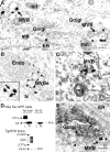
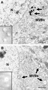
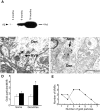
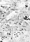
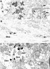
A central question in Alzheimer's disease concerns the mechanism by which beta-amyloid contributes to neuropathology, and in particular whether intracellular versus extracellular beta-amyloid plays a critical role. Alzheimer transgenic mouse studies demonstrate brain dysfunction, as beta-amyloid levels rise, months before the appearance of beta-amyloid plaques. We have now used immunoelectron microscopy to determine the subcellular site of neuronal beta-amyloid in normal and Alzheimer brains, and in brains from Alzheimer transgenic mice. We report that beta-amyloid 42 localized predominantly to multivesicular bodies of neurons in normal mouse, rat, and human brain. In transgenic mice and human Alzheimer brain, intraneuronal beta-amyloid 42 increased with aging and beta-amyloid 42 accumulated in multivesicular bodies within presynaptic and especially postsynaptic compartments. This accumulation was associated with abnormal synaptic morphology, before beta-amyloid plaque pathology, suggesting that intracellular accumulation of beta-amyloid plays a crucial role in Alzheimer's disease.
Figures
Similar articles
-
Oligomerization of Alzheimer's beta-amyloid within processes and synapses of cultured neurons and brain.J Neurosci. 2004 Apr 7;24(14):3592-9. doi: 10.1523/JNEUROSCI.5167-03.2004. J Neurosci. 2004. PMID: 15071107 Free PMC article.
-
Intraneuronal Aβ accumulation, amyloid plaques, and synapse pathology in Alzheimer's disease.Neurodegener Dis. 2012;10(1-4):56-9. doi: 10.1159/000334762. Epub 2012 Jan 21. Neurodegener Dis. 2012. PMID: 22269167
-
Heterogeneous Association of Alzheimer's Disease-Linked Amyloid-β and Amyloid-β Protein Precursor with Synapses.J Alzheimers Dis. 2017;60(2):511-524. doi: 10.3233/JAD-170262. J Alzheimers Dis. 2017. PMID: 28869466 Free PMC article.
-
Plaque formation and the intraneuronal accumulation of β-amyloid in Alzheimer's disease.Pathol Int. 2017 Apr;67(4):185-193. doi: 10.1111/pin.12520. Epub 2017 Mar 5. Pathol Int. 2017. PMID: 28261941 Review.
-
High resolution approaches for the identification of amyloid fragments in brain.J Neurosci Methods. 2019 May 1;319:7-15. doi: 10.1016/j.jneumeth.2018.10.032. Epub 2018 Oct 24. J Neurosci Methods. 2019. PMID: 30367888 Free PMC article. Review.
Cited by
-
Transmissible Endosomal Intoxication: A Balance between Exosomes and Lysosomes at the Basis of Intercellular Amyloid Propagation.Biomedicines. 2020 Aug 4;8(8):272. doi: 10.3390/biomedicines8080272. Biomedicines. 2020. PMID: 32759666 Free PMC article. Review.
-
Enhanced β-secretase processing alters APP axonal transport and leads to axonal defects.Hum Mol Genet. 2012 Nov 1;21(21):4587-601. doi: 10.1093/hmg/dds297. Epub 2012 Jul 27. Hum Mol Genet. 2012. PMID: 22843498 Free PMC article.
-
APOE in the bullseye of neurodegenerative diseases: impact of the APOE genotype in Alzheimer's disease pathology and brain diseases.Mol Neurodegener. 2022 Sep 24;17(1):62. doi: 10.1186/s13024-022-00566-4. Mol Neurodegener. 2022. PMID: 36153580 Free PMC article. Review.
-
Multi-target action of the novel anti-Alzheimer compound CHF5074: in vivo study of long term treatment in Tg2576 mice.BMC Neurosci. 2013 Apr 5;14:44. doi: 10.1186/1471-2202-14-44. BMC Neurosci. 2013. PMID: 23560952 Free PMC article.
-
Analyzing dendritic spine pathology in Alzheimer's disease: problems and opportunities.Acta Neuropathol. 2015 Jul;130(1):1-19. doi: 10.1007/s00401-015-1449-5. Epub 2015 Jun 11. Acta Neuropathol. 2015. PMID: 26063233 Free PMC article. Review.
References
-
- Selkoe DJ: Alzheimer’s disease: genes, proteins, and therapy. Physiol Rev 2001, 81:741-766 - PubMed
-
- Wilson CA, Doms RW, Lee VM: Intracellular APP processing and A beta production in Alzheimer disease. J Neuropathol Exp Neurol 1999, 58:787-794 - PubMed
-
- Hartmann T: Intracellular biology of Alzheimer’s disease amyloid beta peptide. Eur Arch Psychiatry Clin Neurosci 1999, 249:291-298 - PubMed
-
- Holcomb L, Gordon MN, McGowan E, Yu X, Benkovic S, Jantzen P, Wright K, Saad I, Mueller R, Morgan D, Sanders S, Zehr C, O’Campo K, Hardy J, Prada CM, Eckman C, Younkin S, Hsiao K, Duff K: Accelerated Alzheimer-type phenotype in transgenic mice carrying both mutant amyloid precursor protein and presenilin 1 transgenes. Nat Med 1998, 4:97-100 - PubMed
Publication types
MeSH terms
Substances
Grants and funding
LinkOut - more resources
Full Text Sources
Other Literature Sources
Medical
Molecular Biology Databases

