. 2002 Nov 1;30(21):4634-42.
doi: 10.1093/nar/gkf587.

In vivo and in vitro interaction between human transcription factor MOK2 and nuclear lamin A/C

Affiliations

In vivo and in vitro interaction between human transcription factor MOK2 and nuclear lamin A/C

Caroline Dreuillet et al. Nucleic Acids Res. .

Abstract

The human and murine MOK2 proteins are factors able to recognize both DNA and RNA through their zinc finger motifs. This dual affinity of MOK2 suggests that MOK2 might be involved in transcription and post-transcriptional regulation of MOK2 target genes. The IRBP gene contains two MOK2-binding elements, a complete 18 bp MOK2-binding site located in intron 2 and the essential core MOK2-binding site (8 bp of conserved 3'-half-site) located in the IRBP promoter. We have demonstrated that MOK2 can bind to the 8 bp present in the IRBP promoter and repress transcription from this promoter by competing with the CRX activator for DNA binding. In this study, we identify a novel interaction between lamin A/C and hsMOK2 by using the yeast two-hybrid system. The interaction, which was confirmed by GST pull-down assays and co-immunolocalization studies in vivo, requires the N-terminal acidic domain of hsMOK2 and the coiled 2 domain of lamin A/C. Furthermore, we show that a fraction of hsMOK2 protein is associated with the nuclear matrix. We therefore suggest that hsMOK2 interactions with lamin A/C and the nuclear matrix may be important for its ability to repress transcription.


Figures
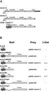
Figure 1
Figure 1
Identification of lamin C as an interaction partner of hsMOK2 by the yeast two-hybrid system and identification of the hsMOK2 interaction domain. (A) Schematic representation of human lamins A and C. Lamins A and C are identical in sequence except that lamin C has a unique 6 amino acid extension at its C-terminus (black box), while lamin A has a 98 amino acid extension (stripped box). Schematic representation of the pGAD-ΔlaminC library vector identified from a HeLa cDNA library on the basis of its ability to activate the LacZ reporter gene in the presence of the LexA binding domain/hsMOK2 hybrid. (B) Constructs expressing full-length or the indicated domains of hsMOK2 and the human polypeptide Δlamin C were co-transformed into yeast. The specificity of the interaction between bait and prey was determined by estimating the degree of color development after incubating the filter for 90 min in the filter lift β-galactosidase assay as described in Materials and Methods. +++, high blue color development; +/–, very low blue color development; –, no color development.
Figure 2
Figure 2
GST pull-down of hsMOK2 by lamin A/C. (A) Schematic representation of the human polypeptide Δlamin C and the three fragments of human lamin A/C expressed in bacteria as fusions with GST. (B) Nuclear extracts (20 µg) from HeLa cells transfected with the expression vector hsMOK2 were incubated with an equal amount (10 µg) of GST or GST–Δlamin C bound to glutathione. Unbound (U) and bound (B) proteins were separated by SDS–PAGE and visualized by immunoblotting with affinity purified anti-hsMOK2 antibody. (C) In vitro 35S-labeled NH2hsMOK2 was incubated with an equal amount (10 µg) of recombinant GST fusion proteins containing the full-length or one of the three different segments of lamin A/C or GST alone bound to glutathione beads. After thoroughly washing the beads, the bound proteins were eluted in SDS sample buffer, resolved by SDS–PAGE and visualized by autoradiography after treatment with 16% sodium salicylate.
Figure 3
Figure 3
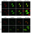
Alteration of the subcellular localization of hsMOK2 by co-transfection with Δlamin C. HeLa cells were transfected with CMV-hsMOK2 (A) or CMV-NH2hsMOK2 (B) or co-transfected with CMV-hsMOK2 and CMV-GST-ΔlaminC (C) or CMV-NH2hsMOK2 and CMV-GST-ΔlaminC (D). Thirty-six hours after transfection, the cells were fixed, permeabilized and double stained sequentially with anti-lamin A/C and anti-hsMOK2 antibodies (A and B) or with anti-GST and anti-hsMOK2 antibodies (C and D). Cells were examined by confocal scanning laser microscopy as described in Materials and Methods. Images correspond to a single confocal section though the middle of the nucleus scanned in the same optical plane. SS overlay corresponds to the single confocal section shown and MP overlay corresponds to maximum projection of all optical sections. In overlay, the co-localization of rhodamine-labeled and fluorescein-labeled structures gives a yellow color. Bar, 10 µm.
Figure 4
Figure 4
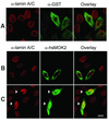
Nuclear translocation of the N-terminal acidic domain of hsMOK2 expressed as a fusion with GST. HeLa cells were transfected with CMV-GST (A) or CMV-GST-NH2hsMOK2 (B) or co-transfected with CMV-GST-NH2hsMOK2 and CMV-GST-ΔlaminC (C). Thirty-six hours after transfection, the cells were fixed, permeabilized and double stained with the indicated antibodies. Cells were examined by confocal scanning laser microscopy as described in Materials and Methods. Images represent a maximum projection of ten 0.284 µm optical sections. In overlay, the co-localization of rhodamine-labeled and fluorescein-labeled structures gives a yellow color. The arrows in (C) show the cells co-transfected with both plasmids. Bar, 10 µm.
Figure 5
Figure 5
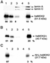
A fraction of hsMOK2 protein is associated with nuclear matrix. HeLa cells transfected with CMV-hsMOK2 (A), CMV-hsMOK2Δ (B) or CMV-NH2hsMOK2 (C) vectors were sequentially extracted as described in Materials and Methods. The various protein fractions correspond to 1% Triton X-100 (fraction 1), DNase I at 37°C and 0.25 M (NH4)2SO4 (fraction 2), 2 M NaCl (fraction 3), RNase A (fraction 4) and, finally, the pellet containing nuclear matrix (fraction 5) resuspended in SDS sample buffer. Four micrograms of proteins from fractions 1–4 and 20 µg of proteins from fraction 5, corresponding to the nuclear matrix, were subjected to SDS–PAGE and western blot analysis. Probing was realized with polyclonal anti-hsMOK2 antibody to detect hsMOK2 and truncated proteins or monoclonal anti-lamin A/C(636) antibody to detect the two nuclear matrix lamins A and C. Western blots were visualized with a Fluor-S Max MultiImager and quantified with Quantity One software (Bio-Rad). The percentages indicated below each panel were normalized at 4 µg of proteins. The molecular weights of the proteins are indicated on the right.
Figure 6
Figure 6
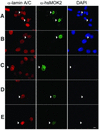
Immunolocalization of hsMOK2 protein in transfected cells following in situ sequential fractionation. HeLa cells were transfected by CMV-hsMOK2 vector. Cells were untreated (A) or submitted to in situ sequential extraction with 0.5% Triton X-100 (B), DNase I at RT (C), 2 M NaCl (D) and RNase A (E) before fixation. Double immunolabeling was performed as described in Materials and Methods using monoclonal anti-lamin A/C and polyclonal anti-hsMOK2 antibodies. All the immunofluorescence photographs were acquired at the same exposure time. Images were printed at the same range of intensities for each color to facilitate comparisons except the image corresponding to untreated cells probed with anti-hsMOK2 antibody (A, green). At the same range of intensities, the image of untreated cells for hsMOK2 was saturated, which is consistent with the observation that a significant fraction of hsMOK2 is released with soluble proteins in western blotting (Fig. 5A). For each field of cells analyzed, DAPI staining is shown. Bar, 10 µm.

Similar articles

Cited by

References

    1. Ernoult-Lange M., Kress,M. and Hamer,D. (1990) A gene that encodes a protein consisting solely of zinc finger domains is preferentially expressed in transformed mouse cells. Mol. Cell. Biol., 10, 418–421. - PMC - PubMed
    1. Ernoult-Lange M., Arranz,V., Leconiat,M., Berger,R. and Kress,M. (1995) Human and mouse Kruppel-like (MOK2) orthologue genes encode two different zinc finger proteins. J. Mol. Evol., 41, 784–794. - PubMed
    1. Arranz V., Harper,F., Florentin,Y., Puvion,E., Kress,M. and Ernoult-Lange,M. (1997) Human and mouse MOK2 proteins are associated with nuclear ribonucleoprotein components and bind specifically to RNA and DNA through their zinc finger domains. Mol. Cell. Biol., 17, 2116–2126. - PMC - PubMed
    1. Fong S.L. and Bridges,C.D. (1990) Interstitial retinol-binding protein: purification, characterization, molecular cloning and sequence. Methods Enzymol., 189, 207–213. - PubMed
    1. Stoykova A. and Gruss,P. (1994) Roles of Pax-genes in developing and adult brain as suggested by expression patterns. J. Neurosci., 14, 1395–1412. - PMC - PubMed

Publication types

MeSH terms