. 2002 Oct 15;21(20):5437-47.
doi: 10.1093/emboj/cdf543.

P21-activated kinase-1 phosphorylates and transactivates estrogen receptor-alpha and promotes hyperplasia in mammary epithelium

Affiliations

P21-activated kinase-1 phosphorylates and transactivates estrogen receptor-alpha and promotes hyperplasia in mammary epithelium

Rui-An Wang et al. EMBO J. .

Abstract

Stimulation of p21-activated kinase-1 (Pak1) induces cytoskeleton reorganization and signaling pathways in mammary cancer cells. Here, we show that inhibition of Pak1 kinase activity by a dominant-negative fragment or by short interference RNA markedly reduced the estrogen receptor-alpha (ER) transactivation functions. To understand the role of Pak1 in mammary glands, we developed a murine model expressing constitutively active Thr423 glutamic acid Pak1 driven by the beta-lactoglobulin promoter. We show that mammary glands from these mice developed widespread hyperplasia associated with apocrine metaplasia and lobuloalveolar hyperdevelopment during lactation. Mammary tissues with active Pak1 also exhibited an increased activation of mitogen-activated protein kinase and stimulated transactivation functions of the ER and expression of endogenous ER target genes. Furthermore, Pak1 directly phosphorylated the activation function-2 domain of the ER at the N-terminal residue Ser305, and its mutation to Ala (S305A) abolished the Pak1-mediated phosphorylation and transactivation functions of the ER, while its mutation to glutamic acid (S305E) promoted transactivation activity of ER. These findings reveal a novel role for the Pak1-ER pathway in promoting hyperplasia in mammary epithelium.


Figures
None
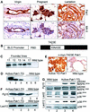
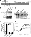
Fig. 1. Pak1 is required for optimum stimulation of the ER pathway. (A) Schematic representation of Pak1. Crib motif, amino acids 70–87 and autoinhibitory domain (AID), amino acids 83–149. (B) Upper three panels show RT–PCR analysis. MCF-7 cells (clones 10 and 17) expressing Pak1 inhibitor, amino acids 83–149, inhibitory fragment. Lower panel shows western blotting and the ER level in MCF-7 cells expressing Pak1 inhibitor. (C) Pak1 inhibitor blocks stimulation of ERE-driven transcription. Cells from clones 10 and 17 were transfected with 0.25 µg of the ERE–luc reporter and treated with or without E2 (1 nM) (n = 4). (D) Dominant-negative Pak1 blocks stimulation of endogenous ER target gene product PR in MCF-7 cells. Stable MCF-7 clones expressing pcDNA or Pak1 inhibitor (clones 10 and 17) were treated with or without E2 for 24 h, and cell lysates were immunoblotted with anti-PR antibodies (n = 3). (E) Growth rates of MCF-7/vector cells and MCF-7 clones 10 and 17 in response to E2 (n = 2).
None
Fig. 2. Knock down of Pak1 by siRNA reduces ER-transactivation functions. (A–D) MCF-7 cells were cotransfected with 10 nM Pak1 siRNA or control Pak1, or GAPDH siRNAs and GFP–Pak1. After 48 h, the expression of exogenous GFP–Pak1 and the endogenous Pak1 was determined by western blotting (A and B) and fluorescence microscopy (C). Upper panel, GFP–Pak1-transfected cells are shown in green (n = 4). (D and E) Reduction in ERE-driven luciferase activity and pS2 gene expression by Pak1-siRNA. MCF-7 cells were cotransfected with 10 nM Pak1 or control siRNA and ERE–luc for 48 h, and treated with or without 1 nM E2 for 16 h before analyzing the status of ERE–luc activity or pS2 mRNA expression (n = 2). Bar = 10 µm.
None
Fig. 3. Generation of active Pak1-TG mice. (A) Expression of Pak1 during various stages of mammary gland development as revealed by immunohistochemistry. The lower panels show myoepithelial cells identified by the mouse keratin-5 marker (MK-5). (B) Structure of the T423E Pak1 transgene. c-myc-tagged T423E-mutated Pak1 was fused to the ovine BLG promoter. PBD, p21-binding domain. (C) Identification of the TG founder mice by Southern blotting. Tail DNA (5 µg) was digested with BamHI, resolved on 0.7% agarose gel and probed with a Pak1 cDNA probe. The transgene band is 2.2 kb. (D) Expression of transgene as determined by RT–PCR followed by Southern blotting. Three Pak1-TG mice on day 12 of lactation and two wild-type mice were analyzed on the same day of lactation. (E) Expression of transgene on day 12 of lactating mammary glands as revealed by immunohistochemistry using anti-c-myc-tag antibody. An age-matched wild-type mammary gland was used as a control. (F) Pak1 kinase activity analysis. Protein lysates from the indicated days of pregnancy or lactation were immunoprecipitated with Pak1 antibody and then in vitro kinase assay was performed using myelin basic protein (MBP) as a substrate. (G) Western blotting of mammary lysates showed an increased level of phosphorylated p38MAPK and MAPK in the Pak1-TG mice, with an unchanged level of total p38 and MAPK. Bar = 40 µm.
None
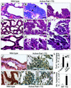
Fig. 4. Active Pak1-TG mice developed mammary gland hyperplasia. H&E staining showing ductal and lobuloalveolar hyperplasia in mammary glands from day 12 lactating Pak1-TG mice. (A, D and G) Smooth, single-layer ductal and alveolar epithelium from wild-type mice. (B and C) Ductal epithelium of Pak1-TG mice showed multi-layered hyperplasia in either a diffuse pattern (B) or a papillar shape (C). Many vacuoles are seen in the ductal epithelial cells. (E, F, H and I) Alveolar epithelium from Pak1-TG mice showing long, often multi-layered protrusions into the lumen. The apical portion of the protrusions often stained bright red. (H and I) Amplifications of the squared portions of (D–F), respectively. (J and M) β-catenin staining of ductal and alveolar epithelium from wild-type mice. (K, N and O) β-catenin staining showing multi-layered hyperplasia in the ductal and alveolar epithelium from Pak1-TG mice. (L and P) are the quantitation of the ductal and alveolar hyperplasia, respectively (n = 6, **P < 0.01). Note the presence of cells with more than one nucleus in Pak1-TG mice. WT, wild type mice; TG, active Pak1-TG mice. Bar = 20 µm.
None
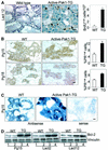
Fig. 5. Abnormal proliferation and expression of ER target genes in transgenic mammary glands. (A) Active Pak1-TG mice show increased BrdU labeling during lactation. Animals on day 12 of lactation were injected with 50 mg/kg BrdU 2 h before being sacrificed, and positive-labeled cells were revealed by immunohistochemistry. Right panel shows the quantitation of the positive cells found per 1000 cells (n = 6). Bar = 10 µm. (B) PR immunostaining of mammary glands obtained from wild-type mice and Pak1-TG mice at days 15 and 18 of pregnancy. Quantitation of the PR-positive cells is shown in the right panels (n = 6). (C) Cathepsin D expression as detected by using in situ hybridization. Pak1-TG mammary glands and wild-type mammary glands at day 15 of pregnancy were probed with antisense or sense control probes. (D) Immunoblot analysis of lysates from day 15 of pregnancy and day 12 of lactation revealed an increase in the Bcl-2 level in transgenic mammary glands compared with the wild-type controls. Bar = 20 µm.
None
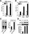
Fig. 6. Pak1 stimulated ER signaling by regulation of the AF2 domain function in MCF-7 human breast cancer cells. (A) Pak1 stimulated ERE activity in the absence of ligand. MCF-7 cells were transfected with 0.25 µg of ERE–luc reporter with or without 0.5 µg of Pak1-T423E or pCMV, treated with or without 1.0 nM E2 for 16 h (n = 4). (B) Pak1 activates AF2 in the absence or presence of E2. MCF-7 cells were cotransfected with 0.25 µg each of gal4–AF2 and gal4–Luc (containing five gal4 binding domains), with or without 0.5 µg of T423E Pak1, wild-type Pak1, or pCMV. As a positive control, cells were treated with 1 × 10⁻⁹ M E2 for 16 h (n = 4). (C) Pak1 stimulates the ER target gene promoter activity. MCF-7 cells were transfected with 0.1 µg of pS2-CAT or myc-luc reporter with or without 0.5 µg of wild-type Pak1 or pCMV. Chloramphenicol acetyltransferase and ERE–luc activities were measured. (D) Pak1 stimulates ER target gene expression. MCF-7 clone expressing HA-tagged T423E Pak1 were treated with 1.0 µg/ml doxycycline to induce the expression of Pak1. Expression of pS2 was assessed by northern blotting (n = 4).
None
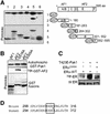
Fig. 7. Pak1 interaction with and phosphorylation of ER. (A) Pak1 phosphorylation of ER. MCF-7 cells were transfected with T7-pcDNA or T7-Pak1, and cell lysates were immunoprecipitated with anti-T7 antibody. Half of the material was used to confirm the expression of Pak1 by western blotting with an anti-T7 mAb (upper panel). The other half of the material was subjected to an in vitro kinase assay using 1 µg of recombinant ER (Affinity Bioreagents Inc.) in the presence of [γ-32P]ATP (lower panel). (B) Interaction of ER with Pak1. GST pull-down assay showing the association of Pak1 with the in vitro-translated ER. (C) GST–Pak1 interacts with T7-ER precipitated from HeLa cells transfected with T7-tagged ER (right lane). (D) Interaction of ER domains with Pak isoforms. Pak1, Pak2 and Pak3 cDNAs were transcribed and translated in vitro, and then incubated with GST–AF1, GST–AF2, or GST and analyzed by the GST pull-down assay. (E) Interaction between endogenous Pak1 and ER. Lysates from virgin mouse uterus (500 µg) were IP-down with an anti-ER antibody or normal mouse IgG, and sequentially immunoblotted with Pak1 and ER antibodies. Purified GST–Pak1 and TNT–T7-ER were used as positive controls for Pak1 and ER, respectively. Residual cleavage of GST–Pak1 resulted in a modest detection of the cleaved Pak1 in lane 4. NS, non-specific. (F) Upper panel, Pak1 fusion proteins were incubated with in vitro translated ER, and binding was analyzed by the GST pull-down assay. The ER binding is within the Nck binding domain amino acids 1–75 (lane 5). Middle panel, the loading control of various ER constructs shown with Ponceau staining; lower panel, a schematic illustration of the various regions of Pak1–GST construct. 1, GST vector alone; 2, full-length Pak1; 3, C-terminal fragment (kinase domain); 4, N-terminal fragment; 5, Nck-binding domain; 6, CRIB domain; and 7, inhibitory domain.
None
Fig. 8. Pak1 regulates ER transactivation functions via Ser305 phosphorylation. (A) Upper panel, GST construct of five various functional domains of ER were incubated with purified Pak1 enzyme in the presence of [32P]ATP; the AF2 domain, which has two consensus Pak1 phosphorylation sites, was strongly phosphorylated. Lower panel, loading control stained with Ponceau, right panel shows a schematic diagram of the various GST–ER domain constructs (Krust et al., 1986). A/B, AF1 domain; C, DNA-binding domain; D, hinge; E, AF2/ligand-binding domain; F, region for modulation of function. (B) S305A mutation (right lane), but not S518A (middle lane), blocked phosphorylation of AF2 by Pak1. Upper panel shows the Pak1 kinase control and the lower panel shows the GST protein loading control. (CIn vivo phosphorylation of ER at S305 by Pak1. HeLa cells were cotransfected with T423E-Pak1 and T7-wild-type or T7-S305A ER for 36 h and incubated with [γ-32P]ATP overnight. Cell lysates were immunoprecipitated with ER antibody, resolved by SDS–PAGE and transferred to membranes. After autoradiography, the membranes were blotted with ER antibody. (D) Comparison of the partial amino acid sequence of mouse and human ER shows that the Pak1 phosphorylation site S305 (Mouse S309), as well as the consensus motif KKXS (boxed), is conserved in human and mouse ER.
None
Fig. 9. Transactivation activity of ER S305 mutants. HeLa cells were cotransfected with 0.25 µg of ERE–luc reporter, 0.5 µg of wild-type Pak1 and 0.5 µg of wild-type ER, S305A ER or S305E ER, with or without 1 nM E2 stimulation (n = 3). (A) S305A mutation of ER blocked activation by Pak1. (B) S305E mutation of ER resulted in stimulation of transactivation activity in the absence of estrogen treatment and was further increased upon 1 nM E2 treatment (n = 3).

Similar articles

Cited by

References

    1. Adam L., Vadlamudi,R., Kondapaka,S.B., Chernoff,J., Mendelsohn,J. and Kumar,R. (1998) Heregulin regulates cytoskeletal reorganization and cell migration through the p21-activated kinase-1 via phosphatidylinositol-3 kinase. J. Biol. Chem., 273, 28238–28246. - PubMed
    1. Adam L., Vadlamudi,R., Mandal,M., Chernoff,J. and Kumar,R. (2000) Regulation of microfilament reorganization and invasiveness of breast cancer cells by p21-activated kinase-1 K299R. J. Biol. Chem., 275, 12041–12050. - PubMed
    1. Ali S., Lutz,Y., Bellocq,J.P., Chenard-Neu,M.P., Rouyer,N. and Metzger,D. (1993) Production and characterization of monoclonal antibodies recognising defined regions of the human oestrogen receptor. Hybridoma, 12, 391–405. - PubMed
    1. Alimandi M., Romano,A., Curia,M.C., Muraro,R., Fedi,P., Aaronson,S.A., Di Fiore,P.P. and Kraus,M.H. (1995) Cooperative signaling of ErbB3 and ErbB2 in neoplastic transformation and human mammary carcinomas. Oncogene, 10, 1813–1821. - PubMed
    1. Bagheri-Yarmand R., Mandal,M., Taludker,A.H., Wang,R.A., Vadlamudi,R.K., Kung,H.J. and Kumar,R. (2001) Etk/bmx tyrosine kinase activates pak1 and regulates tumorigenicity of breast cancer cells. J. Biol. Chem., 276, 29403–29409. - PubMed

Publication types

MeSH terms