

doi: 10.1128/jvi.76.21.11166-11171.2002.
The influenza A virus NS1 protein inhibits activation of Jun N-terminal kinase and AP-1 transcription factors
Affiliations
- PMID: 12368362
- PMCID: PMC136597
- DOI: 10.1128/jvi.76.21.11166-11171.2002
Item in Clipboard
The influenza A virus NS1 protein inhibits activation of Jun N-terminal kinase and AP-1 transcription factors
J Virol.
2002 Nov.
Abstract
The influenza A virus nonstructural NS1 protein is known to modulate host cell gene expression and to inhibit double-stranded RNA (dsRNA)-mediated antiviral responses. Here we identify NS1 as the first viral protein that antagonizes virus- and dsRNA-induced activation of the stress response-signaling pathway mediated through Jun N-terminal kinase.
Figures
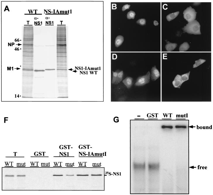
Expression, localization, dimerization, and dsRNA- binding of NS1 proteins encoded by influenza A/PR/8/34 WT and NS-IAmut1 viruses. (A) MDCK cells were infected with WT or NS-IAmut1 viruses. After a 5-h incubation at 37°C, the cells were metabolically labeled with [35S]methionine for 1 h and lysed in RIPA buffer. The NS1 proteins were immunoprecipitated from total-cell lysates with rabbit anti-NS1 serum and analyzed by SDS-gel electrophoresis and autoradiography as described previously (45). The positions of the M1, NP, and NS1 proteins are indicated on the left and right. T, 10% of cell lysate used for immunoprecipitation. (B to E) Intracellular NS1 localization was determined by indirect immunofluorescence analysis of HeLa cells infected with WT (B) or NS-IAmutI (C) virus. Cells were fixed and permeabilized, and the NS1 proteins were detected by the use of rabbit anti-NS1 serum and anti-rabbit immunoglobulin G (IgG)- fluorescein isothiocyanate (FITC) conjugate as described elsewhere (45). The localization of NS1 proteins in the absence of virus infection was determined in MDCK cells after Lipofectamine 2000 (Invitrogen)-mediated transfection with mammalian pcDNA3.1-Myc/His expression plasmids (Invitrogen) encoding NS1 WT (D) or IAmut1 (E) protein. Cells were prepared for microscopic analysis, and the NS1 proteins were visualized by indirect immunofluorescence using the Myc-tag specific antibody 9E10 (Santa Cruz Biotechnology) and secondary FITC-conjugated anti-mouse IgG. The cells were viewed under a Nikon Axiophot 300 microscope equipped with an epifluorescence attachment. Images were recorded by a SPOT RT digital camera and processed using Adobe Photoshop 5.0 software. (F) To assess dimerization of NS1 proteins, equal amounts of GST, GST-NS1, and GST-NS-IAmut1 proteins were immobilized on glutathione-agarose beads and reacted with NS1 WT or NS-IAmut1 (mut) proteins that were synthesized in coupled transcription-translation reactions in the presence of [35S]methionine as described previously (45). The precipitated proteins were separated by SDS-gel electrophoresis and visualized by autoradiography. T, 10% of the total amount used for precipitation. (G) Binding of GST-NS1 fusion proteins (0.4 μM) to dsRNA (approximately 0.5 nM) derived from complementary 32P-labeled transcripts of the polylinker region of plasmid pGEM-11zf (Promega) was examined as described previously (28). Protein-RNA complexes formed in the absence or presence of GST, GST-NS1 WT and GST-NS-IAmutI proteins were separated from free RNA by gel electrophoresis on a nondenaturing 10% polyacrylamide gel. The positions of free and complexed RNAs are indicated to the right.
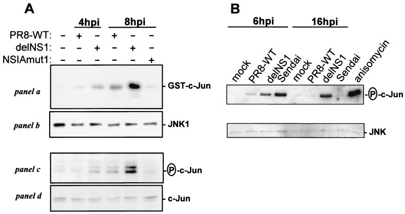
Viral NS1 expression inhibits activation of JNK and phosphorylation of c-Jun. (A) Vero cells were mock infected or infected with PR8 WT, delNS1, or NS-IAmut1 viruses at a multiplicity of infection (MOI) of 5 and lysed 4 or 8 h postinfection (hpi) as described previously (30). Endogenous JNK1 was immunoprecipitated from lysates representing equal number of cells with JNK- specific antiserum (Santa Cruz), and washed immune complexes were analyzed for JNK activity by using recombinant GST-cJun(1-135) as a substrate as described previously (30) (panel a). Equal loadings with precipitated kinase were verified by immunoblot detection with JNK-1 antiserum (panel b). The same lysates were subjected to SDS-polyacylamide gel electrophoresis and analyzed by immunoblotting with antisera specific for phospho-c-Jun and total c-Jun (Cell Signaling) (panels c and d). (B) Human A549 lung epithelial cells were either mock infected, or infected with PR8 WT or delNS1 virus at an MOI of 1. Total-cell extracts were made at 6 and 16 h postinfection, and total JNK was precipitated overnight at 4°C with 2 μg of c-Jun fusion protein coupled to glutathione- agarose beads. Kinase assays were performed for 30 min at 30°C in the presence of 100 μM ATP. The incubated proteins were separated by SDS-gel electrophoresis and analyzed by immunoblotting using phospho-c-Jun-specific antibody (upper panel). In addition, each sample was subjected to anti-JNK immunoblot analysis to determine the total level of JNK protein. A549 cells that were stimulated for 20 min with the common JNK activator anisomycin or infected with Sendai virus for 6 or 16 h were included as positive controls.
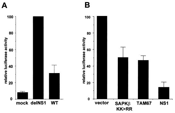
Inhibition of JNK/AP-1 signaling impairs delNS1 virus-induced IFN-β promoter activation. (A) MDCK cells (106) were transfected with 100 ng of the IFN-β promoter luciferase reporter plasmid p125-Luc (47). After 24 h, the cells were mock infected or infected with influenza PR8 WT or delNS virus at an MOI of 1. Cell extracts were prepared at 4 h postinfection in reporter lysis buffer (Promega) and assayed for luciferase activity. For a comparison, enzyme activity induced by delNS1 virus was arbitrarily set to 100%. Average values determined in three independent experiments are shown. (B) Cells were transfected with p125-Luc together with 4 μg of empty pKRSPA vector (vector) or expression plasmids encoding dominant negative JNK/SAPK (SAPKβ KK>>RR), dominant-negative c-Jun (TAM67) (30), or NS1 protein. Cells were infected with delNS1 virus at an MOI of 1, and 4 h later they were lysed and luciferase activity was determined as described for panel A. Error bars indicate standard deviations.
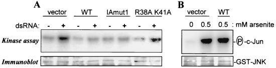
NS1 protein expression inhibits dsRNA- but not arsenite-induced activation of c- Jun. (A) MDCK cells were transfected with a plasmid expressing GST-tagged JNK/SAPKβ together with either empty vector (lanes vector) or derivatives thereof expressing NS1 WT, NS-IAmut1, or the NS1-R38A/K41A RNA-binding mutant protein (lanes WT, IAmut1, and R38A K41A, respectively). At 24 h later, the cells were left untreated or stimulated with synthetic dsRNA (50 μg/ml) for 6 h and extracts were prepared as described previously (30). JNK activity was assessed in the lysates by immune complex kinase assays using GST-c-Jun(1-135) as a substrate (30). The amount of GST-JNK in each sample was determined by immunoblotting with GST-specific antiserum. (B) In parallel reactions, we analyzed c-Jun phosphorylation in cells that were transfected with empty or NS1 expression vector and were mock treated (lane 0) or stimulated with 0.5 mM sodium arsenite, a common JNK activator, for 30 min (lanes 0.5).
Similar articles
-
Double-stranded RNA-induced activation of activating protein-1 promoter is differentially regulated by the non-structural protein 1 of avian influenza A viruses.Viral Immunol. 2012 Feb;25(1):79-85. doi: 10.1089/vim.2011.0059. Epub 2012 Jan 12. Viral Immunol. 2012. PMID: 22239235 Free PMC article.
-
Activation of c-jun N-terminal kinase upon influenza A virus (IAV) infection is independent of pathogen-related receptors but dependent on amino acid sequence variations of IAV NS1.J Virol. 2014 Aug;88(16):8843-52. doi: 10.1128/JVI.00424-14. Epub 2014 May 28. J Virol. 2014. PMID: 24872593 Free PMC article.
-
Inhibition of retinoic acid-inducible gene I-mediated induction of beta interferon by the NS1 protein of influenza A virus.J Virol. 2007 Jan;81(2):514-24. doi: 10.1128/JVI.01265-06. Epub 2006 Nov 1. J Virol. 2007. PMID: 17079289 Free PMC article.
-
The influenza virus NS1 protein as a therapeutic target.Antiviral Res. 2013 Sep;99(3):409-16. doi: 10.1016/j.antiviral.2013.06.005. Epub 2013 Jun 21. Antiviral Res. 2013. PMID: 23796981 Free PMC article. Review.
-
Structure and Function of the Influenza A Virus Non-Structural Protein 1.J Microbiol Biotechnol. 2019 Aug 28;29(8):1184-1192. doi: 10.4014/jmb.1903.03053. J Microbiol Biotechnol. 2019. PMID: 31154753 Review.
Cited by
-
Differential requirement for c-Jun N-terminal kinase 1 in lung inflammation and host defense.PLoS One. 2012;7(4):e34638. doi: 10.1371/journal.pone.0034638. Epub 2012 Apr 13. PLoS One. 2012. PMID: 22514650 Free PMC article.
-
The RNA binding domain of influenza A virus NS1 protein affects secretion of tumor necrosis factor alpha, interleukin-6, and interferon in primary murine tracheal epithelial cells.J Virol. 2007 Sep;81(17):9469-80. doi: 10.1128/JVI.00989-07. Epub 2007 Jun 27. J Virol. 2007. PMID: 17596305 Free PMC article.
-
The Two Sides of the Same Coin-Influenza Virus and Intracellular Signal Transduction.Cold Spring Harb Perspect Med. 2021 Jan 4;11(1):a038513. doi: 10.1101/cshperspect.a038513. Cold Spring Harb Perspect Med. 2021. PMID: 31871235 Free PMC article. Review.
-
The N- and C-terminal domains of the NS1 protein of influenza B virus can independently inhibit IRF-3 and beta interferon promoter activation.J Virol. 2004 Nov;78(21):11574-82. doi: 10.1128/JVI.78.21.11574-11582.2004. J Virol. 2004. PMID: 15479798 Free PMC article.
-
Apoptosis, cytokine and chemokine induction by non-structural 1 (NS1) proteins encoded by different influenza subtypes.Virol J. 2011 Dec 21;8:554. doi: 10.1186/1743-422X-8-554. Virol J. 2011. PMID: 22185562 Free PMC article.
References
-
- Biron, C. A., and G. C. Sen. 2001. Interferons and other cytokines, p. 321-351. In D. M. Knipe, P. M. Howley, D. E. Griffin et al. (ed.), Fields virology, 4th ed. Lippincott, Williams & Wilkins, Philadelphia, Pa.
-
- Chu, W. M., D. Ostertag, Z. W. Li, L. Chang, Y. Chen, Y. Hu, B. Williams, J. Perrault, and M. Karin. 1999. JNK2 and IKKβ are required for activating the innate response to viral infection. Immunity 11:721-731. - PubMed
Publication types
MeSH terms
Substances
LinkOut - more resources
Full Text Sources
Research Materials
Miscellaneous
