. 2002 Oct 1;21(19):5141-50.
doi: 10.1093/emboj/cdf513.

Sendai virus trailer RNA binds TIAR, a cellular protein involved in virus-induced apoptosis

Affiliations

Sendai virus trailer RNA binds TIAR, a cellular protein involved in virus-induced apoptosis

Frédéric Iseni et al. EMBO J. .

Abstract

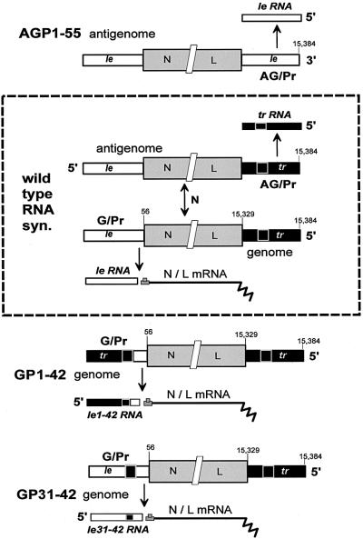
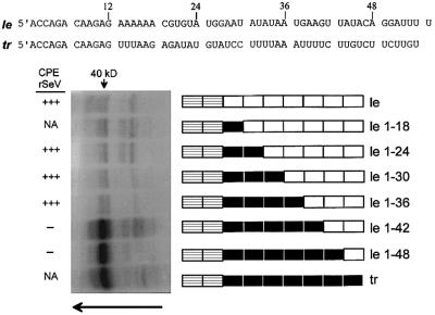
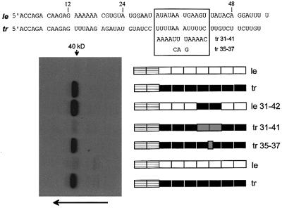
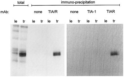
Sendai virus (SeV) leader (le) and trailer (tr) RNAs are short transcripts generated during abortive antigenome and genome synthesis, respectively. Recom binant SeV (rSeV) that express tr-like RNAs from the leader region are non-cytopathic and, moreover, prevent wild-type SeV from inducing apoptosis in mixed infections. These rSeV thus appear to have gained a function. Here we report that tr RNA binds to a cellular protein with many links to apoptosis (TIAR) via the AU-rich sequence 5' UUUUAAAUUUU. Duplication of this AU-rich sequence alone within the le RNA confers TIAR binding on this le* RNA and a non-cytopathic phenotype to these rSeV in cell culture. Transgenic overexpression of TIAR during SeV infection promotes apoptosis and reverses the anti-apoptotic effects of le* RNA expression. More over, TIAR overexpression and SeV infection act synergistically to induce apoptosis. These short viral RNAs may act by sequestering TIAR, a multivalent RNA recognition motif (RRM) family RNA-binding protein involved in SeV-induced apoptosis. In this view, tr RNA is not simply a by-product of abortive genome synthesis, but is also an antigenome transcript that modulates the cellular antiviral response.


Figures

None
Fig. 1. SeV RNA synthesis from wild-type and promoter-variant viruses. The dotted rectangle encloses a flow diagram of viral RNA synthesis. [–] genomes and [+] antigenomes are shown as a series of three boxes that are not drawn to scale; the leader (le) region in white (nucleotides 1–55), the train of six mRNAs in gray (abbreviated as N/L, nucleotides 56–15 329) and the trailer (tr) region in black (nucleotides 15 330–15 384). Nucleotides 31–42 of the black trailer region (or more properly nucleotides 15 342–15 353 of the genome) are highlighted with a white border. Vertical arrows indicate the flow of viral RNA synthesis. Genome replication requires a continuous supply of newly made N protein subunits, in contrast to the synthesis of le RNA, mRNAs and tr RNA. The sequence exchanges in the leader regions of SeV-GP1–42 and SeV-GP31–42, and in the trailer region of SeV-AGP1–55, and their consequences on the promoter-proximal small RNA transcribed from G/Pr and AG/Pr, are also indicated.
None
Fig. 2. UV cross-linking of wild-type and mutant promoter-proximal RNAs to a 40 kDa host protein. The le and tr RNA sequences are shown above in blocks of six nucleotides, which are shown schematically as boxes below. The first two boxes are always striped, as these two hexamers are conserved between le and tr RNA. The subsequent sequences are poorly conserved, and are shown as white boxes for le RNA, and black boxes for tr RNA. The le and tr RNAs and the various chimeric tr/le RNAs (le 1–18 to 1–48) are shown schematically next to the cross-linking gel. The various promoter-proximal RNAs were mixed with HeLa cell cytoplasmic extracts and irradiated with 312 nm light for 10 min [or kept in the dark as a control (not shown), Materials and methods]. The reactions were then digested with RNase A, and separated on 12.5% protein gels to determine the relative abilities of these small RNAs to cross-link to host proteins. The cytopathic nature (CPE) of rSeV expressing these chimeric tr/le sequences in the leader region of G/Pr (Garcin et al., 1998) is indicated on the left; NA, not applicable as these rSeV were not prepared.
None
Fig. 3. The AU-rich sequence 5′ UUUUAAAUUUU mediates promoter-proximal RNA cross-linking to the 40 kDa protein. The tr and le RNA sequences are shown above, and the fourth and fifth hexamers (nucleotides 31–42) are boxed. The various promoter- proximal RNAs examined are shown schematically as for Figure 2, next to the protein gel used to determine their relative abilities to UV cross-link to the host 40 kDa protein. The alternative sequence exchanges (tr 31–41 and tr 35–37) are shown as gray boxes, and their sequences are shown as well in the box above.
None
Fig. 4. rSeV-GP31–42 is non-cytopathic in cell culture, similarly to rSeV-GP1–42. HeLa cells were infected with 20 p.f.u. (or equivalent)/cell of rSeV-wt, rSeV-GP1–42 or rSeV-GP31–42, or mock infected. The cultures were trypsinized at 48 h.p.i. and equal samples of the harvested cells were used to estimate the levels of N and P proteins present intracellularly by western blotting [primary infection, (B)]. Other samples were used to determine the level of annexin V staining by FACS (C). The remaining cells (one-fifth of the total) were replated in Petri dishes and grown for 3 days, and the cultures were stained with methylene blue (A). The second passage cultures were also used to estimate the levels of N and P proteins present intracellularly by western blotting (second passage, B).
None
Fig. 5. TIAR-specific mAb immunoprecipitates the 40 kDa host protein. le or tr RNA cross-linked HeLa cell extracts, intentionally over-reacted to also contain non-specific bands (total), were immunoprecipitated with mAb specific for both TIA-1 and TIAR (TIA/R) or mAb specific for each protein, as indicated above. The precipitates and the starting materials (total) were separated by 12.5% SDS–PAGE, and the gel exposed to film.
None
Fig. 6. TIA/R immunofluoresence of arsenite-treated and SeV-infected HeLa cells. Replicate cultures of HeLa cells were infected with 20 p.f.u. (or equivalent)/cell of the various SeV indicated and examined for TIA/R immunofluorescence by confocal microscopy at 18 h.p.i. (Materials and methods). Some rSeV-AGP1–55-infected cultures were treated with 10 µg/ml of cycloheximide at 18 h.p.i., and fixed for examination at 19 h.p.i. (bottom right). At least 200 cells from each culture in three independent experiments were examined, and the percentages containing SGs (white arrows) are shown in parentheses. Uninfected cells were treated with 500 µM As2O3 for 30 min and then examined, for reference.
None
Fig. 7. Transgenic expression of TIAR during SeV infection. Parallel HeLa cell cultures were infected with 10 infectious units of each of the six rSeV indicated (see text), or mock infected, and harvested at 48 h.p.i. Half of the cells were used to determine the intracellular levels of the transgenes and the SeV N protein by immunoblotting with anti-HA, anti-GFP and anti-N (A). The other half was used to determine the level of annexin V staining by FACS (C). For the three rSeV that express GFP, the level of intracellular replication was determined more accurately by following GFP fluorescence directly by FACS (B). The error bars in (C) indicate the results of infecting duplicate cultures.
None
Fig. 8. Synergy of TIAR overexpression and SeV infection in inducing apoptosis. Duplicate cultures of HeLa cells were transfected with 1 µg of pEBS plasmids expressing TIAR or empty pEBS (as indicated), along with 0.2 µg of pEBS–GFP to follow transfection efficiency, with fuGENE (Roche). The cultures were then infected 24 h later with 10 p.f.u./cell of SeV-wt, or mock infected. All cultures were harvested at 48 h.p.i., and the percentage of apoptotic cells was determined by annexin V staining and FACS. The horizontal line indicates the background level of apoptosis, i.e. independent of TIAR expression and SeV infection, which is subtracted from the other bars to determine the level of synergy. The synergy between TIAR overexpression and SeV infection is indicated by the black segment at the top of the bar.

Similar articles

Cited by

References

    1. Anderson P. (1995) TIA-1: structural and functional studies on a new class of cytolytic effector molecule. Curr. Top. Microbiol. Immunol., 198, 131–143. - PubMed
    1. Anderson P. and Kedersha,N. (2002) Stressful initiations. J. Cell Sci., 115, 3227–3234. - PubMed
    1. Bitzer M., Prinz,F., Bauer,M., Spiegel,M., Neubert,W.J., Gregor,M., Schulze-Osthoff,K. and Lauer,U. (1999) Sendai virus infection induces apoptosis through activation of caspase-8 (FLICE) and caspase-3 (CPP32). J. Virol., 73, 702–708. - PMC - PubMed
    1. Bontron S., Ucla,C., Mach,B. and Steimle,V. (1997) Efficient repression of endogenous major histocompatibility complex class II expression through dominant negative CIITA mutants isolated by a functional selection strategy. Mol. Cell. Biol., 17, 4249–4258. - PMC - PubMed
    1. Brennan C.M. and Steitz,J.A. (2001) HuR and mRNA stability. Cell. Mol. Life Sci., 58, 266–277. - PMC - PubMed

Publication types