. 2002 Oct;55(5):286-93.
doi: 10.1136/mp.55.5.286.

Analysis of apoptotic and antiapoptotic signalling pathways induced by Helicobacter pylori

Affiliations

Analysis of apoptotic and antiapoptotic signalling pathways induced by Helicobacter pylori

S Maeda et al. Mol Pathol. 2002 Oct.

Abstract

Background and aims: Although it is reported that Helicobacter pylori induces apoptosis on gastric epithelial cells, the mechanism remains unknown. Antiapoptotic effects generated by H pylori have not yet been evaluated.

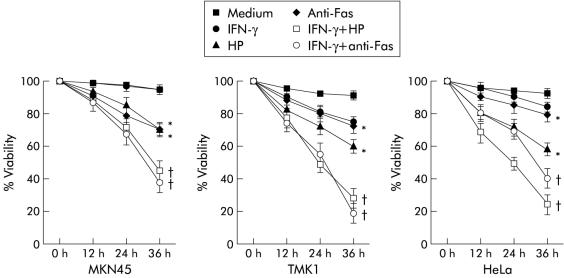
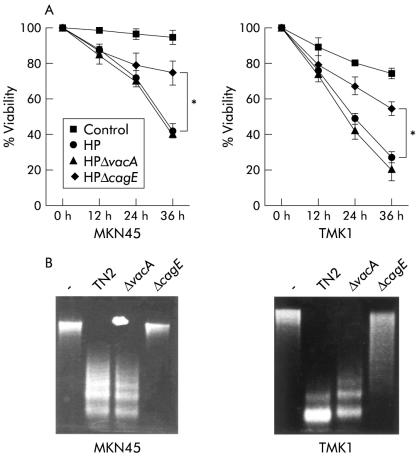
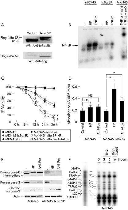
Methods: (1) H pylori strains (type 1 wild, TN2-deltacagE, TN2-deltavacA) were cocultured with MKN45, TMK1, and HeLa cells, and cell viability and apoptosis were assessed by trypan blue exclusion and DNA laddering, respectively. (2) Activation of caspases-3, 7, and 8, cytochrome c release from the mitochondria, and Fas, Fas associated death domain protein (FADD), Bax, Bak, and Bcl-X expression were evaluated by immunoblot analysis. (3) To investigate whether nuclear factor kappa B (NFkappaB) activation induced by cag pathogenicity island (PAI) positive H pylori affects antiapoptosis, MKN45 cells stably expressing super-repressor IkappaBalpha were cocultured with H pylori, and cell viability and caspase activation were evaluated. NFkappaB regulated gene expression was also evaluated by ribonuclease protection assay.

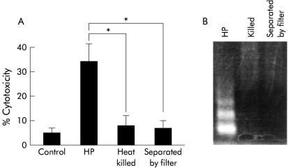
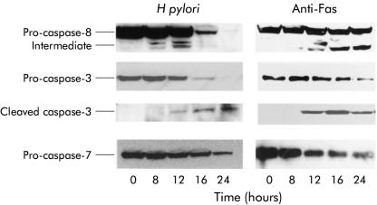
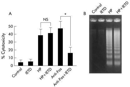
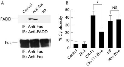
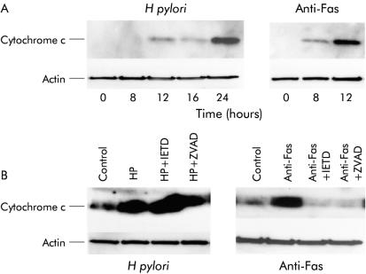
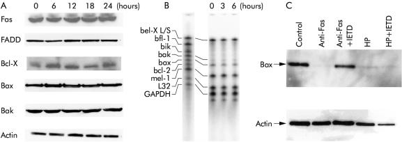
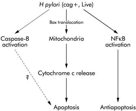
Results: (1) Wild-type and deltavacA mutant H pylori induced apoptosis more potently than the deltacagE mutant. Inhibition of cell contact between H pylori and cancer cells and heat killing H pylori diminished cell death. (2) Caspases-3, 7, and 8 were activated time dependently by H pylori as well as by the agonist anti-Fas. Cytochrome c release from mitochondria was observed and was not inhibited by caspase-8 inhibitor. Although protein expression of Fas, FADD, Bax, Bak, and Bcl-X in the whole cell lysates was not changed by H pylori, Bax was decreased from mitochondria free cytosol suggesting that Bax was translocated into mitochondria. (3) Cell death and the activities of caspases-3 and 8 were promoted in MKN45 cells stably expressing super-repressor IkappaBalpha that inhibits NFkappaB activation. Antiapoptotic proteins c-IAP1 and c-IAP2 were upregulated by the wild-type strains.

Conclusion: cag PAI positive H pylori is capable of inducing apoptotic effects mainly through the mitochondrial pathway. Antiapoptotic effects mediated by NFkappaB activation were also observed.


Figures

Figure 1
Figure 1
Incubation with Helicobacter pylori (HP) cells decreased the viability of cancer cells in the three cancer cell lines. Cell viability was assessed by trypan blue dye exclusion assay at the indicated times by counting 300 cells. Viable cells were quantitated under bright field microscopy. Results are expressed as percentages of viable cells. Values are mean (SD) of three independent experiments. *Percentage viability was significantly (p<0.05) different between the control group at 36 hours and the H pylori coculture or anti-Fas treatment without interferon γ (IFN-γ) group. †Percentage viability was significantly (p<0.05) different between the H pylori coculture or anti-Fas without IFN-γ group and the IFN-γ group.
Figure 2
Figure 2
Attenuation of the apoptotic effect by cagE negative mutant. (A) Cells were treated with interferon γ (IFN-γ 10 ng/ml) for 24 hours and incubated with H pylori (TN2), TN2-ΔcagE, and TN2-ΔvacA. Cell viability was assessed by trypan blue dye exclusion assay at the indicated times by counting 300 cells. Viable cells were quantitated under bright field microscopy. Results are expressed as percentages of viable cells. Values are mean (SD) of three independent experiments. *Percentage viability was significantly (p<0.05) different between the H pylori (HP) wild-type and vacA coculture group and the cagE coculture group. (B) DNA fragmentation was evaluated after 16 hours of infection.
Figure 3
Figure 3
Viable bacteria and direct contact with cells were necessary for inducing apoptosis. MKN45 cells were treated with interferon γ (IFN-γ 10 ng/ml) for 24 hours. Cells and bacteria were separated by membrane filter (Nunc Tissue Culture Inserts No 162138; Nunc, Roskilde, Denmark). We also used heat killed bacteria at 80°C for 30 minutes. Cell viability was assessed by trypan blue dye exclusion assay at 24 hours by counting 300 cells. Viable cells were quantitated under bright field microscopy. Results are expressed as percentages of dead cells. Values are mean (SD) of three independent experiments. *Percentage cytotoxicity was significantly (p<0.05) different between the H pylori (HP) coculture group and the heat killed and separated by filter groups.
Figure 4
Figure 4
Helicobacter pylori induces apoptosis via a caspase dependent pathway. Immunoblot analysis was performed using anti-caspases-8, 3, 7, and cleaved caspase-3 antibodies in MKN45 cells. Incubation with H pylori and anti-Fas resulted in a time dependent degradation of the primary forms of caspases-8, 3, and 7. Processing into two fragments (43 kDa and 41 kDa) of the caspase-8 intermediate form and the 20 kDa active caspase-3 was also observed.
Figure 5
Figure 5
Caspase-8 inhibitor did not inhibit Helicobacter pylori induced apoptosis. (A) MKN45 cells were treated with interferon γ (IFN-γ 10 ng/ml) for 24 hours. Cells were preincubated with or without the caspase-8 inhibitor Ac-IETD-CHO (IETD 100 μM) for one hour and then incubated with H pylori (HP) or anti-Fas (CH-11) for 24 hours. Cell viability was assessed by trypan blue dye exclusion assay by counting 300 cells. Viable cells were quantitated under bright field microscopy. Results are expressed as percentages of dead cells. Values are mean (SD) of three independent experiments. *Percentage cytotoxicity was significantly (p<0.05) different between the anti-Fas treatment group and the anti-Fas treatment with caspase-8 inhibitor treatment group. NS, no significant difference was found. (B) DNA fragmentation was also evaluated after 24 hours of infection.
Figure 6
Figure 6
Fas (CD95) was not associated with Helicobacter pylori induced apoptosis. (A) MKN45 cells were treated with interferon γ (IFN-γ 10 ng/ml) for 24 hours. Cells were then incubated with H pylori (HP) or anti-Fas (CH-11) for 24 hours. Total cell lysates were extracted and immunoprecipitated with anti-Fas antibody and immunoblotted with anti-Fas associated death domain protein (FADD) and anti-Fas antibody. (B) MKN45 cells were treated with IFN-γ (10 ng/ml) for 24 hours, with or without neutralising anti-Fas antibody (ZB-4) for one hour, and incubated with H pylori or anti-Fas (CH-11). Cell viability was assessed by trypan blue dye exclusion assay at the indicated times by counting 300 cells. Viable cells were quantitated under bright field microscopy. Results are expressed as percentages of viable cells. Values are mean (SD) of three independent experiments. *Percentage cytotoxicity was significantly (p<0.05) different between the anti-Fas treatment group and the anti-Fas treatment with ZB-4 treatment group. NS, no significant difference was found.
Figure 7
Figure 7
Helicobacter pylori induced cytochrome c release from the mitochondria. (A) MKN45 cells were treated with interferon γ (IFN-γ 10 ng/ml) for 24 hours, and then with H pylori or anti-Fas (CH-11). At the indicated times, cytosolic fractions and total cell lysates were extracted, separated by electrophoresis, and immunoblotted with anti-cytochrome c and anti-actin, respectively. (B) MKN45 cells were treated with IFN-γ (10 ng/ml) for 24 hours, and 100 μM of the caspase-8 inhibitor Ac-IETD-CHO (IETD), the pan-caspase inhibitor Z-VAD-FMK (ZVAD), or medium alone pretreatment for one hour before exposure to H pylori (HP) or anti-Fas (CH-11). Cells were then incubated with H pylori or anti-Fas (CH-11) for 24 hours. Cytosolic fractions and total cell lysates were extracted, separated by electrophoresis, and immunoblotted with anti-cytochrome c and anti-actin.
Figure 8
Figure 8
Expression of apoptosis related protein. (A) MKN45 cells were incubated with Helicobacter pylori and total cell lysates were extracted at the indicated times. The same amount of extracted protein (20 μg) was separated by electrophoresis and immunoblotted with anti-Fas, Fas associated death domain protein (FADD), Bcl-X, Bax, Bak, and actin. (B) MKN45 cell were incubated with H pylori and total RNA was extracted at the indicated times. The ribonuclease protection assay was performed according to the supplier’s instructions. (C) MKN45 cells were treated with interferon γ (IFN-γ 10 ng/ml) for 24 hours, and 100 μM of the caspase-8 inhibitor Ac-IETD-CHO (IETD) or medium only pretreatment for one hour before exposure to H pylori (HP) or anti-Fas (CH-11). Cells were then incubated with H pylori or anti-Fas (CH-11) for 24 hours. Cytosolic fractions and total cell lysates were extracted, separated by electrophoresis, and immunoblotted with anti-Bax and anti-actin.
Figure 9
Figure 9
(A) Detection of stable MKN45 transfectants expressing IκBα (SS32/36AA). The clone was analysed using a monoclonal antibody to FLAG and polyclonal antibody to IκBα. (B) Specific protein binding activities of nuclear factor kappa B (NFκB) sequences (electrophoretic mobility shift assay). The nuclear extracts were prepared from MKN45 and MKN45 IκBα SR cells. Cells were treated or not treated with Helicobacter pylori (HP) or 10 ng/ml tumour necrosis factor α (TNF-α) for 90 minutes. Nuclear extracts was incubated with 32P labelled oligonucleotide for 30 minutes. Migration of the DNA-protein complex containing NFκB is indicated. This complex was found to be specific, as judged using supershifting antibody against p65 and cold NFκB probes. (C) MKN45 and MKN45 IκBα SR cells were treated with interferon γ (IFN-γ 10 ng/ml) for 24 hours. Cells were incubated with H pylori (HP) or anti-Fas (CH-11) for 24 hours. Cell viability was assessed by trypan blue dye exclusion assay by counting 300 cells. Viable cells were quantitated under bright field microscopy. Results are expressed as percentages of dead cells. Values are mean (SD) of three independent experiments. *Percentage cytotoxicity was significantly (p<0.05) different between the MKN45 treated cells with anti-Fas or cocultured with H pylori and MKN45 IκBα SR treated cells with anti-Fas or cocultured with H pylori. (D) DNA fragmentation was quantified using a commercially available ELISA (Boehringer Mannheim Biochemicals, Mannheim, Germany): 5×10⁴ cells were incubated in triplicate with H pylori (HP), anti-Fas (CH-11), or medium alone for 12 hours and lysed, and the supernatants were used for ELISA. Absorbance was measured at 405 nm. *Absorbance was significantly (p<0.05) different between the control and treated with anti-Fas or cocultured with H pylori in the MKN45 IκBα SR groups; NS, no significant difference was found. (E) MKN45 and MKN-45 IκBαSR cells were treated with IFN-γ (10 ng/ml) for 24 hours. Cells were incubated with H pylori (HP) or anti-Fas (CH-11) for 12 hours. Immunoblot analysis was performed using anti-caspase-8, caspase-3, cleaved caspase-3, and anti-actin antibody. (F) MKN45 and THP-1 cells were incubated with H pylori and total RNA was extracted at the indicated times. The ribonuclease protection assay was performed according to the supplier’s instructions.
Figure 10
Figure 10
Schematic representation of the signalling pathways leading to apoptosis and antiapoptosis in response to Helicobacter pylori in gastric epithelial cells.

Comment in

  • Helicobacter pylori.
    Eguchi H, Moss SF. Eguchi H, et al. Mol Pathol. 2002 Oct;55(5):284-5. doi: 10.1136/mp.55.5.284. Mol Pathol. 2002. PMID: 12354928 Free PMC article. No abstract available.

Similar articles

Cited by

References

    1. Warren JR, Marshall BJ. Unidentified curved bacilli on gastric epithelium in active chronic gastritis. Lancet 1983;1:1273–5. - PubMed
    1. NIH consensus conference. Helicobacter pylori in peptic ulcer disease. NIH consensus development panel on Helicobacter pylori in peptic ulcer disease. JAMA 1994;272:65–9. - PubMed
    1. Graham DY, Lew GM, Klein PD, et al. Effect of treatment of Helicobacter pylori infection on the long-term recurrence of gastric or duodenal ulcer: a randomized, controlled study. Ann Intern Med 1992; 116:705–8. - PubMed
    1. Parsonnet J, Friedman GD, Vandersteen DP, et al. Helicobacter pylori infection and the risk of gastric carcinoma. N Engl J Med 1991;325:1127–31. - PubMed
    1. Nomura A, Stemmermann GN, Chyou PH, et al. Helicobacter pylori infection and gastric carcinoma among Japanese American in Hawaii. N Engl J Med 1991;325:1132–6. - PubMed

Publication types

MeSH terms