. 2002 Sep;110(6):861-70.
doi: 10.1172/JCI15375.

Craniosynostosis in transgenic mice overexpressing Nell-1

Affiliations

Craniosynostosis in transgenic mice overexpressing Nell-1

Xinli Zhang et al. J Clin Invest. 2002 Sep.

Erratum in

  • J Clin Invest 2002 Nov;110(10):1573

Abstract

Previously, we reported NELL-1 as a novel molecule overexpressed during premature cranial suture closure in patients with craniosynostosis (CS), one of the most common congenital craniofacial deformities. Here we describe the creation and analysis of transgenic mice overexpressing Nell-1. Nell-1 transgenic animals exhibited CS-like phenotypes that ranged from simple to compound synostoses. Histologically, the osteogenic fronts of abnormally closing/closed sutures in these animals revealed calvarial overgrowth and overlap along with increased osteoblast differentiation and reduced cell proliferation. Furthermore, anomalies were restricted to calvarial bone, despite generalized, non-tissue-specific overexpression of Nell-1. In vitro, Nell-1 overexpression accelerated calvarial osteoblast differentiation and mineralization under normal culture conditions. Moreover, Nell-1 overexpression in osteoblasts was sufficient to promote alkaline phosphatase expression and micronodule formation. Conversely, downregulation of Nell-1 inhibited osteoblast differentiation in vitro. In summary, Nell-1 overexpression induced calvarial overgrowth resulting in premature suture closure in a rodent model. Nell-1, therefore, has a novel role in CS development, perhaps as part of a complex chain of events resulting in premature suture closure. On a cellular level, Nell-1 expression may modulate and be both sufficient and required for osteoblast differentiation.


Figures
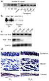
Figure 1
Figure 1
Nell-1 transgenic mice compared with nontransgenic littermates. (a) Transgene copy number. The founders (FA and FB) and their progeny (TF2A1, TF2A2, and TF2B1) have copy numbers between 50 and 100. TF2A1 and TF2A2 are from the founder A line. TF2B1, TF2B2, and TF2B3 are from the founder B line. (b) RT-PCR analyses of Nell-1 RNA expression in both founders. C, control Nell-1 plasmid; M, muscle; H, heart; B, bone; K, kidney; L, liver. (c) Whole body (without head) RNA of newborn progeny. TF2A1 and TF2A2 express different levels of Nell-1. TF2B1 expresses Nell-1 weakly, while TF2B2 and TF2B3 have no Nell-1 expression. (d) Left panels, immunolocalization of Nell-1 protein in newborn NF2 epithelium, muscle, and calvarial bone. There is no detectable Nell-1 expression (brown staining indicates the presence of Nell-1) except some staining in the calvarial bone. Right panels, immunolocalization of Nell-1 protein in TF2A2 epithelium, muscle, and calvarial bone. Abundant Nell-1 expression is present throughout all soft tissue layers as well as in bone. Bar represents 50 μm.
Figure 2
Figure 2
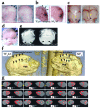
Phenotypic evaluation of Nell-1 transgenic mice. (a and b) Left panels show a newborn Nell-1 phenotype–positive (TF2A1) mouse. Note the protrusion in the frontoparietal area (arrows). Right panels show an NF2 littermate. (c) Left panel, TF2A2 mouse with the scalp removed. The sagittal (yellow arrow) and PF (black arrow) sutures are closed. Right panel, skull of the NF2 littermate with patent sagittal (yellow arrows) and PF (black arrow) sutures and normal vasculature underneath the patent sutures. (d) An infant with craniotelencephalic dysplasia, a severe form of CS. (e) Brain MRI of TF2A1 mouse (left) and NF2 littermate (right). Note the complete absence of ventricles, suggesting elevated intracranial pressure in the TF2A1 mouse (arrows, left) relative to its NF2 littermate (arrows, right). (f) MCT-reconstructed three-dimensional skull views of the newborn Nell-1 phenotype-positive TF2A1 (left) and NF2 (right) littermates. Arrows indicate sagittal and PF suture sites. In TF2A1 mice, the sagittal and PF sutures are largely closed and replaced with an abnormal ridge. In the NF2 littermate, both sagittal and PF sutures are patent. Complete opacity corresponds to greater than 50 mg/cc mineralization. The vertical rods in the background are phantom reference rods corresponding to mineralization densities (from left to right) of 50, 100, 150, and 200 mg/cc. (g) Serial axial MCT sections of the TF2A1 (left) and NF2 littermates (right) shown in f. Yellow arrows indicate the distortion of the cranium. Green arrows indicate increased mineralization of the calvarium in the TF2A mouse (arrow, right).
Figure 3
Figure 3
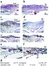
Histologic and immunohistologic evaluation of Nell-1 transgenic mice. (a) Hematoxylin and eosin staining of the sagittal suture of a Nell-1 phenotype-positive TF2A1 mouse. There is closure of the suture, shown by the overlap of calvarial edges (black arrows) and closing osteogenic fronts (red arrows). Lower left panel shows von Kossa staining. Note the close proximity of mineralized calvarial edges. (b) Hematoxylin and eosin staining of the sagittal suture from an NF2 littermate. Note the large distance separating the two calvarial edges (black arrows) at the patent suture site, as well as the advancing osteogenic fronts (red arrows). Lower left panel shows von Kossa staining. Black color indicates mineralization. (c) Immunolocalization of alkaline phosphatase (ALP) in a TF2A1 mouse. Brown staining indicates the presence of alkaline phosphatase (arrows). Lower panel represents the immunolocalization of osteopontin at lower magnification. (d) Upper panel shows immunolocalization of alkaline phosphatase in newborn NF2 cranial suture. Lower panel represents the immunolocalization of osteopontin (OP) at a lower magnification. Bar represents 50 μm. (e) BrdU staining of a TF2 sagittal suture. The nuclei of proliferating cells are stained brown (black arrows). Proliferating cells are significantly decreased relative to those shown for NF2 in d. (f) BrdU staining of a newborn NF2 mouse sagittal suture. Numerous brown-stained cells are proliferating along the calvarial edges (black arrows) of the patent suture, as well as along the advancing osteogenic fronts (red arrows). H&E, hematoxylin and eosin. (g) Number of proliferating cells per field.
Figure 4
Figure 4
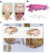
Nell-1 transgenic TF2B1 mouse compared with a nontransgenic littermate. (a) Left, newborn TF2B1 mouse with the scalp removed. Note the abnormal bulging of the occipital area and the relatively narrow width of the cranium. Right, an NF2 littermate. In the TF2B1 transgenic animal, the sagittal suture and several other sutures are closed. (b) Hematoxylin and eosin staining of TF2B1 sagittal suture. Premature closure of the suture is manifest in the severe overlap of calvarial edges (red arrows). The underlying brain tissue has been removed for RNA analysis. (c) Three-dimensional MCT reconstruction of a TF2B1 mouse (left) and its NF2 littermate (right). Note the area of premature midline suture closure in the TF2B1 mouse (arrow, left).
Figure 5
Figure 5
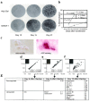
Effects of Nell-1 overexpression on mineralization and bone marker expression. (a) FRCC culture infected with 20 pfu/cell AdNell-1, stained with von Kossa stain. Control cell cultures were infected with Adβ-Gal. Experiments were performed in triplicate. Mineralized nodules are stained black. (b) Quantitation and statistical analysis of mineralized areas. AdNell-1–infected cultures demonstrated significantly greater mineralization than did Adβ-Gal controls. (c) AdNell-1–infected MC3T3 cells grown without ascorbic acid. Typical micronodule appearance is shown. Right panel represents alkaline phosphatase staining of a micronodule. (df) Microarrays of AdNell–infected MC3T3 cells on postinfection days 6, 9, and 12, respectively. Gene expression intensities have been normalized using standardized housekeeping genes (HKGs). Hybridization intensities of AdNell-1–infected cells are represented on the y axis. Hybridization intensities of Adβ-Gal–infected cells are represented on the x axis. HKGs r2 represents the correlation of housekeeping genes (filled squares) between the two samples. ECMs r2 represents the correlation of candidate gene expression (open squares) between the two samples. A photograph of the microarray reading is attached in the upper left corner of each diagram. A twofold or greater upregulation is represented in red, while a twofold or greater downregulation is represented in green (g) Table summarizing genes with a difference in expression that is twofold higher or lower after AdNell-1 infection. The ratio is calculated as Nell-1/β-Gal. Col, collagen.
Figure 6
Figure 6
Effect of Nell-1 downregulation on alkaline phosphatase expression and bone marker expression. (a) Western blot analysis of Nell-1 protein expression in rat FRCCs infected with 20 pfu/cell AdAntiNell-1 or Adβ-Gal control. Downregulation of approximately 60% is observed. (b) Alkaline phosphatase staining (in red) of FRCCs. AdAntiNell-1–infected cells have significantly less staining than do control and AdNell-1–infected cells. (c) Northern analyses of FRCCs on days 3, 6, 9, and 12 after infection. AdAntiNell-1–infected cells have significantly less osteocalcin and osteopontin expression. (d) Expression of osteocalcin (OC) and osteopontin (OP) measured by PhosphorImager and normalized by GAPDH.
Figure 7
Figure 7
Hypothetical model of Nell-1 function in premature suture closure. Dashed line represents potential modulation.

Similar articles

Cited by

References

    1. Cohen, M.M, Jr., and MacLean, R.E. 2000. Craniosynostosis: diagnosis, evaluation and management. 2nd edition. Oxford University Press. New York, New York, USA. 454 pp.
    1. Coffin JD, et al. Abnormal bone growth and selective translational regulation in basic fibroblast growth factor (FGF-2) transgenic mice. Mol Biol Cell. 1995;6:1861–1873. - PMC - PubMed
    1. Carlton MB, Colledge WH, Evans MJ. Crouzon-like craniofacial dysmorphology in the mouse is caused by an insertional mutation at the Fgf3/Fgf4 locus. Dev Dyn. 1998;212:242–249. - PubMed
    1. Jabs EW, et al. A mutation in the homeodomain of the human MSX2 gene in a family affected with autosomal dominant craniosynostosis. Cell. 1993;75:443–450. - PubMed
    1. Liu YH, et al. Premature suture closure and ectopic cranial bone in mice expressing Msx2 transgenes in the developing skull. Proc Natl Acad Sci USA. 1995;92:6137–6141. - PMC - PubMed

Publication types

MeSH terms