. 2002 Sep;161(3):947-56.
doi: 10.1016/S0002-9440(10)64255-1.

Tumor-associated macrophages express lymphatic endothelial growth factors and are related to peritumoral lymphangiogenesis

Affiliations

Tumor-associated macrophages express lymphatic endothelial growth factors and are related to peritumoral lymphangiogenesis

Sebastian F Schoppmann et al. Am J Pathol. 2002 Sep.

Abstract

Formation of lymphatic metastasis is the initial step of generalized spreading of tumor cells and predicts poor clinical prognosis. Lymphatic vessels generally arise within the peritumoral stroma, although the lymphangiopoietic vascular endothelial growth factors (VEGF)-C and -D are produced by tumor cells. In a carefully selected collection of human cervical cancers (stage pT1b1) we demonstrate by quantitative immunohistochemistry and in situ hybridization that density of lymphatic microvessels is significantly increased in peritumoral stroma, and that a subset of stromal cells express large amounts of VEGF-C and VEGF-D. The density of cells producing these vascular growth factors correlates with peritumoral inflammatory stroma reaction, lymphatic microvessel density, and indirectly with peritumoral carcinomatous lymphangiosis and frequency of lymph node metastasis. The VEGF-C- and VEGF-D-producing stroma cells were identified in situ as a subset of activated tumor-associated macrophages (TAMs) by expression of a panel of macrophage-specific markers, including CD68, CD23, and CD14. These TAMs also expressed the VEGF-C- and VEGF-D-specific tyrosine kinase receptor VEGFR-3. As TAMs are derived from monocytes in the circulation, a search in peripheral blood for candidate precursors of VEGFR-3-expressing TAMs revealed a subfraction of CD14-positive, VEGFR-3-expressing monocytes, that, however, failed to express VEGF-C and VEGF-D. Only after in vitro incubation with tumor necrosis factor-alpha, lipopolysaccharide, or VEGF-D did these monocytes start to synthesize VEGF-C de novo. In conclusion VEGF-C-expressing TAMs play a novel role in peritumoral lymphangiogenesis and subsequent dissemination in human cancer.


Figures
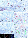
Figure 1.
Figure 1.
Identification of lymphatic vessels (A–F) in consecutive serial sections of a representative case of stage pT1b1 squamous cell carcinoma of the uterine cervix. A: All vessels are immunolabeled (arrowheads) with anti-CD34 antibody that does not discriminate between lymphatic and blood vessel endothelial cells. Besides several vessels, including small arteries (arrows) within the tumor stroma, two vessels are also labeled within the tumor (Tu). B: Consecutive section labeled with rabbit anti-podoplanin antibody, revealing six sections through lymphatic vessels (arrowheads), whereas other vessels (arrows) that were marked by CD34 antibody in the previous section were podoplanin-negative and are blood vessels. C: Next consecutive section immunostained with rabbit anti-LYVE 1 IgG that marks the same lymphatic vessels (arrowheads) as podoplanin in the previous level. Blood vessels are not marked (arrows). D–F: Double immunofluorescence on a squamous cell carcinoma with mouse antibodies to podoplanin (green, D) and rabbit anti-LYVE 1 IgG (red, E) reveals perfect overlap of both markers on the same lymphatic vessel (yellow, F). G–I: Association of peritumoral lymphatic vessels (G), CD68-positive TAMs (H), and VEGF-C-producing cells (I) on consecutive sections of cervix squamous epithelial carcinoma. G: Lymphatic vessels (Ly) are localized rabbit anti-podoplanin antibody within the peritumoral mononuclear infiltrate surrounding infiltrative extensions of the invasive carcinoma (Tu). H: CD68+ TAMs are localized close to the tumor and surround the lymphatic vessels (outlined in green; position was deduced from the preceding section and indicates vessels that were free of erythrocytes in higher magnification in this section). I: VEGF-C is expressed in tumor cells (Tu) in a granular pattern, as well as in peritumoral inflammatory cells (arrows), some of which are associated with the surface of the invading tumor (arrowheads). Original magnifications: ×350 (A–C); ×600 (D–F); ×420 (G–I).
Figure 2.
Figure 2.
Correlations between lymphatic vessel density and clinical parameters. A: There was a clear trend toward higher LMVD in low-grade squamous intraepithelial lesions (LSILs), high-grade squamous intraepithelial lesions (HSILs), and squamous cell cervical cancers (Ca) compared to normal cervical tissue that, however, failed to reach statistical significance. (P = 0.078, Kruskal Wallis test). B: There was a significant correlation between LMVD and the grade of inflammatory stroma reaction (1, absent; 2, moderate; 3, dense homogenous inflammatory infiltrate) (P = 0.012, Kruskal-Wallis test). C: LMVD was significantly increased in cervical cancers with invasion of peritumoral lymphatic vessels by tumor cells (lymphangiosis carcinomatosa) (pos.) when compared to those without (neg.) (11.39 ± 1.62 versus 6.06 ± 1.36) (P = 0.014, Mann-Whitney test). D: The number of VEGF-C-expressing peritumoral cells correlated with the grade of inflammatory stroma reaction in invasive cancers as well as in LSILs (P = 0.043, Kruskal Wallis test). E: LMVD in squamous carcinomas, stage pT1b1, with ≤35 VEGF-C-positive stroma cells was significantly lower than in those cancer samples with >35 VEGF-C-positive cells (6.11 ± 1.53 versus 10.87 ± 1.45) (P = 0.014, Mann-Whitney test), using the median value of 35 VEGF-C-positive stroma cells as cutoff score. F: Lymphatic vessel invasion of tumor cells was highly associated with the presence of lymph node metastases (P = 0.008, chi-square test).
Figure 3.
Figure 3.
Localization of VEGF-C-expressing cells by in situ hybridization (A) and immunofluorescence (B). A: Expression of VEGF-C mRNA is found by in situ hybridization in highest concentration in cells within the peritumoral stroma, some of which directly sit on the surfaces (arrowheads) of the infiltrating tumor extensions (T). Small amounts of VEGF-C mRNA are also expressed by tumor cells (arrows). These results are representative for four different patients. For negative control, hybridization with sense probe was performed and no signal was found (data not shown). B: When VEGF-C and lymphatic vessels are co-localized by double immunofluorescence and confocal laser-scanning microscopy, individual and clusters of VEGF-producing cells (green channel) frequently adjoin the lymphatic vessel wall marked by podoplanin (red channel). Nuclear counterstaining was performed with propidium iodide (blue channel). Original magnifications: ×600 (A); ×2500 (B).
Figure 4.
Figure 4.
Identification of VEGF-C- and VEGF-D-producing peritumoral cells as TAMs. Analysis was performed on paraffin sections of cervical carcinomas by double-labeling confocal laser-scanning microscopy. VEGF-C-expressing cells (green channel) are aligned in the left columns, and the counterlabeling antibodies are displayed in the red channel. This analysis indicates that VEGF-C-expressing cells are also labeled with antibodies specific for CD68, HLA-DR, partially CD16, CD23, CD45, and VEGF-D. Nuclei were counterstained with propidium iodide (blue channel). Original magnifications, ×2500.
Figure 5.
Figure 5.
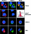
Co-expression of VEGFR-3 and VEGF-C in peritumoral stroma and in CD14 affinity-purified circulating human monocytes. In the peritumoral tissue, both VEGF-C (green channel) and VEGFR-3 (red channel) are co-expressed simultaneously by the same TAMs, and co-localize within the cytoplasm in a granular compartment, presumably in endosomes or lysosomes. By contrast, round CD14 affinity-purified monocytes express VEGFR-3 on their surface membrane, and in a perinuclear compartment, presumably the endoplasmic reticulum, but they do not express VEGF-C. The number of these CD14+ cells that express VEGFR-3 was determined in four different patients by fluorescence-activated cell sorting, and amounted on average to ∼60%. Incubation of these cells with exogenous VEGF-D and TNF-α induced de novo synthesis of VEGF-C. While incubated with VEGF-D (1 μg/ml, 30 minutes, 37°C) they remained round, TNF-α (50 U/ml, 30 minutes, 37°C) induced cell flattening and endocytosis of receptor-ligand complexes, similar to TAMs observed in vivo. Original magnifications, ×2500.
Figure 6.
Figure 6.
A and B: Activation of monocytes with TNF-α and LPS to produce VEGF-C and VEGF-D mRNA, detected by RT-PCR. Monocytes were isolated from peripheral blood by CD14 affinity purification (A) or by elutriation (B) at 4°C to avoid activation. These cells express constitutively VEGFR-3 (787 bp), but not VEGF-C (567 bp) and VEGF-D (525 bp). Incubation with TNF-α (50 U/ml) or LPS (50 U/ml for 30 minutes) causes rapid initiation of VEGF-C and VEGF-D mRNA production. C: The macrophage-related tumor cell line U937 constitutively produces CD68 and CD23, and also VEGF-C and VEGFR-3 without stimulation.
Figure 7.
Figure 7.
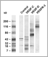
Expression of VEGF-C, VEGF-D, and VEGFR-3 protein in a lysate of isolated, cultured lymphatic endothelial cells (control) and in the human macrophage-related cell line U937. Tissue and cells were lysed in sodium dodecyl sulfate buffer, the proteins were separated by sodium dodecyl sulfate-polyacrylamide gel electrophoresis, transferred onto nitrocellulose, and immunoblotted with polyclonal antibodies specific for VEGF-C, VEGF-D, and VEGFR-3. VEGF-C is detected in the control and in U937 cells as an ∼21-kd protein, along with typical dimers and a degradation products. Also VEGF-D is encountered in U937 cells as an ∼21-kd band, in addition to oligomers. VEGFR-3 is expressed in U937 cells in its mature 195-kd form, and as a proteolytically processed 125-kd product.

Similar articles

Cited by

References

    1. Karpanen T, Alitalo K: Lymphatic vessels as targets of tumor therapy? J Exp Med 2001, 194:37-42 - PMC - PubMed
    1. Kaipainen A, Korhonen J, Mustonen T, van Hinsbergh VW, Fang GH, Dumont D, Breitman M, Alitalo K: Expression of the fms-like tyrosine kinase 4 gene becomes restricted to lymphatic endothelium during development. Proc Natl Acad Sci USA 1995, 92:3566-3570 - PMC - PubMed
    1. Joukov V, Pajusola K, Kaipainen A, Chilov D, Lahtinen I, Kukk E, Saksela O, Kalkkinen N, Alitalo K: A novel vascular endothelial growth factor, VEGF-C, is a ligand for the Flt4 (VEGFR-3) and KDR (VEGFR-2) receptor tyrosine kinases. EMBO J 1996, 15:290-298 - PMC - PubMed
    1. Yamada Y, Nezu J, Shimane M, Hirata Y: Molecular cloning of a novel vascular endothelial growth factor, VEGF-D. Genomics 1997, 42:483-488 - PubMed
    1. Partanen TA, Alitalo K, Miettinen M: Lack of lymphatic vascular specificity of vascular endothelial growth factor receptor 3 in 185 vascular tumors. Cancer 1999, 86:2406-2412 - PubMed

Publication types

MeSH terms

Substances

LinkOut - more resources