. 2002 Oct;76(19):9575-87.
doi: 10.1128/jvi.76.19.9575-9587.2002.

Vaccinia virus J1R protein: a viral membrane protein that is essential for virion morphogenesis

Affiliations

Vaccinia virus J1R protein: a viral membrane protein that is essential for virion morphogenesis

Wen-Ling Chiu et al. J Virol. 2002 Oct.

Abstract

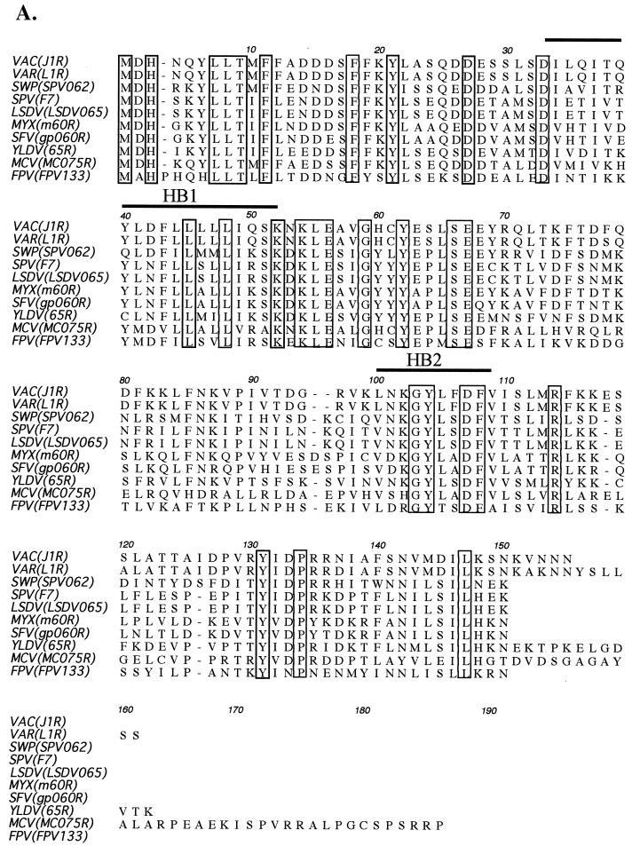
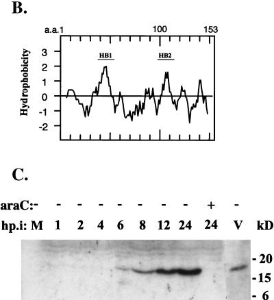
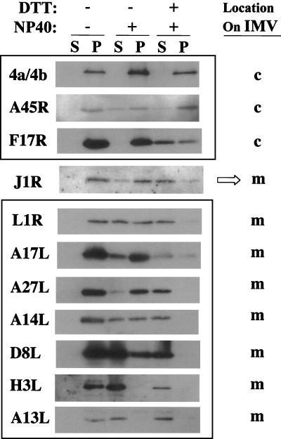
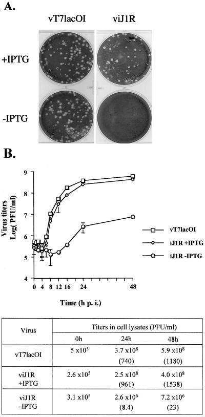
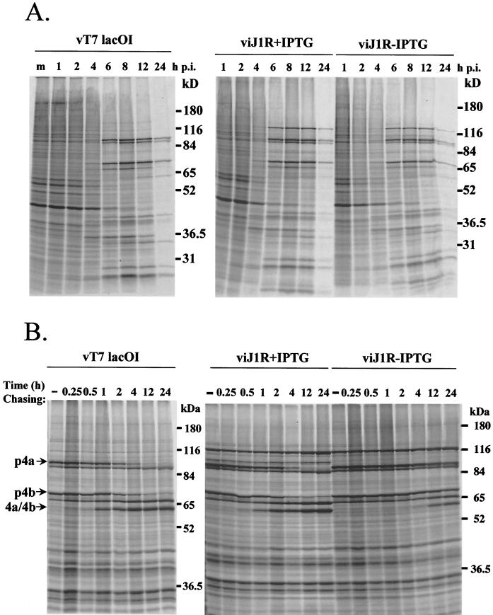
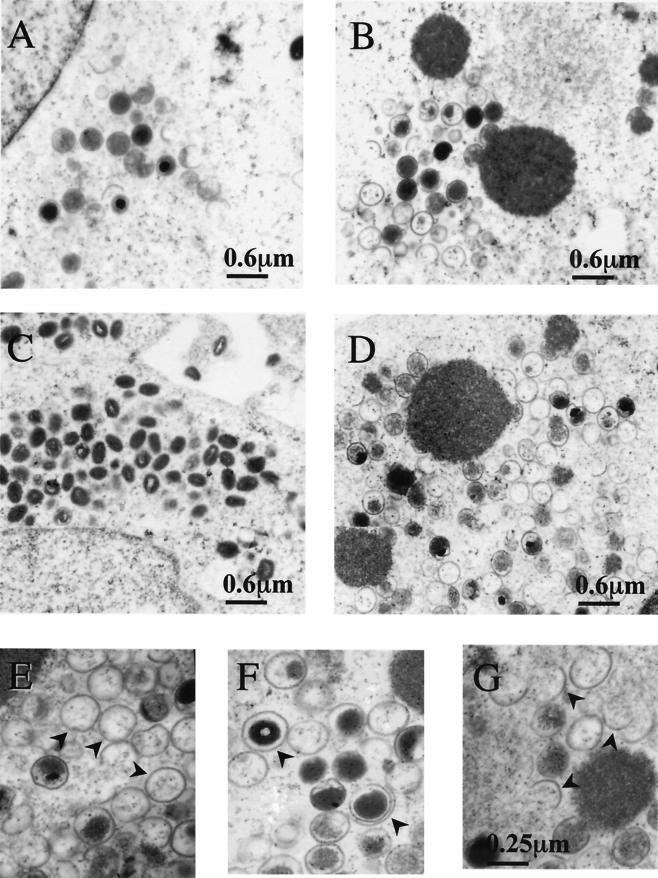
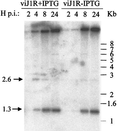
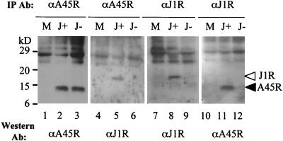
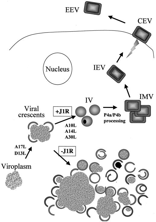
Vaccinia virus, a member of the poxvirus family, contains a conserved J1R open reading frame that encodes a late protein of 17.8 kDa. The 18-kDa J1R protein is associated mainly with the membrane fraction of intracellular mature virus particles. This study examines the biological function of J1R protein in the vaccinia virus life cycle. A recombinant vaccinia virus was constructed to conditionally express J1R protein in an isopropyl-beta-D-galactopyranoside (IPTG)-inducible manner. When J1R is not expressed during vaccinia virus infection, the virus titer is reduced approximately 100-fold. In contrast, J1R protein is not required for viral gene expression, as indicated by protein pulse-labeling. J1R protein is also not required for DNA processing, as the resolution of the concatemer junctions of replicated viral DNA was detected without IPTG. A deficiency of J1R protein caused a severe delay in the processing of p4a and p4b into mature core proteins 4a and 4b, indicating that J1R protein participates in virion morphogenesis. Infected cells grown in the absence of IPTG contained very few intracellular mature virions in the cytoplasm, and enlarged viroplasm structures accumulated with viral crescents attached at the periphery. Abundant intermediate membrane structures of abnormal shapes were observed, and many immature virions were either empty or partially filled, indicating that J1R protein is important for DNA packaging into immature virions. J1R protein also coimmunoprecipited with A45R protein in infected cells. In summary, these results indicate that vaccinia virus J1R is a membrane protein that is required for virus growth and plaque formation. J1R protein interacts with A45R protein and performs an important role during immature virion formation in cultured cells.


Figures

FIG. 1.
FIG. 1.
Vaccinia virus J1R gene encodes a late protein. (A) Alignment of deduced amino acid sequences of vaccinia virus J1R and J1R orthologues in other poxviruses. Accession numbers are from GenBank. VAC, vaccinia virus (strain WR; accession no. P07616); VAR, variola virus (India-1967/isolate IND3; accession no. NP-042122); SWP, swine poxvirus (accession no. NP-570222); SPV, sheep poxvirus (accession no. P19746); LSDV, lumpy skin disease virus (accession no. AAK85026); MYX, myxoma virus (accession no. NP-051774); SFV, Shope fibroma virus (accession no. NP-051949); YLDV, Yaba-like disease virus (accession no. NP-073450); MCV, molluscum contagiosum virus subtype 1 (accession no. NP-044026); FPV, fowl poxvirus (accession no. NP-039096). The boxed sequences are identical amino acids, and the thick lines indicate hydrophobic sequences (HB). (B) Hydropathy plot of J1R protein. a.a., amino acid. (C) Expression of J1R protein in infected cells and IMV. BSC40 cells were infected with wild-type vaccinia virus at an MOI of 5 PFU per cell and harvested at the indicated times. Lysates were separated by SDS-12% PAGE and transferred to nitrocellulose for Western blotting with rabbit antibody against J1R protein. araC, cytosine β-d-arabinofuranoside; hp.i, hours postinfection; V, purified IMV particles.
FIG. 1.
FIG. 1.
Vaccinia virus J1R gene encodes a late protein. (A) Alignment of deduced amino acid sequences of vaccinia virus J1R and J1R orthologues in other poxviruses. Accession numbers are from GenBank. VAC, vaccinia virus (strain WR; accession no. P07616); VAR, variola virus (India-1967/isolate IND3; accession no. NP-042122); SWP, swine poxvirus (accession no. NP-570222); SPV, sheep poxvirus (accession no. P19746); LSDV, lumpy skin disease virus (accession no. AAK85026); MYX, myxoma virus (accession no. NP-051774); SFV, Shope fibroma virus (accession no. NP-051949); YLDV, Yaba-like disease virus (accession no. NP-073450); MCV, molluscum contagiosum virus subtype 1 (accession no. NP-044026); FPV, fowl poxvirus (accession no. NP-039096). The boxed sequences are identical amino acids, and the thick lines indicate hydrophobic sequences (HB). (B) Hydropathy plot of J1R protein. a.a., amino acid. (C) Expression of J1R protein in infected cells and IMV. BSC40 cells were infected with wild-type vaccinia virus at an MOI of 5 PFU per cell and harvested at the indicated times. Lysates were separated by SDS-12% PAGE and transferred to nitrocellulose for Western blotting with rabbit antibody against J1R protein. araC, cytosine β-d-arabinofuranoside; hp.i, hours postinfection; V, purified IMV particles.
FIG. 2.
FIG. 2.
Membrane and core proteins after NP-40-DTT extraction of vaccinia virus IMV. Sucrose-purified vaccinia virus IMV were incubated with buffer containing 1% NP-40 and 50 mM DTT or no DTT. After centrifugation, the supernatant (S) and the insoluble pellet (P) were analyzed with antibodies as described in Materials and Methods. c, core; m, membrane.
FIG. 3.
FIG. 3.
Construction of viJ1R for conditional expression of J1R protein. (A) Schematic diagram of the parental virus vT7lacOI, the intermediate virus vJ1R/iJ1R, and the final mutant virus viJ1R. The J1R, J2R (thymidine kinase [TK]), and A56R (hemagglutinin) loci are indicated. DNA fragments inserted into these loci are shown below the lines. Abbreviations: T7 pol, bacteriophage T7 RNA polymerase gene; lacO, E. coli lac operator; PL, viral late promoter; PE/L, viral early and late promoters; lacI, E. coli lac repressor; EMC, encephalomyocarditis virus cap-independent translation enhancer element; gpt, E. coli guanine phophoribosyltransferase gene; P7.5 and P11, viral promoters; PT7, promoter for T7 RNA polymerase. (B) Expression of J1R protein in cells infected with viJ1R. Cells were infected with vT7lacOI or viJ1R at an MOI of 5 PFU per cell and harvested for Western blotting with antibody against J1R protein. M, mock-infected cells.
FIG. 4.
FIG. 4.
Characterization of viJ1R. (A) viJ1R mutant virus does not form plaques on BSC40 cells. BSC40 cells were infected with vT7lacOI or viJ1R, incubated in medium for 3 days, fixed, stained with crystal violet, and photographed. (B) One-step growth curve analysis of viJ1R virus. BSC40 cells were infected with parental vT7lacOI or viJ1R at an MOI of 5 PFU per cell; incubated in normal medium or medium with IPTG; and harvested at 0, 1, 2, 4, 6, 8, 12, 16, 24, and 48 h p.i. Error bars indicate standard deviations. Virus titers in the lysates were determined by plaque formation assays with BSC40 cells and are listed in the table. Numbers in parentheses are the fold increase in virus titer, determined as the virus titer at 24 or 48 h p.i. divided by the virus titer at 0 h.
FIG. 5.
FIG. 5.
Viral protein synthesis in cells infected by viJ1R. (A) Pulse-labeling of viral proteins. BSC40 cells were infected with vT7lacOI or viJ1R at an MOI of 10 PFU per cell in the presence (+) or absence (−) of 50 μM IPTG. The cells were labeled with [35S]methionine (50 μCi/ml) for 15 min at 1, 2, 4, 6, 8, 12, and 24 h p.i. m, mock infected. Immediately after labeling, the cells were washed and lysed, and the labeled proteins were separated by SDS-10% PAGE. (B) Pulse-chase analysis of precursor p4a and p4b processing. BSC40 cells were infected with either vT7lacOI or viJ1R in the presence (+) or absence (−) of 50 μM IPTG. At 8 h p.i., the cells were pulse-labeled with [35S]methionine for 30 min and either immediately harvested (−) or chased with normal medium for 0.25, 0.5, 1, 2, 4, 12, or 24 h. Proteins were denatured and analyzed by SDS-10% PAGE followed by autoradiography. The mobilities of p4a and p4b and their mature processed forms, 4a and 4b, are shown at the left (68, 69).
FIG. 6.
FIG. 6.
Electron micrographs of vaccinia virion morphogenesis in cells infected with viJ1R. BSC40 cells were infected with viJ1R virus at an MOI of 20 PFU per cell either in the presence (A and C) or in the absence (B and D to G) of IPTG and fixed at 12 h (A and B) or 24 h (C to G) p.i. for electron microscopy. Photos were taken at magnifications of ×12,000 (A to D) and ×30,000 (E to G). Arrowheads in panels E to G represent aberrant membrane structures, such as empty IV (E), double-layer membranes (F), and half-circle membranes (G).
FIG. 7.
FIG. 7.
Processing of viral DNA. BSC40 cells were infected with viJ1R at an MOI of 10 PFU per cell and incubated with complete DMEM containing 10% CS with (+) or without (−) 50 μM IPTG. Cells were harvested at 2, 4, 8, and 24 h p.i. Viral DNA was extracted, digested with BstEII, separated on an 1% agarose gel, and transferred to nitrocellulose paper for hybridization with a 32P-end-labeled 70-mer oligonucleotide as described previously (12). The arrow labeled 1.3 marks the position of the diagnostic 1.3-kb DNA fragment, which indicates the resolution of concatemers to monomeric DNA ends.
FIG. 8.
FIG. 8.
Specific interaction between A45R and J1R proteins. BSC40 cells were either mock infected (M) or infected with viJ1R at an MOI of 5 PFU per cell and then incubated in medium with IPTG (J+) or without IPTG (J−). Cell lysates were harvested at 24 h p.i. and immunoprecipitated (IP) with antibody. Immune complexes were separated by SDS-12% PAGE and analyzed by Western blotting with anti-A45R (1:5,000) or anti-J1R (1:1,000) antibody.
FIG. 9.
FIG. 9.
J1R protein is required for DNA packaging in IV formation during vaccinia virion morphogenesis. Schematic drawing of the stages of vaccinia virus morphogenesis, including crescent formation, IV formation, and IMV formation. IMV could be enveloped to become IEV. J1R protein is not required for crescent formation; instead, it plays a role in DNA packaging in IV formation. Also shown are several other viral proteins (A10L, A14L, A17L, A30L, and D13L) in which a mutation interferes with virion morphogenesis before or during the formation of IV (20, 45, 47, 48, 59, 63, 79, 82). CEV, cell-associated virions; EEV, extracellular enveloped viruses.

Similar articles

Cited by

References

    1. Afonso, C. L., E. R. Tulman, Z. Lu, E. Oma, G. F. Kutish, and D. L. Rock. 1999. The genome of Melanoplus sanguinipes entomopoxvirus. J. Virol. 73:533-552. - PMC - PubMed
    1. Afonso, C. L., E. R. Tulman, Z. Lu, L. Zsak, G. F. Kutish, and D. L. Rock. 2000. The genome of fowlpox virus. J. Virol. 74:3815-3831. - PMC - PubMed
    1. Afonso, C. L., E. R. Tulman, Z. Lu, L. Zsak, F. A. Osorio, C. Balinsky, G. F. Kutish, and D. L. Rock. 2002. The genome of swinepox virus. J. Virol. 76:783-790. - PMC - PubMed
    1. Almazan, F., D. C. Tscharke, and G. L. Smith. 2001. The vaccinia virus superoxide dismutase-like protein (A45R) is a virion component that is nonessential for virus replication. J. Virol. 75:7018-7029. - PMC - PubMed
    1. Antoine, G., F. Scheiflinger, F. Dorner, and F. G. Falkner. 1998. The complete genomic sequence of the modified vaccinia Ankara strain: comparison with other orthopoxviruses. Virology 244:365-396. - PubMed

Publication types

LinkOut - more resources