. 2002 Nov 8;277(45):43216-23.
doi: 10.1074/jbc.M207662200. Epub 2002 Aug 29.

Targeted deletion of the Tsg101 gene results in cell cycle arrest at G1/S and p53-independent cell death

Affiliations

Targeted deletion of the Tsg101 gene results in cell cycle arrest at G1/S and p53-independent cell death

Andrea Krempler et al. J Biol Chem. .

Abstract

The tumor susceptibility gene 101 (Tsg101) was originally discovered in a screen for potential tumor suppressors using insertional mutagenesis in immortalized fibroblasts. To investigate essential functions of this gene in cell growth and neoplastic transformation, we derived primary mouse embryonic fibroblasts from Tsg101 conditional knockout mice. Expression of Cre recombinase from a retroviral vector efficiently down-regulated Tsg101. The deletion of Tsg101 caused growth arrest and cell death but did not result in increased proliferation and cellular transformation. Inactivation of p53 had no influence on the deleterious phenotype, but Tsg101(-/-) cells were rescued through expression of exogenous Tsg101. Fluorescence-activated cell sorting, proliferation assays, and Western blot analysis of crucial regulators of the cell cycle revealed that Tsg101 deficiency resulted in growth arrest at the G(1)/S transition through inactivation of cyclin-dependent kinase 2. As a consequence, DNA replication was not initiated in Tsg101-deficient cells. Our results clearly demonstrate that Tsg101 is not a primary tumor suppressor in mouse embryonic fibroblasts. However, the protein is crucial for cell proliferation and cell survival.


Figures
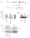
Fig. 1
Fig. 1. Cre-mediated excision and transcriptional down-regulation of the Tsg101 gene in MEFs, which were derived from Tsg101fl/fl embryos
A, diagram of the Cre-mediated deletion of the Tsg101 gene. X, XbaI. B, verification of the recombination event by XbaI Southern analysis in Tsg101fl/fl cells (pBabe or pBabe-Cre) as illustrated in A. DNA from a tail biopsy of a Tsg101 heterozygous knockout animal served as a positive control (+/−). The null allele was generated previously by transmitting the floxed allele through the female germ-line of MMTV-Cre (line A) transgenic mice (Footnote 2). The 2.1-kb XbaI fragment represents the recombined Tsg101 null allele. Both the wild type and unrecombined floxed allele are represented by a 2.7-kb XbaI restriction fragment. C, Northern blot analysis. Tsg101 mRNA was almost undetectable in Tsg101fl/fl MEFs expressing Cre recombinase (pBabe-Cre), whereas the infection with a control virus had no effect on Tsg101 transcription (pBabe). D, Western blot analysis. Tsg101 protein was almost undetectable in MEFs expressing Cre recombinase (pBabe-Cre). The infection with a retroviral control vector (pBabe) had no significant effect on Tsg101 translation or its stability compared with untreated control cells (no virus). The SL6 cell line and its parental NIH3T3 cell line (1) were used as negative and positive controls for Tsg101 protein expression. SL6 cells still express significant amounts of the Tsg101 protein. β-Actin (ActB) served as a loading control.
Fig. 2
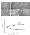
Fig. 2. Reduced cell growth and cell death associated with Tsg101 deficiency
A: i, untreated Tsg101fl/fl MEFs in culture. ii, 24 h after infection with a retroviral vector expressing Cre recombinase. iii–v, 3, 5, and 7 days after infection with a Cre vector and selection with puromycin. vi, MEFs derived from wild type embryos (Tsg101+/+) were infected with the same Cre expressing viral vector and selected for 7 days with puromycin. Low level expression of Cre recombinase and selection with puromycin did not result in significant growth inhibition in wild type control cells. B, growth curves of pBabe and pBabe-Cre-infected Tsg101fl/fl and Tsg101+/+ MEFs as determined by MTT color assay. The A570 nm values corresponding to cell numbers for Tsg101 knockout cells in the assay decreased, whereas cell numbers in the controls increased within 4 days. Error bars correspond to standard deviations.
Fig. 3
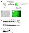
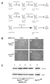
Fig. 3. Restoration of cell proliferation and survival of cells lacking the endogenous Tsg101 gene through expression of a HA-tagged Tsg101 protein from a retroviral vector
A, experimental design. Homozygous floxed MEFs were simultaneously infected with retroviral vectors expressing Cre recombinase (puromycin resistance) and an HA-tagged mouse Tsg101 protein (GFP selection marker). B and C, puromycin-resistant clones expressing the green fluorescent protein were isolated, expanded, and subjected to Western analysis. D, Western blot analysis of Tsg101 and β-actin (ActB) as loading control on puro-mycin and GFP positive clones (numbers 1, 3, 6, 7, 9, and 10). C1, control cells expressing Tsg101 from the endogenous locus. The HA-tagged Tsg101 protein is slightly larger than the wild type protein. E, XbaI Southern blot analysis to verify the presence of two Tsg101 knockout alleles on the genomic level. C1, control cells with two floxed alleles; C2, Tsg101fl/fl controls infected with pBabe-Cre. Two selected clones numbered 6 and 10 completely lack endogenous Tsg101 expression.
Fig. 4
Fig. 4. Effects of Tsg101 deficiency on cell cycle progression
A, fluorescence-activated cell sorting analysis of the DNA content. The number of Tsg101-deficient cells (Tsg101fl/fl, pBabe-Cre) in S-phase is significantly reduced compared to untreated cells (Tsg101fl/fl). Expression of Cre recombinase had no significant effect on cell cycle progression in wild type control MEFs (Tsg101+/+). B, percentages of cells in different stages of the cell cycle over time. pBabe-Cre-infected Tsg101fl/fl cells contained only a few cells in S-phase (bold). The distribution of cells in Tsg101 knockout cells resembled the numbers observed in serum-starved MEFs.
Fig. 5
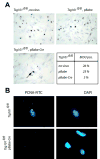
Fig. 5. Analysis of cell proliferation in Tsg101-deficient MEFs
A, BrdUrd labeling of untreated Tsg101fl/fl cells (no virus) as well as Tsg101fl/fl cells infected with the pBabe control vector or pBabe-Cre. The arrow points to one of the few BrdUrd-labeled cells in the Tsg101 knockout MEFs. B, immunocytochemistry of DNA-bound PCNA protein using a fluorescein isothiocyanate (FITC)-labeled antibody against PCNA in Tsg101fl/fl controls and Tsg101fl/fl MEFs expressing Cre re-combinase. DNA bound PCNA was virtually absent from the Tsg101 knockout MEFs. 4,6-Diamidino-2-phenylindole (DAPI) staining was used to visualize nuclei of Tsg101-deficient cells and their controls.
Fig. 6
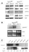
Fig. 6. Analysis of cyclin expression and cdk2 activity
A, Western blot analysis of G1, G1/S, S, and M phase cyclins. The S and M phase cyclins A2 and B1 were markedly down-regulated in Tsg101-deficient cells. B, Northern blot showing the expression of cyclin A2. Cyclin A2 mRNA was not detectable in Tsg101-deficient cells. Equal loading of RNA from Tsg101fl/fl and Cre-expressing Tsg101fl/fl cells was demonstrated by visualization of 28 S and 18 S RNA in the agarose gel used for blotting. C, cdk2 kinase assay using histone H1 as a substrate. Tsg101fl/fl MEFs infected with pBabe-Cre vector showed reduced cdk2 activity. Equal protein loading was confirmed by reprobing the blot with the cdk2 antibody used for immunoprecipitation.
Fig. 7
Fig. 7. Analysis of cell cycle regulator expression
A, Western blot analysis of Mdm2, p19ARF, and the main cdk inhibitory proteins p21waf1/cip1 and p27Kip1. Blots were reprobed with β-actin (ActB) to confirm protein loading. Expression of cdk inhibitory proteins was largely unaffected in Tsg101 knockout cells.
Fig. 8
Fig. 8. Cell death in Tsg101-deficient cells with impaired p53 function
A, different strategies used to obtain p53-deficient Tsg101fl/fl cells. In all three approaches (mutation of p53 through immortalization of Tsg101fl/fl MEFs; infection of Tsg101fl/fl MEFs with human papilloma virus E6 virus; Tsg101fl/flp53−/− MEFs) deletion of Tsg101 by Cre-mediated recombination resulted in cell death. B, lack of proliferation and cell death in Tsg101−/− p53-deficient cells. i-iv, 4 and 7 days after pBabe-Cre infection and puromycin selection of Tsg101fl/fl/human papilloma virus E6 and Tsg101fl/flp53−/− MEFs. Both cell types underwent rapid cell death after deletion of Tsg101. C, Western blot analysis of four surviving colonies in Tsg101fl/flp53−/− cell cultures that were infected with pBabe-Cre and selected with puromycin. The four samples showed normal expression of Tsg101 when compared with a noninfected Tsg101fl/fl cell lysate (C).

Similar articles

Cited by

References

    1. Li L, Cohen SN. Cell. 1996;85:319 –329. - PubMed
    1. Li L, Li X, Francke U, Cohen SN. Cell. 1997;88:143–154. - PubMed
    1. Ali IU, Lidereau R, Theillet C, Callahan R. Science. 1987;238:185–188. - PubMed
    1. Reeve AE, Sih SA, Raizis AM, Feinberg AP. Mol Cell Biol. 1989;9:1799 –1803. - PMC - PubMed
    1. Steiner P, Barnes DM, Harris WH, Weinberg RA. Nat Genet. 1997;16:332–333. - PubMed

Publication types

MeSH terms

Substances