. 2002 Aug 19;158(4):659-68.
doi: 10.1083/jcb.200204081. Epub 2002 Aug 19.

Rab32 is an A-kinase anchoring protein and participates in mitochondrial dynamics

Affiliations

Rab32 is an A-kinase anchoring protein and participates in mitochondrial dynamics

Neal M Alto et al. J Cell Biol. .

Abstract

A-kinase anchoring proteins (AKAPs) tether the cAMP-dependent protein kinase (PKA) and other signaling enzymes to distinct subcellular organelles. Using the yeast two-hybrid approach, we demonstrate that Rab32, a member of the Ras superfamily of small molecular weight G-proteins, interacts directly with the type II regulatory subunit of PKA. Cellular and biochemical studies confirm that Rab32 functions as an AKAP inside cells. Anchoring determinants for PKA have been mapped to sites within the conserved alpha5 helix that is common to all Rab family members. Subcellular fractionation and immunofluorescent approaches indicate that Rab32 and a proportion of the cellular PKA pool are associated with mitochondria. Transient transfection of a GTP binding-deficient mutant of Rab32 promotes aberrant accumulation of mitochondria at the microtubule organizing center. Further analysis of this mutant indicates that disruption of the microtubule cytoskeleton results in aberrantly elongated mitochondria. This implicates Rab32 as a participant in synchronization of mitochondrial fission. Thus, Rab32 is a dual function protein that participates in both mitochondrial anchoring of PKA and mitochondrial dynamics.


Figures

Figure 1.
Figure 1.
Identification of Rab32 as a putative AKAP. Murine RIIα 1–45/LexA fusion was used as bait to screen a human brain cDNA library using the yeast two-hybrid assay. (A) Detection of positive colonies expressing Rab32 and other AKAP fragments were detected by β-galactosidase. The name of each AKAP is indicated. (B) Control and bacterial extracts expressing the Rab32 106–225 fragment (indicated above each lane) were separated on SDS-PAGE (4–15%) and electrotransferred to nitrocellulose. RII binding was assessed by overlay using 32P-labeled RIIα and detected by autoradiography. Molecular weight markers are indicated.
Figure 2.
Figure 2.
Mapping the RII binding domain of Rab32. (A) Schematic representation of the mapping strategy to identify the RII binding domain of Rab32. A family of 20-mer peptides (each offset by three residues) spanning the region 153–224 of Rab32 were synthesized and immobilized to a membrane support. (B) Solid-phase binding of RII was assessed by the RII overlay procedure. The sequence of the RII binding peptide is indicated using the one letter amino acid code (*). (C) Ribbon diagram of the Rab3a backbone based upon coordinates provided by Ostermeier and Brunger (1999). Top (left) and side (right) views are presented. The α5 helix is marked (red). (D) Helical wheel alignment of residues 181–193 of Rab32. Hydrophobic (white circles) and hydrophilic (yellow circles) residues are indicated. (E) Purified recombinant GST–Rab32 fusion protein (2 μg) was separated by SDS-PAGE and electrotransferred to nitrocellulose. RII overlays were performed in the absence of competitor peptide (left), in the presence of 10 μM Rab32 175–205 peptide (middle), or in the presence of 10 μM Rab32 175–205 L188P peptide (right). Molecular weight markers are indicated. (F) Solution binding of recombinant RII and Rab32. [32P]RIIα was incubated with either GST–Rab32 or GST–Rab32L188P mutant followed by glutathione-Sepharose purification of the complex. RII binding was assessed by measuring the counts per minute (cpm) corresponding to [32P]RIIα bound to Rab32 (top). Equal amounts of GST fusion proteins were used in these experiments (bottom).
Figure 3.
Figure 3.
Rab32 can interact with the holoenzyme of PKA in mammalian cells. HEK-293 cells were transiently transfected with Flag–Rab32 or Flag–Rab32L188P cDNA. Triton X-100–soluble extracts were immunoprecipitated with Sepharose-conjugated antiFlag antibody. (A) Copurification of the PKA holoenzyme was measured by assaying for PKA catalytic subunit activity stimulated by exogenous cAMP. PKA activity was measured as pmol/min/mg of 32P incorporated into the PKA substrate kemptide using a filter paper binding assay (Corbin and Reimann, 1974). Specific PKA activity was blocked by 10 μM of the inhibitor PKI (5–24) peptide. (B) AntiFlag immunoprecipitates were separated on SDS-PAGE after the catalytic subunit had been eluted from the complex with exogenous cAMP and electrotransferred to nitrocellulose. RII overlay assays (top) were performed to determine if Rab32 was the only AKAP present in these fractions. Control experiments confirmed that equal levels of protein were immunoprecipitated in these experiments (bottom). A representative example of three independent experiments is shown.
Figure 4.
Figure 4.
Only Rab32 and RabRP-1 contain determinants for PKA anchoring. (A) The sequences of Rab32 and nine other Rab proteins are aligned within the α5 helix region. The first and last residues of each sequence and the name of each Rab protein are indicated. Boxed and shaded regions depict sequence identity and similarity. Star indicates the conserved phenylalanine in most Rab proteins. (B) A solid-phase peptide array of these Rab sequences was screened for RII binding by the overlay assay. Binding of [32P]RIIα was detected by autoradiography. (C) Quantitation of RII binding was assessed by densitometry. Arbitrary units were normalized to one, indicating the highest level of RII binding. The data presented are an amalgamation of two independent experiments. (D) Yeast two-hybrid analysis was used to determine if RII interacts with selected, full-length Rab family members. Two-hybrid crosses of RIIα 1–45 fragment with full-length cDNAs for Rab32, Rab32L188P, Rab5, Rab6, or Rab7 fused to the LexA DNA binding domain. Interactions were assayed by growth on minimal media plates in the absence of histidine (left). As a toxicity control, all cotransformed yeast strains were able to grow in the presence of histidine (right). (E) HEK-293 cell extracts expressing Flag-tagged Rab32 and mutants (indicated above each lane) were separated by SDS-PAGE on 4–15% gels and electrotransferred to nitrocellulose. Binding of [32P]RIIα was assessed by the overlay assay. (Top) RII binding was detected by autoradiography. Equal loading of recombinant Rab32 proteins was confirmed by Western blot using antiFlag antibodies. (F) Sequence alignment of Rab32 and its most closely related family members. The first and last residues of each sequence and the name of each Rab protein are indicated. Boxed and shaded regions depict sequence similarity and identity. Star indicates the position of alanine 185 in the Rab32 sequence. (G) A solid-phase peptide array of these closely related Rab sequences was screened for RII binding by the overlay assay. Binding of [32P]RIIα was detected by autoradiography.
Figure 5.
Figure 5.
The cellular and subcellular distribution of Rab32. The tissue distribution of Rab32 mRNA was assessed by Northern blot analysis. (A) A human multi-tissue (tissue sources indicated above each lane) was screened using a 113-bp cDNA probe that corresponds to the COOH-terminal hypervariable region of Rab32. Hybridization was detected by autoradiography. The sizes of DNA markers are indicated. (B) Triton X-100–soluble extracts from the WI-38 human fetal lung fibroblasts were immunoblotted with affinity-purified anti-Rab32 antibody (left) or preimmune sera (right). Signals were detected by chemiluminescence. Molecular weight standards are indicated. Confocal immunofluorescence microscopy of WI-38 fibroblasts triple labeled with polyclonal antibodies against Rab32 (C, green), cell-permeable dye MitoTracker RedTM (D, red), and a monoclonal antibody against α-tubulin (E, blue). A merged image (F) indicates the cellular distribution of all three signals. (G) Subcellular fractionation of WI-38 cells was performed according to the Materials and methods. Protein (20 μg each) from whole cell (W), nuclear (P1), and mitochondria-enriched (P2) fractions were subjected to SDS-PAGE and electrotransferred to nitrocellulose. Membranes were immunoblotted using affinity-purified polyclonal anti-Rab32 antibody (top) and a monoclonal anticytochrome oxidase subunit I (bottom) as a marker for mitochondria. Immunocytochemistry and confocal analysis were used to demonstrate the subcellular location of RII (H) and Rab32 (I). A merged image shows a significant overlap of the signals (J).
Figure 6.
Figure 6.
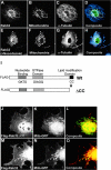
Mitochondrial targeting of Rab32. Immunofluorescence analysis of Rab 32 location upon depolymerization of microtubules in Cos7 cells. Control (A–D) or Cos7 cells treated with 5 μM nocodazole for 30 min (E–H) were fixed and stained. Immunocytochemistry was performed using anti-Rab32 (A and E, green), MitoTracker RedTM (B and F, red), and a monoclonal antibody against α-tubulin (C and G, blue). Composite images are shown for each sample (D and H). (I) Schematic representation of a nucleotide binding site, GTPase domain, and sites of putative lipid modification are indicated. Arrows indicate an amino acid substitution. (J) Flag-tagged wild-type or Rab32ΔCC mutant were expressed in Cos7 cells. The subcellular distribution of recombinant Rab32ΔCC (J) or Rab32 (M) were analyzed using confocal fluorescence microscopy with an antiFlag monoclonal antibody and Texas red–conjugated secondary (J and M). Mitochondria were visualized by using Mito–GFP targeting construct (K and N). Merged images are presented (L and O).
Figure 7.
Figure 7.
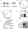
Nucleotide binding properties and GTPase activity of Rab32. Bacterially purified GST fusion proteins were assayed for guanine nucleotide binding. (A) Schematic diagram depicting a mutation in the nucleotide binding domain of Rab32. A nucleotide binding site, GTPase domain, and sites of putative lipid modifications are indicated. (B) GST–Rab32 (2 μg) was preloaded with 2 μCi of [3H]GDP. GDP off rate is presented as percent GDP bound to Rab32 over time. (C) Nucleotide-depleted Rab32 was incubated with [γ32P]GTP. The GTP on rate for GST–Rab32 was measured and is presented as the percentage of GTP bound over time. Amalgamated data from three experiments are presented. (D) Biochemical characterization of guanine nucleotide binding to recombinant wild-type GST–Rab32 and GST–Rab32T39N mutant. [3H]GDP binding (gray bars) and [γ32P]GTP binding (black bars) to Rab32 and Rab32T39N are indicated. Results are presented as the percent nucleotide bound normalized to 1. 2 μg of GST recombinant proteins were used and shown in the inset. A representative experiment from three independent analyses is presented. (E) GST pulldowns of GDP-loaded Rab32 or GTPγS-loaded Rab32 in the presence of recombinant RII (1 μg). Proteins were separated by SDS-PAGE and transferred to nitrocellulose. (Top) Coprecipitation of RII was detected by Western blot using an anti-RII monoclonal antibody. (Bottom) Control experiments showing equal loading of proteins. (F) Yeast coexpressing pLexA Rab32 mutants and pACT2 RII 1–45 were assayed for growth on minimal media without histidine (left). As a toxicity control, yeast harboring these plasmids can grow in media supplemented with histidine (right).
Figure 8.
Figure 8.
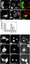
The GTP binding mutant Rab32T39N induces mitochondrial collapse around the microtubule organizing center. Cos7 cells were transiently transfected with Flag–Rab32 or Flag–Rab32T39N. Cells were labeled with MitoTracker RedTM for 30 min, fixed, and permeabilized before the Rab proteins were detected with a FITC-conjugated antiFlag antibody. (A) Rab32T39N mutant or (D) Rab32 and (B and E) mitochondria are shown. (C and F) Composite images are presented. A mitochondrial collapse phenotype is observed in Rab32T39N-expressing cells (B, inset). (G) Mitochondrial collapse was scored in cells expressing various Rab32 mutants (indicated below each column). Data are presented as the number of cells exhibiting the phenotype per number of cells transfected. 50 Rab-expressing cells were analyzed in three independent experiments for each construct. Control experiments were performed to determine if expression of Rab32T39N (H, K, and N) alters the morphology or distribution of other organelles. (I, L, and O) Mitochondria were visualized with MitoTracker RedTM. (J) Endosomes were stained with antibodies against the marker protein EEA-1. (M) Lysosomes were stained with antibodies against the marker protein LAMP-1. (P) Endoplasmic reticulums were stained with antibodies against the marker protein calnexin.
Figure 9.
Figure 9.
Expression of Rab32T39N leads to aberrant mitochondrial fusion. Analysis of mitochondrial morphology was performed on cells expressing various levels of Rab32T39N (A–H) and the wild-type protein (I–L). (A) Analysis of collapsed mitochondrial morphology in Cos7 cells expressing low levels of Flag–Rab32T39N. (B) Mitochondria were detected with MitoTracker RedTM stain. (C) A 3× enlargement of the boxed region shown in B (yellow box). (D) Composite image of A and B is presented. (E) Analysis of collapsed mitochondrial morphology in Cos7 cells expressing Flag–Rab32T39N treated with 5 μM nocodazole for 1 h. (F) Mitochondria were detected with MitoTracker RedTM stain. (G) A 3× enlargement of the boxed region shown in F (yellow box). (H) Composite image of E and F is presented. (I) Analysis of dispersed mitochondrial morphology in Cos7 cells expressing Flag–Rab32 treated with 5 μM nocodazole for 1 h. (J) Mitochondria were detected with MitoTracker RedTM stain. (K) A 3× enlargement of the boxed region shown in J (yellow box). (L) Composite image of I and J is presented.

Similar articles

Cited by

References

    1. Bao, X., A.E. Faris, E.K. Jang, and R.J. Haslam. 2002. Molecular cloning, bacterial expression and properties of Rab31 and Rab32. Eur. J. Biochem. 269:259–271. - PubMed
    1. Bock, J.B., H.T. Matern, A.A. Peden, and R.H. Scheller. 2001. A genomic perspective on membrane compartment organization. Nature. 409:839–841. - PubMed
    1. Bourne, H.R., D.A. Sanders, and F. McCormick. 1991. The GTPase superfamily: conserved structure and molecular mechanism. Nature. 349:117–121. - PubMed
    1. Carr, D.W., and J.D. Scott. 1992. Blotting and band-shifting: techniques for studying protein-protein interactions. Trends Biochem. Sci. 17:246–249. - PubMed
    1. Carr, D.W., R.E. Stofko-Hahn, I.D.C. Fraser, S.M. Bishop, T.S. Acott, R.G. Brennan, and J.D. Scott. 1991. Interaction of the regulatory subunit (RII) of cAMP-dependent protein kinase with RII-anchoring proteins occurs through an amphipathic helix binding motif. J. Biol. Chem. 266:14188–14192. - PubMed

Publication types

Substances