. 2002 Aug 19;196(4):517-27.
doi: 10.1084/jem.20011608.

The CD16(+) (FcgammaRIII(+)) subset of human monocytes preferentially becomes migratory dendritic cells in a model tissue setting

Affiliations

The CD16(+) (FcgammaRIII(+)) subset of human monocytes preferentially becomes migratory dendritic cells in a model tissue setting

Gwendalyn J Randolph et al. J Exp Med. .

Erratum in

  • J Exp Med 2002 Sep 16;196(6):869

Abstract

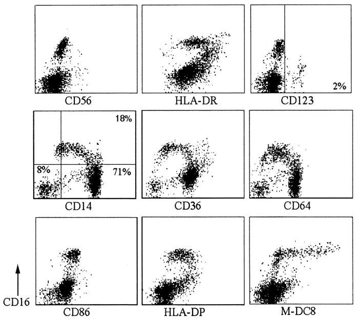
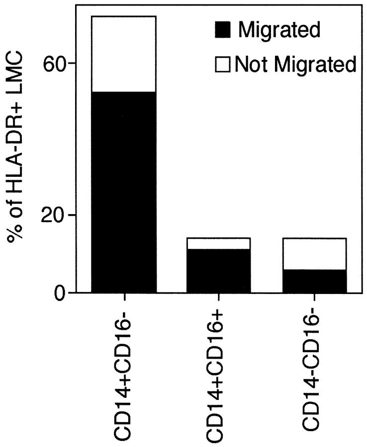
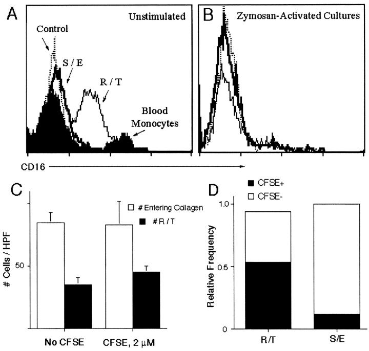
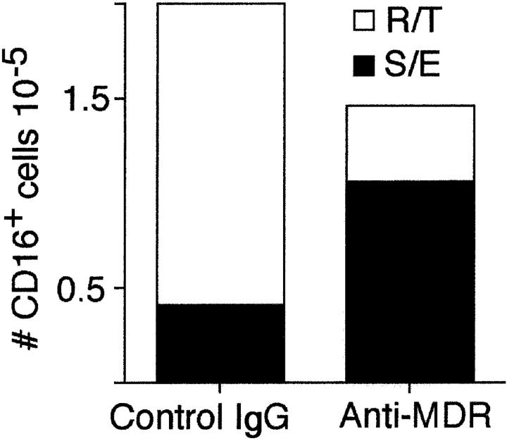
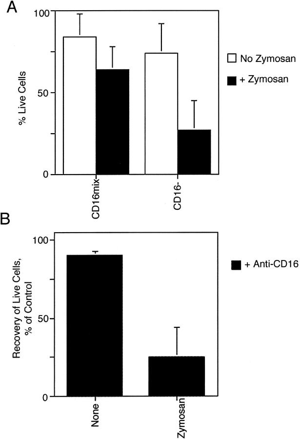
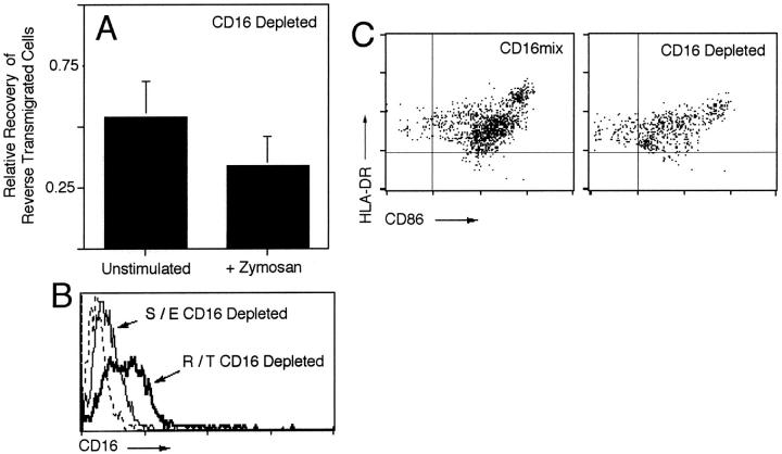
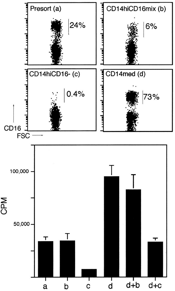
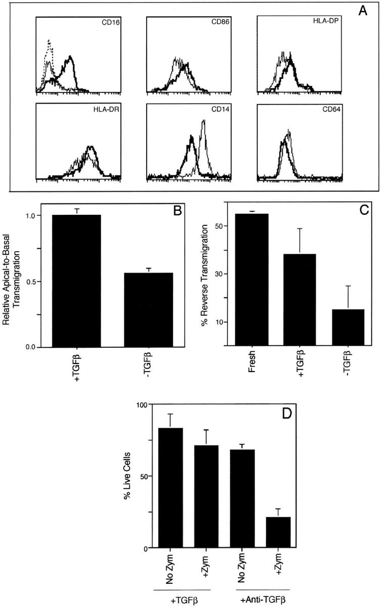
Much remains to be learned about the physiologic events that promote monocytes to become lymph-homing dendritic cells (DCs). In a model of transendothelial trafficking, some monocytes become DCs in response to endogenous signals. These DCs migrate across endothelium in the ablumenal-to-lumenal direction (reverse transmigration), reminiscent of the migration into lymphatic vessels. Here we show that the subpopulation of monocytes that expresses CD16 (Fcgamma receptor III) is predisposed to become migratory DCs. The vast majority of cells derived from CD16(+) monocytes reverse transmigrated, and their presence was associated with migratory cells expressing high levels of CD86 and human histocompatibility leukocyte antigen (HLA)-DR, and robust capacity to induce allogeneic T cell proliferation. A minority of CD16(-) monocytes reverse transmigrated, and these cells stimulated T cell proliferation less efficiently. CD16 was not functionally required for reverse transmigration, but promoted cell survival when yeast particles (zymosan) were present as a maturation stimulus in the subendothelial matrix. The cell surface phenotype and migratory characteristics of CD16(+) monocytes were inducible in CD16(-) monocytes by preincubation with TGFbeta1. We propose that CD16(+) monocytes may contribute significantly to precursors for DCs that transiently survey tissues and migrate to lymph nodes via afferent lymphatic vessels.


Figures

Figure 1.
Figure 1.
Cell surface markers distinguish CD16+ and CD16HLA-DR+ PBMCs. Freshly isolated PBMCs were depleted of CD56+ NK cells. Two-color flow cytometry was conducted with a gate set to exclude smaller PBMCs of the lymphocyte lineage. Expression of macrophage-associated markers CD14, CD36, and CD64 or DC-associated markers HLA-DR, HLA-DP, and CD86 (all x-axis) were examined. Cells were counterstained with a mAb to CD16 (y-axis). Some cells were stained with M-DC8 mAb to identify a subset of CD16+ cells (reference 11). Quadrant markers (shown in CD14 and CD123 panels) are positioned according to the level of fluorescence observed in cells stained with nonbinding isotype-matched control mAbs (lower left quadrant, negative staining). The phenotype illustrated was similarly observed among four donors examined.
Figure 2.
Figure 2.
Transendothelial migration of blood DC precursors across unstimulated endothelium. The entire fraction of freshly isolated PBMCs were incubated with endothelial cell/collagen cultures for 1.5 h to permit transmigration. The apical surface of the cultures was washed to collect nonmigrated cells, and the migrated population was recovered from the subendothelial collagen using collagenase D. Cells considered for quantitative evaluation were large mononuclear cells (LMC) uniformly positive for HLA-DR and negative for the B cell marker CD19 or T cell marker CD3. The plot shows the percent distribution of three distinct populations: CD14+CD16, CD14+CD16+, and CD14CD16 cells. The total height of each bar represents the relative distribution of these populations in freshly isolated PBMCs. The filled portion of each bar indicates the fraction of each population that emigrated beneath the endothelium, and the open portion of each bar represents the portion of the population that was recovered in the nonmigrated fraction. These data are representative of results obtained using PBMCs from three different blood donors.
Figure 3.
Figure 3.
Distribution of CD16+ monocyte-derived cells after coculture with endothelial cells grown on collagen. (A) Expression of CD16 was monitored in monocyte/endothelial cocultures at 48 h, when the majority of cells that will reverse transmigrate have done so. Assessments were made in reverse transmigrated (R/T, thin-lined profile) and subendothelial (S/E, bolded line profile) monocyte-derived cells from cultures that received no activation stimuli such as exogenous cytokines or phagocytic particles. Dotted line demarcates the staining intensity of cells incubated with isotype-matched mAbs to an irrelevant antigen. Filled profile represents the expression of CD16 in freshly isolated monocytes. (B) In other cultures, zymosan was included in the collagen to promote DC maturation. In some unstimulated endothelial cultures, the whole fraction of PBMCs (C) or CD16+ monocytes sorted using flow cytometry (D), were labeled with CFSE. Transendothelial migration into the collagen and subsequent reverse transmigration was evaluated to assess the distribution of CFSE+ cells. CFSE-labeled CD16+ monocytes were remixed with CD16 PBMCs so that CD16+ CFSE+ cells represented 15% of the total population (D).
Figure 4.
Figure 4.
Effect of a reverse transmigration antagonist on the distribution of CD16+ cells in endothelial cell cultures. PBMCs were incubated with endothelial/collagen cultures for 1.5 h, then washed to remove nonadherent, nonmigrated cells. Cultures were fed with medium containing mAb to MDR-1 or isotype-matched control mAb UPC10, and incubation was continued for 48 h to allow reverse transmigration. The presence of CD16+ cells in reverse-transmigrated and subendothelial leukocytes was monitored after 48 h by flow cytometry. The total number of CD16+ cells recovered from cultures in the presence and absence of anti-MDR-1 is shown. The fraction of such cells that had reverse transmigrated (R/T) is shown by the open portion of the bars, whereas the fraction that remained in the subendothelium (S/E) is shown by the filled portion of the bars.
Figure 5.
Figure 5.
Viability of reverse-transmigrated cells in the presence or absence of functional CD16. The viability of reverse-transmigrated cells was assessed by trypan blue staining after recovery from cultures lacking or containing zymosan. (A) Viability assessment from cultures in which CD16-depleted monocytes (CD16) or the full fraction of monocytes containing both CD16+ and CD16 subpopulations (CD16mix) were applied to the endothelium. (B) Recovery of live CD16mix monocytes in the presence of neutralizing anti-CD16 mAb 3G8 was calculated in relation to the number of live cells recovered in the presence of isotype-matched control antibody UPC10 under each condition.
Figure 6.
Figure 6.
Evaluation of reverse transmigration and expression of CD16 after depletion of peripheral blood CD16+ monocytes. CD56+ NK cells were depleted from the starting PBMC fraction, leaving a fraction of PBMCs that included both CD16 and CD16+(CD16mix) CD14+ monocytes. In some samples, the remaining CD16+ cells were depleted, leaving CD56CD16 PBMCs. CD16mix or CD16 PBMCs were applied to endothelial/collagen cultures at the same starting density. The number of reverse transmigrated cells in the CD16 fraction was evaluated after 2 d and compared in five independent experiments to the number of reverse transmigrated cells in the control CD16mix population of PBMCs (A). The relative recovery was calculated by setting equal to 1.0 the number of reverse transmigrated cells recovered per well of cultured endothelium after application of CD56CD16mix PBMCs and then determining the fractional recovery in each experiment when CD16-depleted PBMCs were applied. (B) The possibility that CD16 might be upregulated on peripheral blood cells that originally lacked CD16 was tested by examining the expression of CD16 in reverse-transmigrated and subendothelial populations after full depletion of CD16+ blood cells. Flow cytometric evaluation of CD16 expression in reverse transmigrated (R/T) and subendothelial (S/E) cells that arose from CD16-depleted PBMCs is plotted as a histogram. (C) Phenotyping for CD86 and HLA-DR expression was conducted in reverse-transmigrated populations derived in the presence or absence of CD16+ monocytes. The dot plots shown in panel C represent the number and phenotype of cells recovered from equal numbers of zymosan-containing endothelial cell microtiter cultures to which equivalent densities of CD16mix and CD16 monocytes were applied.
Figure 7.
Figure 7.
Capacity of reverse-transmigrated cells derived from monocyte subsets to stimulate allogeneic T cell proliferation. Monocytes were sorted according to their level of expression of CD14. First lymphocytes were removed from the PBMC fraction to generate a “presort” population of monocytes (a). Monocytes were then sorted into CD14hi (b, c) and CD14med (d) expressing cells. Some of the CD14hi monocytes were then treated with anti-CD16 miniMACs beads to remove any remaining CD16+ cells (c). These populations were added to endothelial cultures in which zymosan was present in the subendothelial matrix, and then live reverse transmigrated cells collected 2 d later were tested for their ability to induce allogeneic T cell proliferation at a ratio of 1 antigen-presenting cell: 80 T cells. In some conditions, a 1:1 mix of some reverse-transmigrated populations (d + b, or d + c) were used as APCs. Background counts for T cells cultured alone were <300. This pattern of responses was observed in 2 independent analyses.
Figure 8.
Figure 8.
Effect of TGFβ1 on the phenotype, migratory ability, and survival of CD16 monocytes. PBMCs were depleted of CD16+ cells using miniMACS magnetic selection. Then remaining cells were cultured for up to 3 d in TGFβ1 (bold lines) or anti-TGFβ (thin lines) to block activity of endogenous TGFβ. (A) The cell surface phenotype of these cultured cells was assessed by flow cytometry. Control mAb staining is depicted as a dashed line in top left histogram. (B) Monocytes cultured overnight in TGFβ1 or anti-TGFβ were applied to endothelial monolayers grown on collagen gels lacking zymosan and apical-to-basal transendothelial migration was quantified after a 1.5 incubation. (C) Reverse transmigration was quantified at 48 h as the percent of cells that originally migrated across the endothelium in each condition and then later retraversed the endothelium in ablumenal-to-lumenal direction. (D) Percentage of live cells in the reverse transmigrated populations derived from TGFβ1 or anti-TGFβ treated monocytes incubated with endothelial cultures lacking or containing zymosan within the subendothelium was assessed by trypan blue exclusion.

Similar articles

Cited by

References

    1. O'Doherty, U., R.M. Steinman, M. Peng, P.U. Cameron, S. Gezelter, I. Kopeloff, W.J. Swiggard, M. Pope, and N. Bhardwaj. 1993. Dendritic cells freshly isolated from human blood express CD4 and mature into typical immunostimulatory dendritic cells after culture in monocyte-conditioned medium. J. Exp. Med. 178:1067–1076. - PMC - PubMed
    1. Thomas, R., L.S. Davis, and P.E. Lipsky. 1993. Isolation and characterization of human peripheral blood dendritic cells. J. Immunol. 150:821–834. - PubMed
    1. Siegal, F.P., N. Kadowaki, M. Shodell, P.A. Fitzgerald-Bocarsly, K. Shah, S. Ho, S. Antonenko, and Y.J. Liu. 1999. The nature of the principal type 1 interferon-producing cells in human blood. Science. 284:1835–1837. - PubMed
    1. Randolph, G.J., S. Beaulieu, S. Lebecque, R.M. Steinman, and W.A. Muller. 1998. Differentiation of monocytes into dendritic cells in a model of transendothelial trafficking. Science. 282:480–483. - PubMed
    1. Sallusto, F., and A. Lanzavecchia. 1994. Efficient presentation of soluble antigen by cultured human dendritic cells is maintained by granulocyte/macrophage colony-stimulating factor plus interleukin 4 and downregulated by tumor necrosis factor alpha. J. Exp. Med. 179:1109–1118. - PMC - PubMed

Publication types

MeSH terms