

Vascular endothelial growth factor (VEGF) stimulates neurogenesis in vitro and in vivo
- PMID: 12181492
- PMCID: PMC129374
- DOI: 10.1073/pnas.182296499
Vascular endothelial growth factor (VEGF) stimulates neurogenesis in vitro and in vivo
Abstract
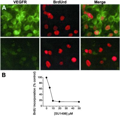
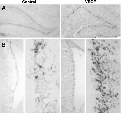
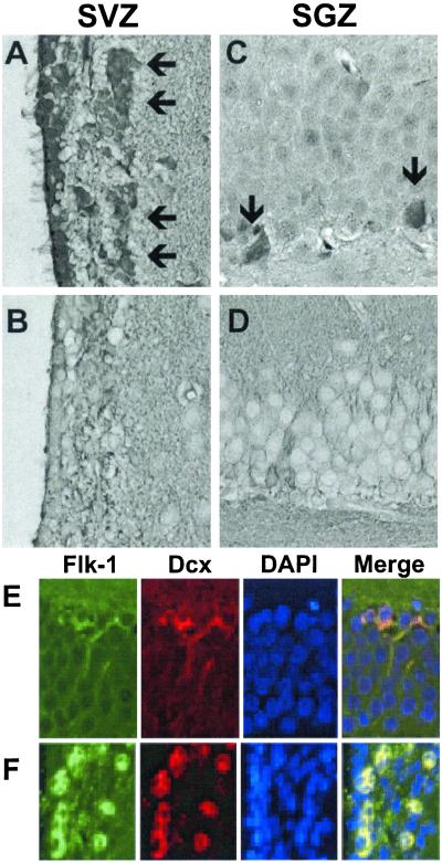
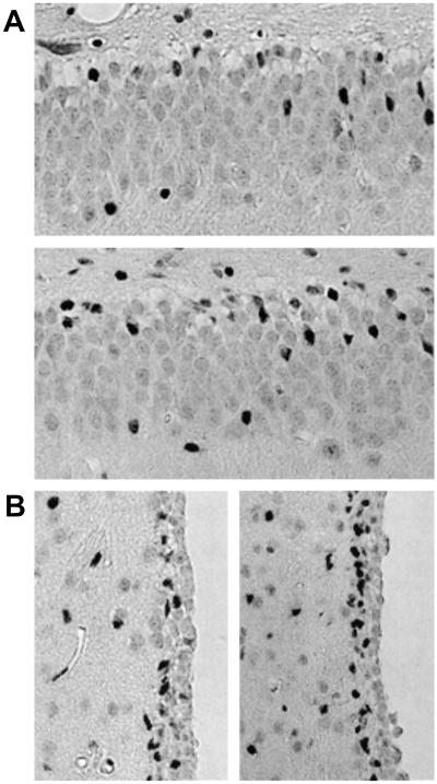
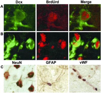
Vascular endothelial growth factor (VEGF) is an angiogenic protein with neurotrophic and neuroprotective effects. Because VEGF promotes the proliferation of vascular endothelial cells, we examined the possibility that it also stimulates the proliferation of neuronal precursors in murine cerebral cortical cultures and in adult rat brain in vivo. VEGF (>10 ng/ml) stimulated 5-bromo-2'-deoxyuridine (BrdUrd) incorporation into cells that expressed immature neuronal marker proteins and increased cell number in cultures by 20-30%. Cultured cells labeled by BrdUrd expressed VEGFR2/Flk-1, but not VEGFR1/Flt-1 receptors, and the effect of VEGF was blocked by the VEGFR2/Flk-1 receptor tyrosine kinase inhibitor SU1498. Intracerebroventricular administration of VEGF into rat brain increased BrdUrd labeling of cells in the subventricular zone (SVZ) and the subgranular zone (SGZ) of the hippocampal dentate gyrus (DG), where VEGFR2/Flk-1 was colocalized with the immature neuronal marker, doublecortin (Dcx). The increase in BrdUrd labeling after the administration of VEGF was caused by an increase in cell proliferation, rather than a decrease in cell death, because VEGF did not reduce caspase-3 cleavage in SVZ or SGZ. Cells labeled with BrdUrd after VEGF treatment in vivo include immature and mature neurons, astroglia, and endothelial cells. These findings implicate the angiogenesis factor VEGF in neurogenesis as well.
Figures

Similar articles
-
Inhibition of vascular endothelial growth factor-associated tyrosine kinase activity with SU5416 blocks sprouting in the microvascular endothelial cell spheroid model of angiogenesis.Microvasc Res. 2002 May;63(3):304-15. doi: 10.1006/mvre.2001.2383. Microvasc Res. 2002. PMID: 11969307
-
Neuroprotective effect of exogenous vascular endothelial growth factor on rat spinal cord neurons in vitro hypoxia.Chin Med J (Engl). 2005 Oct 5;118(19):1644-50. Chin Med J (Engl). 2005. PMID: 16232350
-
Coordinate expression of vascular endothelial growth factor receptor-1 (flt-1) and its ligand suggests a paracrine regulation of murine vascular development.Dev Dyn. 1995 Nov;204(3):228-39. doi: 10.1002/aja.1002040303. Dev Dyn. 1995. PMID: 8573716
-
The potential role of vascular endothelial growth factor in the central nervous system.Rev Neurosci. 2004;15(4):293-307. doi: 10.1515/revneuro.2004.15.4.293. Rev Neurosci. 2004. PMID: 15526553 Review.
-
The role of vascular endothelial growth factor in neurogenesis in adult brain.Mini Rev Med Chem. 2006 Jun;6(6):667-9. doi: 10.2174/138955706777435742. Mini Rev Med Chem. 2006. PMID: 16787377 Review.
Cited by
-
Endothelial cell secreted VEGF-C enhances NSC VEGFR3 expression and promotes NSC survival.Stem Cell Res. 2021 May;53:102318. doi: 10.1016/j.scr.2021.102318. Epub 2021 Apr 1. Stem Cell Res. 2021. PMID: 33836422 Free PMC article.
-
Signaling through the vascular endothelial growth factor receptor VEGFR-2 protects hippocampal neurons from mitochondrial dysfunction and oxidative stress.Free Radic Biol Med. 2013 Oct;63:421-31. doi: 10.1016/j.freeradbiomed.2013.05.036. Epub 2013 May 31. Free Radic Biol Med. 2013. PMID: 23732519 Free PMC article.
-
Neurogenesis After Stroke: A Therapeutic Perspective.Transl Stroke Res. 2021 Feb;12(1):1-14. doi: 10.1007/s12975-020-00841-w. Epub 2020 Aug 29. Transl Stroke Res. 2021. PMID: 32862401 Free PMC article. Review.
-
Association between Genetic Variants in NOS2 and TNF Genes with Congenital Zika Syndrome and Severe Microcephaly.Viruses. 2021 Feb 20;13(2):325. doi: 10.3390/v13020325. Viruses. 2021. PMID: 33672623 Free PMC article.
-
Ethanol extract of Oenanthe javanica increases cell proliferation and neuroblast differentiation in the adolescent rat dentate gyrus.Neural Regen Res. 2015 Feb;10(2):271-6. doi: 10.4103/1673-5374.152382. Neural Regen Res. 2015. PMID: 25883627 Free PMC article.
References
Publication types
MeSH terms
Substances
Grants and funding
LinkOut - more resources
Full Text Sources
Other Literature Sources
Molecular Biology Databases
Research Materials
