. 2002 Aug 1;16(15):1977-89.
doi: 10.1101/gad.996502.

The lytic switch protein of KSHV activates gene expression via functional interaction with RBP-Jkappa (CSL), the target of the Notch signaling pathway

Affiliations

The lytic switch protein of KSHV activates gene expression via functional interaction with RBP-Jkappa (CSL), the target of the Notch signaling pathway

Yuying Liang et al. Genes Dev. .

Abstract

The RTA protein of the Kaposi's sarcoma (KS)-associated herpesvirus (KSHV) is responsible for the switch from latency to lytic replication, a reaction essential for viral spread and KS pathogenesis. RTA is a sequence-specific transcriptional activator, but the diversity of its target sites suggests it may act via interaction with host DNA-binding proteins as well. Here we show that KSHV RTA interacts with the RBP-Jkappa protein, the primary target of the Notch signaling pathway. This interaction targets RTA to RBP-Jkappa recognition sites on DNA and results in the replacement of RBP-Jkappa's intrinsic repressive action with activation mediated by the C-terminal domain of RTA. Mutation of such sites in target promoters strongly impairs RTA responsiveness. Similarly, such target genes are induced poorly or not at all by RTA in fibroblasts derived from RBP-Jkappa(-/-) mice, a defect that can be reversed by expression of RBP-Jkappa. In vitro, RTA binds to two adjacent regions of RBP-Jkappa, one of which is identical to the central repression domain that binds the Notch effector fragment. These results indicate that KSHV has evolved a ligand-independent mechanism for constitutive activation of the Notch pathway as a part of its strategy for reactivation from latency.


Figures
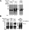
Figure 1
Figure 1
Coimmuniprecipitation of RBP-Jκ and RTA in vivo. (A) 293T cells were transfected with pV5-50ΔSTAD (lanes 1,4,7) or pFLAG-RBPJκ (lanes 2,5,8), or both (lanes 3,6,9). Cell extracts were immunoprecipitated with either anti-Flag mAb (lanes 13) or anti-V5 mAb (lanes 46), and analyzed by Western blot with either anti-V5 mAb (lanes 16) or anti-Flag mAb (lanes 79). (B) BCBL-1 cells were either mock-induced (labeled U, lanes 1,3,5) or TPA-induced (labeled I, lanes 2,4,6). Cell extracts were immunoprecipitated with anti-RBPJκ (lanes 1,2), anti-NIK (NFκB-inducing kinase) (lanes 3,4), or anti-RTA (lanes 5,6) antibodies. As a control, RBPJκ null cells (OT11) were transfected with pcDNA3-Flc50. Cell extracts were immunoprecipitated with either anti-RBPJκ (lane 7) or anti-RTA (lane 8) Ab. All immunoprecipitates were analyzed by Western blot with anti-RTA antibody.
Figure 2
Figure 2
RTA activates promoters containing RBP-Jκ recognition sequences in WT (SLK) but not RBPJκ-null cell line (OT11). (A) SLK cells were cotransfected with 0 to 1 μg of pcDNA3-Flc50 and 0.5 μg of luciferase reporter constructs driven by promoters containing tandem repeats (indicated in number) of either WT or mutant (mut) RBPJκ binding site in either orientation (forward or reverse). (B) OT11 cells were cotransfected with 0 to 0.5 μg of pcDNA3-Flc50 and 0.5 μg of luciferase reporter constructs containing three copies of WT or mutant (mut) RBPJκ binding site in forward direction, with (+) or without (−) 1 μg of pcDNA3.1-RBPJκ. Luciferase assay was performed as described in Materials and Methods. The maximal transactivation by RTA was plotted for each promoter, with the error bars representing standard deviations of the results from at least two independent experiments.
Figure 3
Figure 3
RTA/RBP-Jκ interactions in authentic viral promoters. (A) Promoters of MTA (ORF57) and SSB contain RBPJκ recognition sites. The sequences of MTA and SSB promoters are shown, with the RBPJκ recognition site outlined by an empty box (orientation: reverse in MTA promoter and forward in SSB promoter). The previously identified 52-bp RTA-response element (50RE) between −106 and −54 nt of MTA promoter is underlined. (B) RTA transactivation of MTA or SSB promoters (WT or RBPJκ site-mutated) in OT13 (WT) or OT11 (RBPJκ−/−) cell line. OT13 or OT11 cells were cotransfected with 0 to 0.5 μg of pcDNA3-Flc50 and 0.5 μg of luciferase reporter constructs driven by MTA promoter, MTA promoter with mutations at RBPJκ recognition site (see Materials and Methods) (MTAmut), SSB promoter, or SSB promoter with mutations at RBPJκ recognition site (SSBmut). In OT11 cells, RTA transactivation was tested in the absence (−) or presence (+) of 1 μg of cotransfected pcDNA3.1-RBPJκ. (C) Schematic representation of mutants of PAN promoter and their responsiveness to RTA transactivation in SLK and OT11 cells. The RTA-response element (50RE) identified by Song et al. (2001) extends from −69 to −38 bp upstream of RNA start site. Mutation on 50RE, represented by a hatched box, was created by replacing the 12-nt sequence (−67 to −56) with linker sequence TATCATATGATA. RBPJκ recognition site was from −818 to −824 in reverse direction. Mutation of RBPJκ recognition site (changes of TTCCCACGG to TTCCAAGCC) is represented by the gray box. RTA transactivation on the WT and mutant PAN promoters in SLK and OT11 cells was measured in the same way as in (B).
Figure 3
Figure 3
RTA/RBP-Jκ interactions in authentic viral promoters. (A) Promoters of MTA (ORF57) and SSB contain RBPJκ recognition sites. The sequences of MTA and SSB promoters are shown, with the RBPJκ recognition site outlined by an empty box (orientation: reverse in MTA promoter and forward in SSB promoter). The previously identified 52-bp RTA-response element (50RE) between −106 and −54 nt of MTA promoter is underlined. (B) RTA transactivation of MTA or SSB promoters (WT or RBPJκ site-mutated) in OT13 (WT) or OT11 (RBPJκ−/−) cell line. OT13 or OT11 cells were cotransfected with 0 to 0.5 μg of pcDNA3-Flc50 and 0.5 μg of luciferase reporter constructs driven by MTA promoter, MTA promoter with mutations at RBPJκ recognition site (see Materials and Methods) (MTAmut), SSB promoter, or SSB promoter with mutations at RBPJκ recognition site (SSBmut). In OT11 cells, RTA transactivation was tested in the absence (−) or presence (+) of 1 μg of cotransfected pcDNA3.1-RBPJκ. (C) Schematic representation of mutants of PAN promoter and their responsiveness to RTA transactivation in SLK and OT11 cells. The RTA-response element (50RE) identified by Song et al. (2001) extends from −69 to −38 bp upstream of RNA start site. Mutation on 50RE, represented by a hatched box, was created by replacing the 12-nt sequence (−67 to −56) with linker sequence TATCATATGATA. RBPJκ recognition site was from −818 to −824 in reverse direction. Mutation of RBPJκ recognition site (changes of TTCCCACGG to TTCCAAGCC) is represented by the gray box. RTA transactivation on the WT and mutant PAN promoters in SLK and OT11 cells was measured in the same way as in (B).
Figure 4
Figure 4
Mapping the RTA-interaction domains in RBPJκ. The repressor domain, represented by the hatched box, was previously identified (Hsieh and Hayward 1995) to extend from aa 179 to 360. Truncated versions of RBPJκ (AF, AG, AH, AB, AC, AG, BE, BF, BG, EE, and CE) are shown schematically along with the positions of the start and end residues. All constructs (directed under T7 promoter) were 35S-labeled by in vitro translation (1/50 loading; lanes 1,4,7,10,13,16,19,22,25,28,31,34), bound to either GST (lanes 2,5,8,11,14,17,20,23,26,29,32,35) or GST-50ΔSTAD (lanes 3,6,9,12,15,18,21,24,27,30,33,36) beads, washed with NETN buffer, and separated by SDS-PAGE.
Figure 5
Figure 5
Mapping the RBP-Jκ-interaction domains in RTA. The conserved domains of RTA are shown schematically: the basic region (1 to 237 aa), leu repeats (247 to 269 aa), activation domain (AD), and two nuclear localization sites (NLS-1 and NLS-2). Truncated versions of RBP-Jκ (50ΔSTAD, RTA-AF, RTA-AG, RTA-BE, RTA-BF, RTA-DE, and RTA-CE) are shown schematically along with the positions of the start and end residues. All constructs (directed under T7 promoter) were 35S-labeled by in vitro translation (1/50 loading, lanes 1,4,7,10,13,16,19), bound to either GST (lanes 2,5,8,11,14,17,20) or GST-50ΔSTAD (lanes 3,6,9,12,15,18,21) beads, washed with NETN buffer, and separated by SDS-PAGE.
Figure 6
Figure 6
Binding of RBP-Jκ and RBP-Jκ/RTA complexes to an RBP-Jκ recognition site. The indicated proteins were cloned into a T7 transcription vector and expressed in a rabbit reticulocyte lysate (RRL). The RRL extracts programmed with the indicated vectors were added to a 32P-labeled oligonucleotide corresponding to a canonical RBP-Jκ site, and complexes were analyzed by EMSA. Arrow indicates the position of RBP-Jκ/DNA complex. “Vector” refers to lysate programmed with an empty T7 transcription vector. WT, wild-type RBP-Jκ site; MT, mutant RBP-Jκ site.

Similar articles

Cited by

References

    1. Ambroziak JA, Blackbourn DJ, Herndier BG, Glogau RG, Gullett JH, McDonald AR, Lennette ET, Levy JA. Herpes-like sequences in HIV-infected and uninfected Kaposi's sarcoma patients. Science. 1995;268:582–583. - PubMed
    1. Ansieau S, Strobl LJ, Leutz A. Activation of the Notch-regulated transcription factor CBF1/RBP-Jκ through the 13SE1A oncoprotein. Genes & Dev. 2001;15:380–385. - PMC - PubMed
    1. Aster J, Pear W. Notch signaling in leukemia. Curr Opin Hematol. 2001;8:237–244. - PubMed
    1. Bais C, Santomasso B, Coso O, Arvanitakis L, Raaka EG, Gutkind JS, Asch AS, Cesarman E, Gershengorn MC, Mesri EA, et al. G-protein-coupled receptor of Kaposi's sarcoma-associated herpesvirus is a viral oncogene and angiogenesis activator. Nature. 1998;391:86–89. - PubMed
    1. Blackbourn DJ, Lennette E, Klencke B, Moses A, Chandran B, Weinstein M, Glogau RG, Witte MH, Way DL, Kutzkey T, et al. The restricted cellular host range of human herpesvirus 8. AIDS. 2000;14:1123–1133. - PubMed

Publication types

MeSH terms

LinkOut - more resources