. 2002 Aug 1;16(15):1950-63.
doi: 10.1101/gad.1002002.

Smad3 allostery links TGF-beta receptor kinase activation to transcriptional control

Affiliations

Smad3 allostery links TGF-beta receptor kinase activation to transcriptional control

Bin Y Qin et al. Genes Dev. .

Abstract

Smad3 transduces the signals of TGF-betas, coupling transmembrane receptor kinase activation to transcriptional control. The membrane-associated molecule SARA (Smad Anchor for Receptor Activation) recruits Smad3 for phosphorylation by the receptor kinase. Upon phosphorylation, Smad3 dissociates from SARA and enters the nucleus, in which its transcriptional activity can be repressed by Ski. Here, we show that SARA and Ski recognize specifically the monomeric and trimeric forms of Smad3, respectively. Thus, trimerization of Smad3, induced by phosphorylation, simultaneously activates the TGF-beta signal by driving Smad3 dissociation from SARA and sets up the negative feedback mechanism by Ski. Structural models of the Smad3/SARA/receptor kinase complex and Smad3/Ski complex provide insights into the molecular basis of regulation.


Figures

Figure 1
Figure 1
Crystal structures of unliganded Smad3, Smad3/SARA complex, and S4AF. (A) Crystal structure of unliganded Smad3 shown by ribbon representation. The Cα trace of a symmetry-related subunit is shown. (B) Crystal structure of Smad3/SARA complex. The SARA SBD is in dark yellow. All proline residues are displayed. Smad3 structures that are ordered upon SARA binding are circled. (C) Crystal structure of S4AF (Qin et al. 1999). The Smad4 activation domain (SAD) is in dark yellow, in which the disordered sequence, GHYWPVHNELA, has a helical propensity. The three-helix bundle and β-sandwich subdomains are in red and cyan, respectively. The disordered regions of the structure are represented by dots and colored according to the subdomain color. The side chains are in dark blue.
Figure 2
Figure 2
Trimerization-induced tilting of the three-helix bundle subdomain relative to the β-sandwich core. (A) Superposition of the R-Smad MH2 domains that were crystallized in the monomeric form. (B) Superposition of the R-Smad MH2 domains that were crystallized in the trimeric form. (C) Superposition of the monomeric and trimeric form of the R-Smad MH2 domain. Superpositions were performed by aligning the β-sandwich subdomain. The amino termini of the unliganded Smad3 and Smad1 are shorter due to disordering of the structures in the crystal.
Figure 3
Figure 3
Proposed structural model of the dimeric Smad3/SARA complex. (A) The dimeric SARA FYVE domain model. The model includes residues 590 to 657 of SARA. The two zinc ions within each subunit are shown in spheres. Ins(1,3)P2 is shown using stick presentation. (B) Crystal structure of the dimeric arrangement of the Smad3/SARA complex. The two copies of the complex are related by a vertical twofold crystallographic axis. The coloring is based on Fig. 1B. (C) Close-up view of the dimer interface of the SARA FYVE domain dimer. (D) Model of the Smad3/SARA complex containing the FYVE domain. The linker between the FYVE domain and the SBD contains 14 residues not included in the model (residues 658–670) and are shown by dots.
Figure 4
Figure 4
Proposed structural model of Smad3/SARA/receptor kinase complex. (A) Proposed structure of the Smad3/SARA/receptor kinase complex. The type I receptor kinase model is shown in green, with the exception of the GS domain, which is in gray. The kinase active site has an ATP molecule. The type II receptor (ectodomain and transmembrane helix only) is shown in pink. The TGF-β dimer is in dark blue. Smad3 and SARA are colored based on Fig. 3D. The specificity determinants in the kinase L45 loop and Smad3 L3 loop are shown by blue spheres (left to right, N267, D269, and N270) and red spheres (top to bottom, R384 and T387), respectively. The GS loop phosphorylation sites are shown using pink spheres. The structural consequence of GS domain phosphorylation is depicted by a cartoon and is labeled active GS domain. The carboxy-terminal tail of Smad3, modeled using the Smad1 crystal structure, is shown in pink. The ligand/ectodomain structure is based on the crystal structures of the bone morphogenetic protein (BMP) in complex with the BMP type I receptor and TGF-β in complex with type II receptor (Kirsch et al. 2000; Hart et al. 2002). (B) The kinase/Smad3 interface contains residues that are mostly conserved or subtype specific in the type I receptor kinases and the R-Smads. Subtype-specific residues are boxed. Smad3 and kinase residues are labeled in black and green, respectively. (C) The cytoplasmic organization of the receptor complex is consistent with the ligand/ectodomain complex. Top views of cytoplasmic section (top) and extracellular section (bottom) are shown. The coloring is based on Fig. 4A. The type II kinase, which phosphorylates the GS loop, is shown in a cartoon.
Figure 5
Figure 5
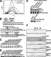
Biochemical characterization of Smad3–SARA and Smad3–Ski interactions. (A) DCDT+ analysis of a sedimentation velocity experiment conducted on a 1:1 mixture of S3LC and SARA SBD conducted at 42K rpm, 24.7°C and total protein concentrations of 4.0, 8.6, and 17.5 μM complex. Fitting with DCDT+ (and SVEDBERG) gives an average S20,w = 2.698 ± 0.012 (2.700 ± 0.037) and an average MW = 42,183 ± 1264 (39,407 ± 1287). The absence of a shift in the peak position, especially relative to the obvious shift upon trimerization by S3LC alone (reproduced from Correia et al. 2001), and the similarity of the sedimentation coefficient to that of monomeric Smad4 (2.46 S20,w), corrected for the size of SBD, are consistent with this being a 1:1 complex. (B) Phosphorylated Smad3 has a weaker affinity for SARA SBD. (Top) GST–SARA_SBD was used to detect interaction with either the unphosphorylated or phosphorylated S3LC by use of the GST pull-down assay. The effect of S4AF was performed by including S4AF in the first two washes, followed by regular washes. (first lane) No S4AF; (second lane) 0.5 mg/mL S4AF; (third lane) 2 mg/mL S4AF. The bound S3LC in the absence of S4AF reflects ∼20% to 30% of the input. (Middle and bottom gels) Size exclusion chromatography of S3LC(2P)/SARA complex and S3LC(V276D)/SARA complex. The loading sample contains 0.5 mg/mL of GST–SARA–SBD and 1 mg/mL of S3LC. The positions of the eluted species are indicated above the SDS–polyacrylamide gels. The fraction numbers are marked below the bottom gel. (C) Ski binds to subsite of the SARA-binding site. The GST–Ski(17–45) was used to detect interaction with the S3LC mutants. The bound S3LC in the first lane reflects ∼20% to 30% of the input. (D) Hydrophobic residues in Ski mediate direct interaction with Smad3. The GST–Ski(17–45) mutants were used to detect interaction with S3LC. The bound Ski(WT) reflects ∼20% to 30% of the input. (E) Smad3–Ski interaction requires trimerization of Smad3. (Top) The GST–Ski(17–45) and GST–SARA_SBD were used to detect interaction with the trimer interface mutants of S3LC. The bound S3LC in the first lane reflects ∼20% to 30% of the input. (Bottom) S3LC(2P) binds Ski better than the unphosphorylated S3LC after extensive washing. In addition to the standard conditions, the beads were equilibrated with the washing buffer for 60 min between washes. (F) Ski interacts with the Smad3/Smad4 heterotrimeric complex. Protein compositions as indicated to the right of each gel panel were injected into the size exclusion column. The eluted fractions were analyzed by SDS-PAGE. The molecular weight standards are shown at top. The fraction numbers are shown at bottom. All gels in this figure were stained with Coomassie blue.
Figure 6
Figure 6
Proposed structural model of the Smad3/Ski complex. (A) Sequence and secondary structural comparison between SARA SBD and Ski(16–40). The secondary structures of SARA SBD from the crystal structure are shown above the SARA sequence. The predicted secondary structures of Ski(16–40) based on the Garnier-Osguthorpe-Robson method in the GCG program are shown below the Ski sequence. Ski mutations that weaken Smad3–Ski interaction are boxed in black. Ski mutations that have no effect on Smad3–Ski interaction are boxed in green. The three-proline turn in SARA SBD and the corresponding sequence in Ski are boxed in red. (B, right) Proposed structure of the Smad3/Smad4/Ski complex. The Smad4 subunit contains the SAD domain. (Left) Close-up view of the Smad3/Ski subunit model. The coloring is based on Fig. 1.
Figure 7
Figure 7
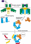
Trimerization-dependent Smad signaling in the TGF-β pathway.

Comment in

Similar articles

Cited by

References

    1. Abdollah S, Macias-Silva M, Tsukazaki T, Hayashi H, Attisano L, Wrana JL. TbetaRI phosphorylation of Smad2 on Ser465 and Ser467 is required for Smad2-Smad4 complex formation and signaling. J Biol Chem. 1997;272:27678–27685. - PubMed
    1. Akiyoshi S, Inoue H, Hanai J, Kusanagi K, Nemoto N, Miyazono K, Kawabata M. c-Ski acts as a transcriptional co-repressor in transforming growth factor-β signaling through interaction with Smads. J Biol Chem. 1999;274:35269–35277. - PubMed
    1. Attisano L, Wrana JL. Smads as transcriptional co-modulators. Curr Opin Cell Biol. 2000;12:235–243. - PubMed
    1. Blobe GC, Schiemann WP, Lodish HF. Role of transforming growth factor β in human disease. New Eng J Med. 2000;342:1350–1358. - PubMed
    1. Brunger AT, Adams PD, Clore GM, DeLano WL, Gros P, Grosse-Kunstleve RW, Jiang JS, Kuszewski J, Nilges M, Pannu NS, et al. Crystallography & NMR system: A new software suite for macromolecular structure determination. Acta Crystallogr D Biol Crystallogr. 1998;54:905–921. - PubMed

Publication types

MeSH terms