. 2002 Aug 1;21(15):3989-4000.
doi: 10.1093/emboj/cdf398.

Differential sorting and fate of endocytosed GPI-anchored proteins

Affiliations

Differential sorting and fate of endocytosed GPI-anchored proteins

Marc Fivaz et al. EMBO J. .

Abstract

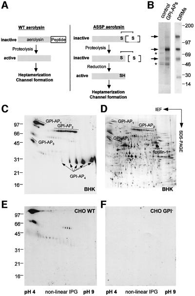
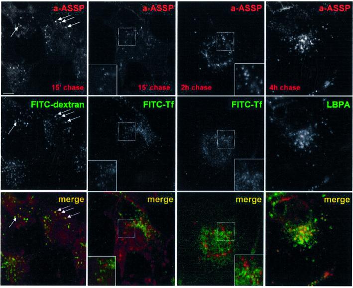
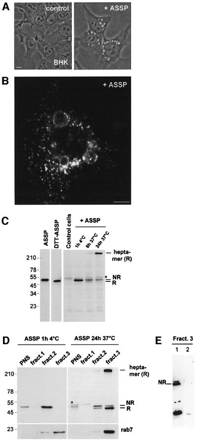
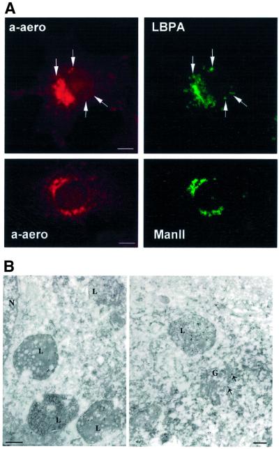
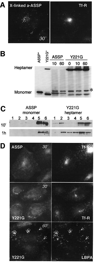
In this paper, we studied the fate of endocytosed glycosylphosphatidyl inositol anchored proteins (GPI- APs) in mammalian cells, using aerolysin, a bacterial toxin that binds to the GPI anchor, as a probe. We find that GPI-APs are transported down the endocytic pathway to reducing late endosomes in BHK cells, using biochemical, morphological and functional approaches. We also find that this transport correlates with the association to raft-like membranes and thus that lipid rafts are present in late endosomes (in addition to the Golgi and the plasma membrane). In marked contrast, endocytosed GPI-APs reach the recycling endosome in CHO cells and this transport correlates with a decreased raft association. GPI-APs are, however, diverted from the recycling endosome and routed to late endosomes in CHO cells, when their raft association is increased by clustering seven or less GPI-APs with an aerolysin mutant. We conclude that the different endocytic routes followed by GPI-APs in different cell types depend on the residence time of GPI-APs in lipid rafts, and hence that raft partitioning regulates GPI-APs sorting in the endocytic pathway.


Figures

None
Fig. 1. Mapping endogenous GPI-APs using aerolysin. (A) Activation of the wild-type precursor is triggered by proteolytic removal of a C-terminal peptide. The resulting mature toxin is able to oligomerize into a heptameric ring that forms a pore in the target membrane. The ASSP aerolysin mutant is inactive, even after C-terminal proteolysis, due to an engineered disulfide bridge that links the propeptide to the mature toxin (van der Goot et al., 1994). Disulfide reduction is required for ASSP to oligomerize and form pores. (B) BHK GPI-APs (indicated by arrows) were purified from 35S-labeled cells (lane labeled GPI-APs; see Materials and methods). The control lane corresponds to non-GPI-APs released from the Triton X-114 detergent phase. Contaminants are labeled with asterisks. In parallel, an aerolysin overlay assay was performed on BHK DRMs (30 µg of protein). (CF) 2D maps of endogenous GPI-APs were generated by performing aerolysin overlays on DRMs of BHK (C), wild-type (E) and GPI-deficient CHO (F) cells (50 µg of protein per gel). (D) The protein profile revealed by silver stain of a Triton X-114 detergent phase prepared from BHK DRMs (150 µg of protein). The most abundant BHK GPI-APs are labeled in (C) and (D).
None
Fig. 2. Endocytosis of GPI-APs in BHK cells. BHK cells were incubated with the GPI-AP-specific probe Alexa–ASSP (a-ASSP, 10 nM) for 1 h at 4°C, rinsed, and further incubated at 37°C for various times. Early endosomes were labeled by 5-min internalized FITC–dextran, recycling endosomes by 15-min internalized FITC–transferrin and late endosomes by immunolabeling against LBPA. Arrows indicate examples of co-localization. Scale bar, 10 µm. Note that cell surface labeling with transferrin was low due to the fact that 70% of the receptor is intracellular on these cells and that the cell surface receptors are diluted over a large surface.
None
Fig. 3. ASSP is activated in late endosomes in BHK cells. (A) BHK cells were incubated or not with ASSP (10 nM, 37°C). After 6 h, large cytoplasmic vacuoles were observed by phase-contrast microscopy in ASSP-treated cells but not in control cells. Scale bar, 10 µm. (B) The ASSP-induced vacuoles were immunostained with an anti-lamp-1 antibody. (C) BHK cells were treated with trypsin-nicked ASSP (10 nM) or left untreated (control cells) and then harvested after various incubation times at 37°C. Total cell extracts were analyzed by SDS–PAGE under non-reducing conditions (30 µg per lane) followed by western blotting using an anti-aerolysin antibody. The migration patterns of pure trypsin-nicked ASSP in the absence and presence of DTT are shown as markers of the non-reduced (NR) and reduced (R) forms. Note that the aerolysin heptamer, which can only be formed after disulfide reduction of ASSP, is resistant to SDS and therefore migrates at the top of the gel. The asterisk indicates the presence of a cellular protein cross-reacting with the anti-aerolysin antibody. (D) BHK cells were incubated with trypsin-nicked ASSP (10 nM) for 1 h at 4°C or 24 h at 37°C, and fractionated on a sucrose step gradient to separate late endosomes from other membranes. The three membrane fractions of the gradient were analyzed by non-reducing SDS–PAGE and assayed for the presence of ASSP and rab7 by western blotting. (E) BHK cells were incubated with trypsin-nicked biotin–ASSP (10 nM) for 24 h at 37°C, and fractionated on a sucrose step gradient to separate late endosomes from other membranes. Two forms of biotinylated ASSP were used: biotin–ASSP (lane 1) and the cleavable counterpart biotin-SS–ASSP (lane 2). The late endosomal fractions of the gradients were analyzed by non-reducing SDS–PAGE, transferred onto a nitrocellulose membrane, and assayed for the presence of biotin–ASSP using streptavidin–HRP. Note that biotinylation inhibits oligomerization and thereby allows degradation of the toxin.
None
Fig. 4. Steady-state distribution of GPI-APS in BHK cells. (A) BHK cells were fixed/permeabilized and double labeled with 7.5 nM Alexa–aerolysin (a-aero) and anti-LBPA or anti-mannosidase II (ManII) antibodies. In the upper panel, examples of co-localization are shown with arrows. Scale bar, 10 µm. (B) BHK cells were fixed in paraformaldehyde/glutaraldehyde and processed for frozen sectioning. Ultrathin sections were incubated with biotinylated aerolysin followed by 10 nm anti-biotin–gold. Specific labeling is associated with multi-vesicular late endocytic structures (L) and with the Golgi complex (G; arrows show gold particle in the Golgi); N, nucleus. Scale bar, 200 nm.
None
Fig. 5. DRM domains in late endosomes of BHK cells. (A) DRMs were prepared from BHK cells treated for 24 h with trypsin-nicked ASSP (10 nM), by flotation on an OptiPrep gradient using a Beckman SW60 rotor. Fractions of 200 µl were collected. SDS–PAGE was performed under non-reducing conditions. The gel was loaded by yield. (B and C) DRMs were prepared from purified late endosomes by flotation on an OptiPrep gradient using a TLS-55 Beckman rotor and 400 µl samples were collected. (B) The total cholesterol content of each fraction of the gradient was determined by 1D TLC and quantified using the ScanAnalysis software. Cholesterol contents were expressed as a percentage of the total cholesterol content of the gradient. Values correspond to the mean of four experiments. Error bars represent the standard deviation. (C) The total content of each fraction was loaded on a 15% SDS gel. The distributions of lamp-1, flotillin-1 and rab7 were analyzed by western blotting and that of GPI-APs by aerolysin overlay. Note that we are here analyzing the late endosomal lamp-1 population and not lysosomal lamp-1, which is found in heavy organelles and not low buoyancy fractions. (D) Subcellular fractionation of BHK cells. BHK cells were fractionated on a sucrose density gradient to separate late endosomes for other cellular membranes. Three membrane fractions were collected at the sucrose interfaces, and probed for caveolin-1 and flotillin-1 by western blotting (10 µg of proteins per lane). Fraction 3 is highly enriched in late endosomes (Kobayashi et al., 1998). Fraction 2 contains plasma membrane rafts (van der Goot, 1997) and early endosomes (Aniento et al., 1996). (E) BHK cells were fixed/permeabilized and double labeled for flotillin-1 and LBPA. The anti-flotillin-1 antibody also stained the plasma membrane; however, brief exposure times were chosen so as to highlight the intracellular staining.
None
Fig. 6. GPI-APs in CHO cells are delivered to the recycling endosome. CHO were incubated with aerolysin–ASSP (10 nM) for 1 h at 4°C followed by various times at 37°C and processed for fluorescence microscopy. (A) Alexa–ASSP was allowed to internalize for 15 min. During the last 5 min, FITC–dextran (upper panels) was internalized to label early endosomes. Arrows indicate examples of co-localization. In a parallel experiment, ASSP was internalized for 15 min in the presence of FITC–transferrin (lower panels) in order to label the recycling endosome (indicated by an arrowhead in the upper panel and an arrow in the lower). (B) Quantification of Alexa–ASSP uptake. More than 20 cells from two independent experiments were analyzed for each condition, and standard deviations of the mean were calculated. (C) CHO cells were treated with trypsin-nicked ASSP (10 nM) for 24 h and imaged by phase-contrast microscopy. (D) Total cell extracts of wild-type and GPI-deficient CHO cells treated or not with trypsin-nicked ASSP (10 nM) were analyzed by non-reducing SDS–PAGE and western blotting using anti-aerolysin antibodies (30 µg/lane). The arrow indicates an aerolysin degradation product. The asterisk indicates the presence of a protein cross-reacting with the anti-aerolysin antibody. (E) CHO cells were fixed 3% paraformaldehyde and permeabilized with 0.1% saponin. A triple-labeling experiment was performed using Alexa–aerolysin (7.5 nM), filipin (specific probe for cholesterol) and antibodies against the transferrin receptor (Tf-R). Scale bar, 10 µm. (F) CHO cells were fixed in paraformaldehyde/glutaraldehyde and processed for frozen sectioning. Ultrathin sections were incubated with the biotinylated aerolysin probe followed by 10 nm anti-biotin–gold. Specific labeling (arrows) is associated with the Golgi complex (G; upper panel) and with tubulo-vesicular profiles in close proximity to the centriole (C; lower panel), presumed to be reycling endosomes. Scale bars, 200 nm.
None
Fig. 7. Differential endocytic fate of CD14 in CHO and BHK cells. CHO (A) or BHK (B) cells, transiently transfected with human CD14, were incubated with FITC-conjugated monoclonal antibodies against human CD14 (1 µg/ml) at 4°C, shifted to 37°C for 60 min and incubated with rhodamine-transferrin during the last 30 min. To better resolve intracellular staining of CD14, the surface-bound anti-CD14 monoclonal antibody was removed by a brief acid wash. BHK cells were also incubated with polyclonal anti-CD14 antibodies (2 µg/ml), revealing endogenous CD14, for 15 h (37°C). Cells were fixed/permeabilized and a double immunostaining was performed against CD14 (secondary antibody only) and LBPA.
None
Fig. 8. Detergent solubility of intracellular GPI-APs in CHO cells. CHO cells were incubated with biotin-SS–ASSP (10 nM) at 4°C, shifted or not at 37°C for 45 min, then treated or not with 30 mM MesNa (15 min at 4°C) and PNSs were prepared. Thirty micrograms of protein were analyzed by SDS–PAGE (10% gel), blotted on nitrocellulose and the membrane was probed with [125I]streptavidin. (A) A representative experiment and (B) quantification by phosphoimager analysis (n = 4; bars represent standard deviation of the mean). The percentage of MesNa-resistant biotin-SS–ASSP was derived from the ratio of signal intensities obtained with and without MesNa treatment and is a direct measure of biotin-SS–ASSP internalization. (C and D) Cells were incubated with biotin-SS–ASSP (10 nM) at 4°C, shifted to 37°C for 45 min, treated or not with 30 mM MesNa (15 min at 4°C) and then extracted in 2% cold Triton for 1 h. DRMs were isolated on a sucrose density gradient using a Beckman SW60 rotor. Fractions of 700 µl were collected, precipitated, analyzed on a 12.5% gel and probed with streptavidin–HRP (C) or [125I]streptavidin (D) for quantification. In (D) the percentage of detergent-resistant biotin-SS–ASSP (expressed as the ratio of the signal in the top two fractions over the sum of the signals in all fractions) was plotted (n = 4; bars represent standard deviation of the mean).
None
Fig. 9. Clustering of GPI-APs inhibits trafficking to the recycling endosome of CHO cells. (A) CHO cells were sequential incubated at 4°C with Alexa–ASSP (10 nM) for 1 h, anti-aerolysin polyclonal antibody (1 h) and anti-chicken antibody (1 h) and then warmed to 37°C for 30 min in the presence of FITC–transferrin. (BD) CHO cells were incubated at 4°C with trypsin-nicked ASSP or Y221G (10 nM) and then warmed up at 37°C for various times. Cells were lysed in 1% Triton X-100, analyzed by SDS–PAGE and western blotting for the presence of the various forms of the toxin (non-reducing 10% gel, 40 µg of protein per lane except for the standards) (B) or fractionated on a sucrose density gradient to isolate DRMs (C). The asterisk in (B) indicates the presence of a protein cross-reacting with the anti-aerolysin antibody. In (C), DRMs were prepared by a short centrifugation on sucrose gradients in order to visualize the difference in behavior between ASSP and Y221G. (D) ASSP and Y221G were detected with anti-aerolysin antibodies followed by an anti-chicken secondary antibody. We used anti-aerolysin antibodies since Alexa labeling of Y221G inhibited oligomerization. The anti-aerolysin antibody gives a background staining in particular in the nucleus. Cells were incubated for the last 30 min with FITC–transferrin or labeled with an anti-LBPA antibody. Scale bar, 10 µm.

Similar articles

Cited by

References

    1. Abrami L. and van der Goot,F.G. (1999) Plasma membrane micro domains act as concentration platforms to facilitate intoxication by aerolysin. J. Cell Biol., 147, 175–184. - PMC - PubMed
    1. Abrami L., Fivaz,M., Glauser,P.E., Parton,R.G. and van der Goot,F.G. (1998) A pore-forming toxin interact with a GPI-anchored protein and causes vacuolation of the endoplasmic reticulum. J. Cell Biol., 140, 525–540. - PMC - PubMed
    1. Abrami L., Fivaz,M. and van der Goot,F.G. (2000) Adventures of a pore-forming toxin at the target cell surface. Trends Microbiol., 8, 168–172. - PubMed
    1. Abrami L., Fivaz,M., Kobayashi,T., Kinoshita,T., Parton,R.G. and van der Goot,F.G. (2001) Cross-talk between caveolae and glycosylphosphatidylinositol-rich domains. J. Biol. Chem., 276, 30729–30736. - PubMed
    1. Abrami L., Velluz,M.C., Hong,Y., Ohishi,K., Mehlert,A., Ferguson,M., Kinoshita,T. and Gisou van der Goot,F. (2002) The glycan core of GPI-anchored proteins modulates aerolysin binding but is not sufficient: the polypeptide moiety is required for the toxin-receptor interaction. FEBS Lett., 512, 249–254. - PubMed

Publication types