. 2002 Jul 1;21(13):3296-306.
doi: 10.1093/emboj/cdf349.

Involvement of the mitogen-activated protein kinase SIMK in regulation of root hair tip growth

Affiliations

Involvement of the mitogen-activated protein kinase SIMK in regulation of root hair tip growth

Jozef Samaj et al. EMBO J. .

Abstract

Mitogen-activated protein kinases (MAPKs) are involved in stress signaling to the actin cytoskeleton in yeast and animals. We have analyzed the function of the stress-activated alfalfa MAP kinase SIMK in root hairs. In epidermal cells, SIMK is predominantly nuclear. During root hair formation, SIMK was activated and redistributed from the nucleus into growing tips of root hairs possessing dense F-actin meshworks. Actin depolymerization by latrunculin B resulted in SIMK relocation to the nucleus. Conversely, upon actin stabilization with jasplakinolide, SIMK co-localized with thick actin cables in the cytoplasm. Importantly, latrunculin B and jasplakinolide were both found to activate SIMK in a root-derived cell culture. Loss of tip-focused SIMK and actin was induced by the MAPK kinase inhibitor UO 126 and resulted in aberrant root hairs. UO 126 inhibited targeted vesicle trafficking and polarized growth of root hairs. In contrast, overexpression of gain-of-function SIMK induced rapid tip growth of root hairs and could bypass growth inhibition by UO 126. These data indicate that SIMK plays a crucial role in root hair tip growth.


Figures
None
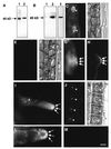
Fig. 1. Immunoblot and immunofluorescence detection of total and active SIMK. (A) Root extracts were prepared and immunoblotted with actin antibody (lane 1) or with SIMK antibody M23 (lane 2). (B) Salt treatment of roots for 10 min activated SIMK as revealed by immunoblotting crude root extracts with phospho-specific SIMK antibody N103 (lane 2) and SIMK-specific antibody M23 (lane 3). Active SIMK is hardly detected in control roots with N103 (lane 1). (C) Immunofluorescence microscopy of SIMK in elongating root cells of M.sativa L. using the Steedman’s wax embedding technique. Note that SIMK is localized predominantly to nuclei (indicated by arrowheads), but depleted from nucleoli (indicated by stars). (D) DIC image of (C). (E) Immunodepletion control of epidermal root cells (shown in F) with M23 after pre-incubation with FNPEYQQ heptapeptide. (F) Corresponding DIC image for (E). (G) Trichoblast before root hair initiation showing cell periphery-associated spot-like SIMK labeling at the outer tangential cell wall (arrows). (H) Trichoblast at the bulging stage: SIMK labeling appears at the outermost domain of the developing bulge (arrows). (I) Growing root hair showing SIMK labeling focused to the tip (arrows) and in spot-like structures along the root hair tube. SIMK is depleted from the nucleus and nucleoli (arrowhead and star, respectively). (J) Root epidermal cells showing very low levels of active SIMK labeled with N103 antibody. (K) Corresponding DIC image for (J). Nuclei and nucleoli in (J) and (K) are indicated by arrowheads and stars. (L) Tip of a growing root hair showing accumulation of active SIMK in spot-like structures at the root hair tip (arrows). (M) Immunodepletion control of root hair with N103 after pre-incubation with CTDFMTpEYpVVTRWC peptide. Bar = 15 µm for (C–F), 10 µm for (G–K) and 5 µm for (L) and (M).
None
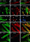
Fig. 2. Co-immunolocalization of tubulin and SIMK (A–C) or actin and SIMK (D–O) in root hairs using the freeze-shattering technique. (A) Microtubules are organized in longitudinal and net-axially arranged arrays in non-growing parts of the root hair tube and are much less abundant in subapical and apical zones of growing root hair apices. (B) SIMK accumulates in root hair apices and in distinct spots. (C) Merged image indicating no significant co-localization (yellow color) of microtubules and SIMK at root hair tips and within root hair tubes. Arrows indicate root hair tip. (D–F) Control growing root hairs. (G–I) Growing root hairs treated with 10 µM latrunculin B (LB) for 30 min. (J–L) Growing root hairs treated with 5 µM jasplakinolide (JK) for 60 min. (M–O) Growing root hairs treated with 50 µM brefeldin A (BFA) for 60 min. (D) Dense actin meshworks are present at root tips, and F-actin organizes in the form of longitudinal bundles further away from the root hair tip. (E) SIMK accumulation in root hair apices and in distinct spots further away from the hair tip. (F) Co-localization (yellow color) of actin and SIMK at root hair tips (indicated by arrowheads). Nuclei are indicated by arrows in (E) and (F). (G) LB disrupts F-actin in growing root hairs and depletes actin from root hair tips (arrowheads). (H) SIMK relocates from tips (arrowheads) to nuclei (arrows) upon LB treatment. (I) DAPI staining of (H). Nuclei are indicated by arrows. (J) JK induces F-actin stabilization and the appearance of thick actin cables protruding to root hair tips. (K) SIMK is located to thick cables and to round-shaped cytoplasmic spots after JK treatment. (L) Extensive co-localization (yellow) of SIMK with thick F-actin cables in JK-treated hairs. (M) BFA causes the disappearance of the F-actin meshwork from the tip (arrowhead) while F-actin filaments deeper in the cytoplasm remain intact. (N) SIMK is relocated from the tip and concentrates in patches within the cytoplasm in BFA-treated hairs. (O) SIMK patches are associated with actin filaments (arrows) in BFA-treated hairs. Bar = 25 µm for (D–F), 30 µm for (G–I) and 15 µm for (A), (B) and (J–O).
None
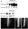
Fig. 3. Immunokinase analysis of the jasplakinolide (JK)- and the latrunculin B (LB)-induced activation of SIMK in root-derived suspension-cultured cells. Alfalfa cells were treated with 5 µM JK or 10 µM LB for the indicated times. Extracts from treated cells, containing 100 µg of total protein, were immunoprecipitated with 5 µg of protein A-purified SIMK antibody. Kinase reactions were performed with 1 mg/ml MBP as a substrate, 0.1 mM ATP and 2 µCi of [γ-32P]ATP. Phosphorylation of MBP was analyzed by autoradiography after SDS–PAGE. Corresponding controls with DMSO [DMSO was used at the same concentrations as for dilution of jasplakinolide (0.25%) and latrunculin B (0.1%), respectively] showing no kinase activity at 0, 10, 30 and 60 min are presented in the lower panels. An immunoblot showing constant levels of SIMK protein during treatments with actin drugs is presented in the lowest panel.
None
Fig. 4. Effect of overexpression of SIMK-GOF and SIMK-LOF on kinase activity and root hair formation. (A) MAPK activity and protein levels in control (c) tobacco SR1 plants and transformed SIMK-GOF (1–4) and SIMK-LOF (1 and 2) SR1 lines using M23 recognizing both SIMK and its tobacco homolog SIPK/Ntf4. (B) Root of a SIMK-GOF tobacco plant showing a very short root hair formation zone (indicated by a bracket) and much longer root hairs (indicated by arrow) as compared with (C and D). (C and D) Roots of non-transformed and SIMK-LOF plants, respectively, showing normal root hair formation zones (brackets) and regular length of root hairs (arrows). Bar = 400 µm for (B–D).
None
Fig. 5. Morphological changes on emerging (A) and growing root hairs (D) induced by 2 h treatment with 10 µM UO 126, a MAPKK inhibitor. (A) UO 126 causes swelling of emerging root hairs (arrows). (B and C) Emerging root hairs are not affected by 2 h treatment with 10 µM UO 124 or 0.1% DMSO, respectively. (D) UO 126 induces vacuolation and swelling of apices of growing root hairs (arrows). (E and F) UO 124 (10 µM) and (0.1%) DMSO have no morphological effect on growing root hairs. Bar = 30 µm for (A), (D) and (F), and 40 µm for (B), (C) and (E).
None
Fig. 6. Immunolocalization of actin (green, A and C) and SIMK (red, B and D) in root hairs treated with 10 µM UO 126, a MAPKK inhibitor (A and B), or with 10 µM UO 124, an inactive analog of UO 126 (C–E) for 60 min. (A) Note the vacuolation at the tips (arrows) and F-actin depletion from root hairs. The remaining actin labeling is evenly distributed. (B) SIMK is distributed evenly in root hair cytoplasm and nuclei, except holes represented by vacuoles (arrows). (C) The tip actin meshwork (arrows) and filamentous actin are preserved in hairs treated with UO 124. (D) SIMK is tip focused in hairs treated with UO 124 (arrows). (E) Co-localization of SIMK with actin meshworks at the tip (arrows, yellow color). Thick F-actin bundles within trichoblasts are indicated by arrowheads. Bar = 30 µm for (A) and (B), 18 µm for (C) and (D) and 23 µm for (E–G).
None
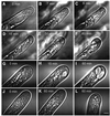
Fig. 7. Video-enhanced microscopy of growing root hairs treated with UO 126. (A) An untreated growing alfalfa root hair with a vesicle-rich apical dome. (BF) Alfalfa root hair treated with 10 µM UO 126. Note that UO 126 markedly changes the cytoarchitecture and shape of the root hair apex causing inhibition of root hair tip growth. (GI) Tobacco growing root hair treated with 10 µM UO 126. This concentration of UO 126 caused growth arrest within 5 min and similar changes in the cytoarchitecture and shape of the root hair apex to those in alfalfa (A–F). (JL) Tobacco SIMK-GOF root hair treated with 10 µM UO 126 (J and K) or 100 µM UO 126 (L) for the indicated time points. Note that 10 µM UO126 did not cause growth arrest and changes in root hair cytoarchitecture and shape within 60 min, while 100 µM UO 126 inhibits root hair growth within 60 min, causing minor changes in the cytoarchitecture. Bar = 7 µm for (A–F) and 5 µm for (G–L).

Similar articles

Cited by

References

    1. Baier R., Schiene,K., Kohring,B., Flaschel,E. and Niehaus,K. (1999) Alfalfa and tobacco cells react differently to chitin oligosaccharides and Sinorhizobium meliloti nodulation factors. Planta, 210, 157–164. - PubMed
    1. Baluška F. and Volkmann,D. (2002) Actin-driven polar growth of plant cells. Trends Cell Biol., 12, 14. - PubMed
    1. Baluška F., Salaj,J., Mathur,J., Braun,M., Jasper,F., Šamaj,J., Chua, N.-H., Barlow,P.W. and Volkmann,D. (2000a) Root hair formation: F-actin-dependent tip growth is initiated by local assembly of profilin-supported F-actin meshworks accumulated within expansin-enriched bulges. Dev. Biol., 227, 618–632. - PubMed
    1. Baluška F., Ovecka,M. and Hirt,H. (2000b) Salt stress- and cell cycle phase-dependent changes in expression and subcellular localisation of the alfalfa mitogen-activated protein kinase SIMK. Protoplasma, 212, 262–267.
    1. Benndorf R., Hayess,K., Ryazantsev,S., Wieske,M., Behlke,J. and Lutsch,G. (1994) Phosphorylation and supramolecular organization of murine small heat shock protein HSP25 abolish its actin polymerization-inhibiting activity. J. Biol. Chem., 269, 20780–20784. - PubMed

Publication types

MeSH terms