Role of NF kappa B activator Act1 in CD40-mediated signaling in epithelial cells
- PMID: 12089335
- PMCID: PMC123150
- DOI: 10.1073/pnas.142294499
Role of NF kappa B activator Act1 in CD40-mediated signaling in epithelial cells
Abstract
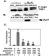
CD40, a cell surface receptor in the tumor necrosis factor receptor family, first identified and functionally characterized on B lymphocytes, is also expressed on epithelial and other cells and is now thought to play a more general role in immune regulation. Overexpression of the NF kappa B activator 1 (Act1) leads to the activation of both NF kappa B and Jun kinase in epithelial cell lines. Endogenous Act1 is recruited to the CD40 receptor in human intestinal (HT29) and cervical (HeLa) epithelial cells upon stimulation with CD40 ligand, indicating that Act1 is involved in this signaling pathway. Act1 also interacts with tumor necrosis factor receptor-associated factor 3, a component involved in CD40-activated pathway. Furthermore, transfection of Act1 into C33A cervical epithelial cells, which do not express it, renders these cells sensitive to CD40 ligand-induced NF kappa B activation and protects them from CD40 ligand-induced apoptosis. We conclude that Act1 plays an important role in CD40-mediated signaling in epithelial cells.
Figures


Similar articles
-
Act1 modulates autoimmunity through its dual functions in CD40L/BAFF and IL-17 signaling.Cytokine. 2008 Feb;41(2):105-13. doi: 10.1016/j.cyto.2007.09.015. Epub 2007 Dec 3. Cytokine. 2008. PMID: 18061473 Review.
-
IFN regulatory factor-1 is required for the up-regulation of the CD40-NF-kappa B activator 1 axis during airway inflammation.J Immunol. 2003 Jun 1;170(11):5674-80. doi: 10.4049/jimmunol.170.11.5674. J Immunol. 2003. PMID: 12759449
-
Act1, a negative regulator in CD40- and BAFF-mediated B cell survival.Immunity. 2004 Oct;21(4):575-87. doi: 10.1016/j.immuni.2004.09.001. Immunity. 2004. PMID: 15485634
-
Flagellin acting via TLR5 is the major activator of key signaling pathways leading to NF-kappa B and proinflammatory gene program activation in intestinal epithelial cells.BMC Microbiol. 2004 Aug 23;4:33. doi: 10.1186/1471-2180-4-33. BMC Microbiol. 2004. PMID: 15324458 Free PMC article.
-
Immune regulation by CD40-CD40-l interactions - 2; Y2K update.Front Biosci. 2000 Nov 1;5:D880-693. doi: 10.2741/kooten. Front Biosci. 2000. PMID: 11056083 Review.
Cited by
-
Act1 mediates IL-17-induced EAE pathogenesis selectively in NG2+ glial cells.Nat Neurosci. 2013 Oct;16(10):1401-8. doi: 10.1038/nn.3505. Epub 2013 Sep 1. Nat Neurosci. 2013. PMID: 23995070 Free PMC article.
-
New insight into the functions of the interleukin-17 receptor adaptor protein Act1 in psoriatic arthritis.Arthritis Res Ther. 2012 Oct 31;14(5):226. doi: 10.1186/ar4071. Arthritis Res Ther. 2012. PMID: 23116200 Free PMC article. Review.
-
Astrocyte-restricted ablation of interleukin-17-induced Act1-mediated signaling ameliorates autoimmune encephalomyelitis.Immunity. 2010 Mar 26;32(3):414-25. doi: 10.1016/j.immuni.2010.03.004. Epub 2010 Mar 18. Immunity. 2010. PMID: 20303295 Free PMC article.
-
Function of Act1 in IL-17 family signaling and autoimmunity.Adv Exp Med Biol. 2012;946:223-35. doi: 10.1007/978-1-4614-0106-3_13. Adv Exp Med Biol. 2012. PMID: 21948371 Free PMC article. Review.
-
Downregulation of Macrophage-Specific Act-1 Intensifies Periodontitis and Alveolar Bone Loss Possibly via TNF/NF-κB Signaling.Front Cell Dev Biol. 2021 Mar 4;9:628139. doi: 10.3389/fcell.2021.628139. eCollection 2021. Front Cell Dev Biol. 2021. PMID: 33748112 Free PMC article.
References
-
- Banchereau J, Bazan F, Blanchard D, Briere F, Galizzi J P, van Kooten C, Liu Y J, Rousset F, Saeland S. Annu Rev Immunol. 1994;12:881–922. - PubMed
-
- van Kooten C, Banchereau J. Int Arch Allergy Immunol. 1997;113:393–399. - PubMed
-
- Young L S, Eliopoulos A G, Gallagher N J, Dawson C W. Immunol Today. 1998;19:502–506. - PubMed
-
- Young L S, Dawson C W, Brown K W, Rickinson A B. Int J Cancer. 1989;43:786–794. - PubMed
Publication types
MeSH terms
Substances
Grants and funding
LinkOut - more resources
Full Text Sources
Molecular Biology Databases
Research Materials
Miscellaneous

