Comparative Study
. 2002 Jun 24;157(7):1257-65.
doi: 10.1083/jcb.200202067. Epub 2002 Jun 24.

Proximal location of mouse prostate epithelial stem cells: a model of prostatic homeostasis

Affiliations
Comparative Study

Proximal location of mouse prostate epithelial stem cells: a model of prostatic homeostasis

Akira Tsujimura et al. J Cell Biol. .

Abstract

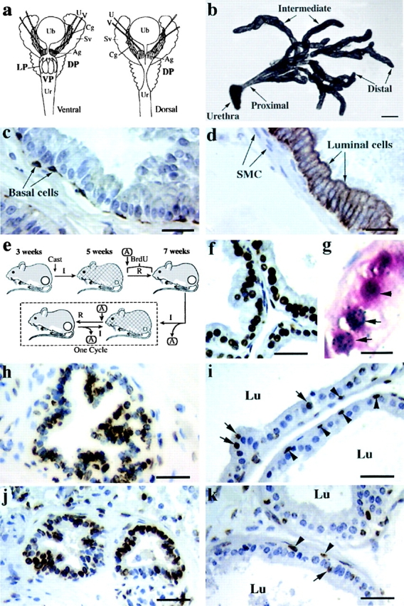
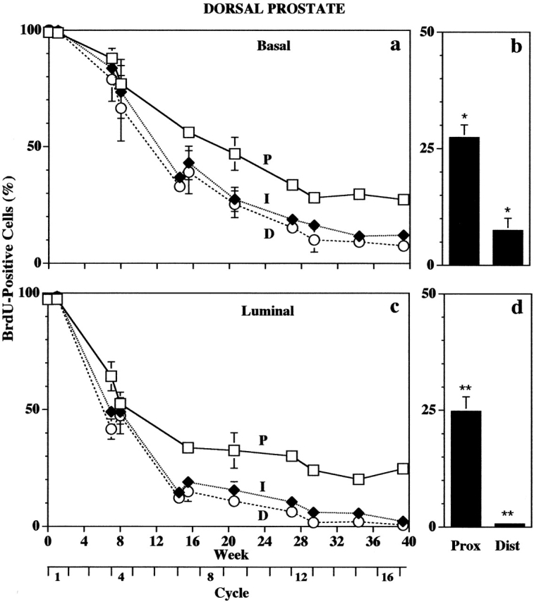
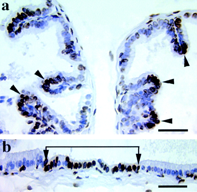
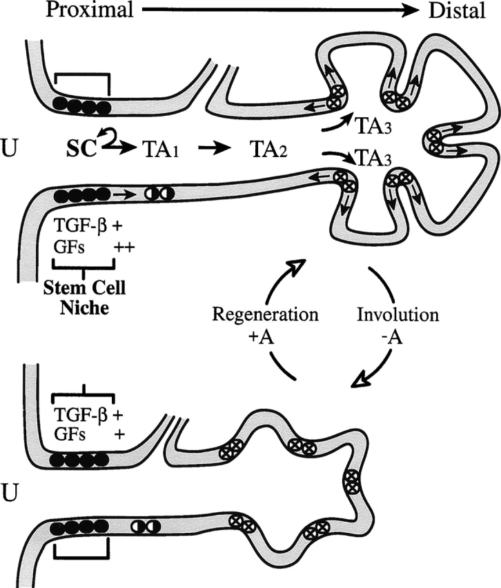
Stem cells are believed to regulate normal prostatic homeostasis and to play a role in the etiology of prostate cancer and benign prostatic hyperplasia. We show here that the proximal region of mouse prostatic ducts is enriched in a subpopulation of epithelial cells that exhibit three important attributes of epithelial stem cells: they are slow cycling, possess a high in vitro proliferative potential, and can reconstitute highly branched glandular ductal structures in collagen gels. We propose a model of prostatic homeostasis in which mouse prostatic epithelial stem cells are concentrated in the proximal region of prostatic ducts while the transit-amplifying cells occupy the distal region of the ducts. This model can account for many biological differences between cells of the proximal and distal regions, and has implications for prostatic disease formation.


Figures

Figure 1.
Figure 1.
The proximal region of mouse prostatic ducts contains a high density of slow-cycling stem cells. (a) Schematic diagrams showing the ventral and dorsal views of the urogenital organs of a male mouse. Ag, ampullary gland; Cg, coagulating gland; DP, dorsal prostate; LP, lateral prostate; Sv, seminal vesicle; U, ureter; Ub, urinary bladder; Ur, urethra; V, vas deferens; VP, ventral prostate. (b) A segment from a microdissected mouse ventral prostate illustrating the distal, intermediate, and proximal regions of prostatic ducts. Bar, 1 mm. (c) A paraffin section of a prostatic duct showing basal cells (arrows) immunohistochemically stained using an antibody against K14 keratin. Bar, 125 μm. (d) A paraffin section of a prostatic duct showing luminal cells (arrows) immunohistochemically stained using an antibody against K8 keratin. SMC, smooth muscle cells. Bar, 125 μm. (e) A schematic diagram illustrating the BrdU labeling protocol used for the identification of the slow-cycling label-retaining cells. In this protocol, almost all prostatic epithelial cells are initially labeled with BrdU. The BrdU label of the more rapidly proliferating transit-amplifying cells is then diluted out during the subsequent involution–regeneration cycles so that the label is retained only by the slow-cycling stem cells that can thus be identified as the label-retaining cells. A, testosterone pellet; Cast, castrate; I, involute; R, regenerate. Each cycle comprised 10 d of regeneration and 7 d of involution. (f) A paraffin section of a prostatic duct indicating that almost all epithelial cells are BrdU labeled (black cells) after the labeling procedure depicted in Fig. 1 e. Bar, 50 μm. (g) A paraffin section of the proximal region of a prostatic duct from the dorsal prostate of a mouse that had been BrdU labeled as described in panel e, followed by androgen withdrawal to involute the prostate. 40 h after a pulse of androgen, animals were given 3H-thymidine and were killed 1 h later. Note that two of the red BrdU-labeled cells (arrow head) contain silver grains (arrows) due to 3H-thymidine incorporation, indicating that slow-cycling cells can divide after androgen administration. Bar, 150 μm. (h) A paraffin section of the proximal region of a prostatic duct from the ventral prostate after 11 cycles of involution–regeneration. Note the high concentration of label-retaining cells in this region. Bar, 50 μm. (i) A paraffin section of the distal region of a prostatic duct from the ventral prostate after 11 cycles. Note the small number of label-retaining cells in this region. Arrowheads, labeled basal cells; arrows, labeled luminal cells. Lu, lumen of duct. Bar, 50 μm. (j) A paraffin section of the proximal region of prostatic ducts from the dorsal prostate after 11 cycles. Note the high concentration of label-retaining cells in this region. Bar, 50 μm. (k) A paraffin section of the distal region of prostatic ducts from the dorsal prostate after 11 cycles. Note the low number of label-retaining cells. Bar, 50 μm.
Figure 2.
Figure 2.
Cell kinetic data showing that the proximal region of the mouse dorsal prostate contains the highest percentage of label-retaining cells. (a) Quantification of BrdU-labeled basal cells in the distal (D), intermediate (I), and proximal (P) regions of ducts examined after various periods of chase. (b) Quantification of BrdU-labeled basal cells in the proximal (Prox) and distal (Dist) region after a 39-wk chase (16 cycles). *P < 0.0001. (c) Quantification of BrdU-labeled luminal cells in the distal (D), intermediate (I), and proximal (P) regions of ducts after various periods of chase. (d) Quantification of BrdU-labeled luminal cells in the proximal (Prox) and distal (Dist) region after a 39-wk chase (16 cycles). **P < 0.0001. The absence of error bars reflects an error range too small to be evident on the graph.
Figure 3.
Figure 3.
Discrete clusters of (intermediate stage) label-retaining cells occur in the intermediate and distal regions of the ducts. Paraffin sections of the ventral prostate after a 20-wk chase showing that clusters of label-retaining cells (a, arrowheads) are associated with ridges protruding into the lumen of a duct, and (b) occur along the ducts (between arrows). Bars, 50 μm.
Figure 4.
Figure 4.
Rapidly cycling cells are located in the distal regions of the prostatic ducts. The percentage of BrdU-labeled basal (a) and luminal cells (b) in the dorsal prostate after a single pulse of BrdU. Distal (Dist), intermediate (Int), and proximal (Prox) regions of the ducts of 5-, 17-, and 34-wk-old mice.
Figure 5.
Figure 5.
The proliferative capacity of proximal cells is greater than that of distal cells. Cells isolated from the proximal (a) and distal (b) regions of the dorsal prostate were seeded at 4 × 10³ cells/well in duplicate wells of eight-well chamber slides. Colonies were fixed and stained 5 d later. Bars, 175 μm. Cells isolated from the proximal (P) and distal (D) regions of the dorsal prostate (DP) (c) were seeded at 2 × 10³ cells/well in duplicate wells in 96-well plates. The total cell output from 2 × 10³ cells from each region was determined at the end of the serial culture when their growth capacity was exhausted (see Materials and methods). *P < 0.0001.
Figure 6.
Figure 6.
Proximal cells form large, branched glandular structures in collagen gels. Proximal (Prox) and distal (Dist) cells (8 × 10³ cells) from the dorsal prostate as well as cells from the entire dorsal prostate (Tot) were seeded in collagen gels and cultured for 6 d. The numbers of ductal structures (a), surface area of structures (b), and branch points/structure (c) in each population were measured. (d and g) Phase contrast microscopy showing the morphology of ductal structures from the proximal (d) and distal (g) regions of ducts. Bars, 150 μm. (e and h) Paraffin sections of proximal (e) and distal (h) ductal structures showing immunofluorescence of basal (PE, red fluorescence) and luminal (FITC, green fluorescence) cells stained using antibodies against K14 (basal) and K8 (luminal) keratins. Bars, 150 μm. (f and i) Paraffin sections of proximal (f) and distal (i) ductal structures stained with hematoxylin and eosin. Bars, 150 μm. (j and k) Paraffin sections of proximal ducts immunohistochemically stained with antibodies specific for prostatic secretory products (j, brown stain). Control (k) section stained with nonspecific antibodies. Sections were counterstained with hematoxylin. Bars, 50 μm. (l) Paraffin section of a branched ductal structure obtained from a single proximal cell stained with antibodies against K14 (basal) and K8 (luminal) keratins showing that it is comprised of both basal (PE, red immunofluorescence) and luminal (FITC, green immunofluorescence) cells. An asterisk indicates the lumen. Bar, 150 μm.
Figure 7.
Figure 7.
A model of prostatic stem cell homeostasis. A schematic diagram showing the location of prostatic epithelial stem cells (SC) in the proximal region of the prostatic duct. The stem cell “niche” is encircled by a thick band of smooth muscle cells that express a high level of TGF-β. It is hypothesized that TGF-β maintains the stem cells in a quiescent state that can be overcome by an androgen-induced increase in the production of growth factors (GFs). The stem cells give rise to young transit-amplifying cells (TA1) that in turn give rise to further generations of transit-amplifying cells (TA2, TA3, etc.) with progressively diminished proliferative potential. Young transit-amplifying cells are positioned in clusters within the ducts where they are available to respond rapidly to androgen and to repopulate the ducts after a surge in the androgen (A) level. U, urethra.

Similar articles

Cited by

References

    1. Baldwin, B.H., B.C. Tennant, T.J. Reimers, R.G. Cowan, and P.W. Concannon. 1985. Circannual changes in serum testosterone concentrations of adult and yearling woodchucks (Marmota monax). Biol. Reprod. 32:804–812. - PubMed
    1. Banerjee, P.P., S. Banerjee, B.R. Zirkin, and T.R. Brown. 1998. Telomerase activity in normal adult Brown Norway rat seminal vesicle: regional distribution and age-dependent changes. Endocrinology. 139:1075–1081. - PubMed
    1. Barrandon, Y., and H. Green. 1987. Three clonal types of keratinocyte with different capacities for multiplication. Proc. Natl. Acad. Sci. USA. 84:2302–2306. - PMC - PubMed
    1. Beauchamp, J.R., L. Heslop, D.S. Yu, S. Tajbakhsh, R.G. Kelly, A. Wernig, M.E. Buckingham, T.A. Partridge, and P.S. Zammit. 2000. Expression of CD34 and Myf5 defines the majority of quiescent adult skeletal muscle satellite cells. J. Cell Biol. 151:1221–1234. - PMC - PubMed
    1. Berardi, A.C., A. Wang, J.D. Levine, P. Lopez, and D.T. Scadden. 1995. Functional isolation and characterization of human hematopoietic stem cells. Science. 267:104–108. - PubMed

Publication types