. 2002 Jun;129(12):2891-903.
doi: 10.1242/dev.129.12.2891.

beta8 integrins are required for vascular morphogenesis in mouse embryos

Affiliations

beta8 integrins are required for vascular morphogenesis in mouse embryos

Jiangwen Zhu et al. Development. 2002 Jun.

Abstract

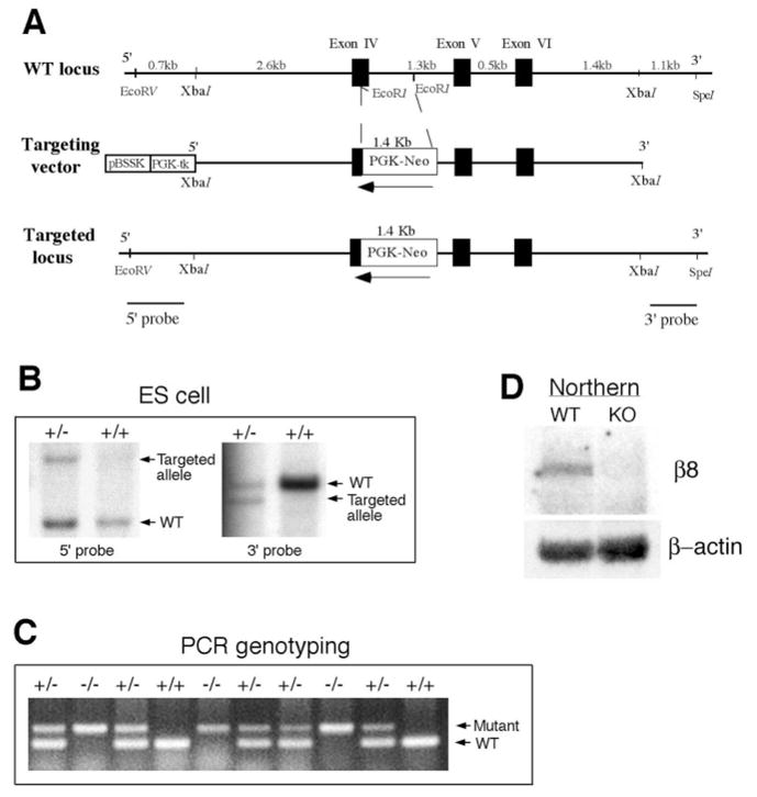
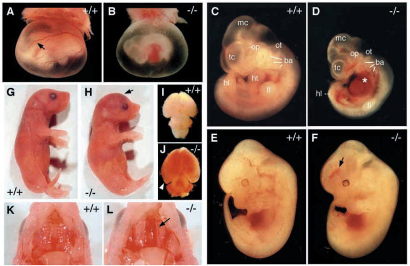
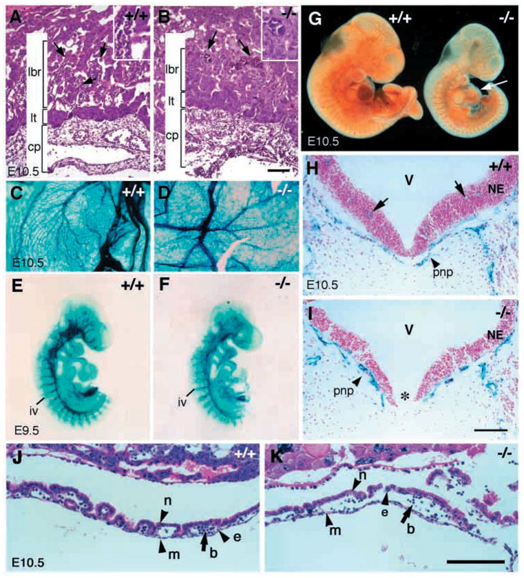
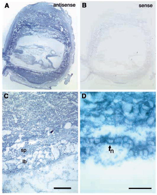
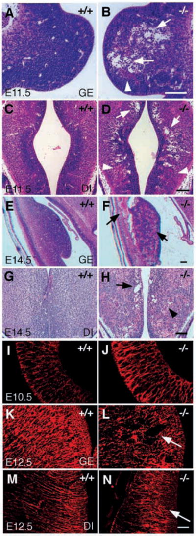
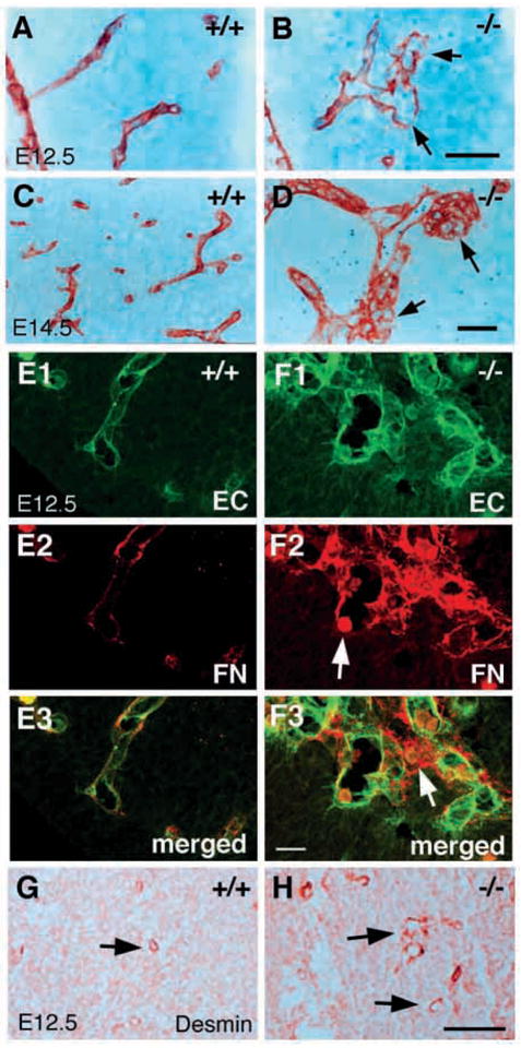
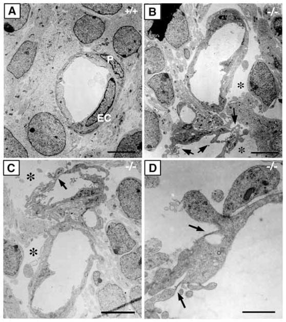
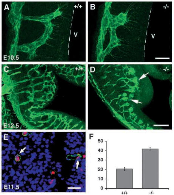
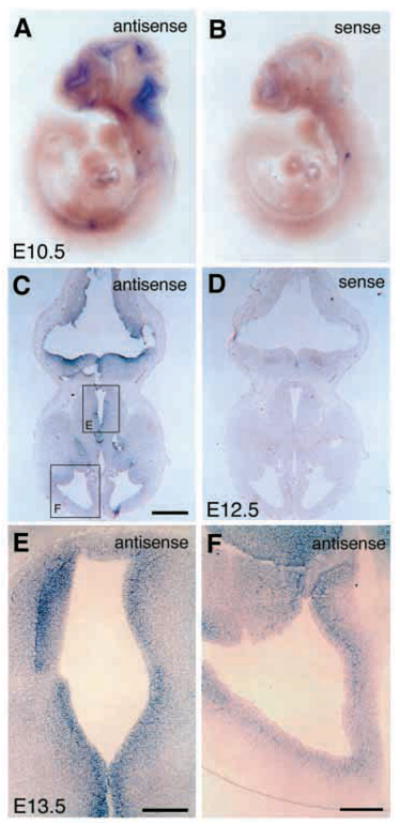
In order to assess the in vivo function of integrins containing the beta8 subunit, we have generated integrin beta8-deficient mice. Ablation of beta8 results in embryonic or perinatal lethality with profound defects in vascular development. Sixty-five percent of integrin beta8-deficient embryos die at midgestation, with evidence of insufficient vascularization of the placenta and yolk sac. The remaining 35% die shortly after birth with extensive intracerebral hemorrhage. Examination of brain tissue from integrin beta8-deficient embryos reveals abnormal vascular morphogenesis resulting in distended and leaky capillary vessels, as well as aberrant brain capillary patterning. In addition, endothelial cell hyperplasia is found in these mutant brains. Expression studies show that integrin beta8 transcripts are localized in endodermal cells surrounding endothelium in the yolk sac and in periventricular cells of the neuroepithelium in the brain. We propose that integrin beta8 is required for vascular morphogenesis by providing proper cues for capillary growth in both yolk sac and embryonic brain. This study thus identifies a molecule crucial for vascular patterning in embryonic yolk sac and brain.


Figures

Fig. 1
Fig. 1
Generation of integrin β8-deficient mice. (A) Schematic drawing of integrin β8 genomic region encompassing Exons IV, V and VI in the wild type and mutant. Black boxes represent exons and white box represents the PGK-neor-cassette. Arrows indicate the transcriptional orientation of the cassette. 5′ and 3′ probes for Southern blot analyses are indicated. (B) Identification of ES clones containing a mutated β8 allele with 5′ and 3′ probes by Southern blot analyses. The wild-type and mutant alleles are labeled. (C) PCR analysis of genotypes from a heterozygous intercross. Mutant and wild-type amplification products are indicated. (D) Northern blot analysis showing the absence of integrin β8 transcripts in homozygous mutant mice. Comparable amounts of wild-type and mutant total RNA were loaded, as indicated by the presence of equal amounts of β-actin RNA. RT-PCR analysis further verified that no functional transcript is expressed in the mutant (data not shown).
Fig. 2
Fig. 2
Phenotypic defects in integrin β8-deficient mice. (A,B) E10.5 yolk sacs. The vasculature is less prominent and often pale in mutant embryos (B) when compared with wild type (A, arrow). (C,D) Side views of E10.5 embryos. The class A mutant embryo exhibits a smaller body size (D) compared with a wild-type littermate (C). An example of enlarged pericardiac cavities is shown in D (*). (E,F) Side views of E12.5 embryos. Intracerebral hemorrhage in a class B mutant embryo is shown (F, arrow) when compared with its wild-type littermate (E). (G,H) Side views of P0 wild-type (G) and mutant (H) pups. Severe hemorrhage in the mutant head is obvious (H, arrow). (I,J) The P0 mutant brain (J) shows characteristics of hydrocephalus (arrowhead) and exhibits visible hemorrhage compared with a wild type (I). (K,L) A cleft palate is obvious in a mutant neonate (L, arrow), but is absent in a wild-type littermate (K). mc, metencephalon; tc, telencephalon; op, optic vesicle; ot, otic pit; ba, bronchial arches; he, heart; fl, forelimb; hl, hindlimb.
Fig. 3
Fig. 3
Angiogenesis defects in class A integrin β8-deficient mutants. (A,B) Hematoxylin and Eosin staining of transverse sections of placentas from an E10.5 wild type (A) and a mutant (B). While the chorionic plate (cp) and labyrinthine trophoblast layer (lt) are comparable in mutant and wild-type littermates, the labyrinthine layer (lbr) is reduced in the mutant. While the interdigitation of fetal blood vessels (A, arrows) and maternal blood vessels in wild-type embryos is elaborate, only a few fetal blood vessels have penetrated into the labyrinthine layer in the mutant (B, arrows). Inserts in A,B provide higher magnification photos of the vasculature. (C,D) Vasculature of E10.5 yolk sac in wild-type (C) and mutant (D) as depicted by X-gal staining of a Tie2:lacZ reporter gene in these mice. (E–G) Vascular patterns revealed by whole-mount staining of β-galactosidase activity in E9.5 embryos expressing the Tie2:lacZ reporter gene (E,F) and whole-mount immunohistochemistry with anti- PECAM antibody in E10.5 embryos (G). An E9.5 mutant embryo (E) shows no obvious abnormalities in vascular pattern compared with a wild-type littermate (F) (the tails of the embryos in both E and F were used for genotyping). E10.5 mutant embryos (G, right) have similar vascular patterns as wild-type littermates (G, left) except for reduced vasculature development in the heart (G, arrow). (H,I) X-gal staining of transverse sections of E10.5 neural tubes in wild-type (H) and mutant embryos (I) with the Tie2:lacZ reporter gene reveals the vasculature (blue) and cell nuclei labeled with Nuclear Fast Red (pink). While perineural plexuses are present in both the wild type and the mutant (H,I, arrowheads), there is no apparent penetration of vessels into the neural tube in the mutant when compared with the wild-type embryo (H, arrows). Note that the floor plate in the mutant (I, *) is absent. cp, chorionic plate; lt, labyrinthine trophoblast; lbr, labyrinthine layer; iv, intersomitic vessel; pnp, perineural plexus; V, ventricle; NE, neuroepithelium. (J,K) Hematoxylin and Eosin staining of E10.5 yolk sac showing presence of endothelial cells (e), mesothelial cells (m), blood cells (b) and endoderm cells (n) in both wild type (J) and mutant (K). Scale bars: 100 μm in A,B,H,I (500 μm in insets); 150 μm in J,K.
Fig. 4
Fig. 4
Integrin β8 expression in E9.5 placenta tissue. (A–D) Sections of E9.5 placenta hybridized with integrin β8 antisense (A,C,D) or sense (B) probes showing localization of integrin β8 to most of placenta tissues, notably in the trophoblast giant cells (A), labyrinthine layer and spongiotrophoblast layer (C). In the yolk sac, integrin β8 appears to be expressed specifically in the endoderm cells (D, arrow). sp, spongiotrophoblast layer; lb, labyrinthine layer. Scale bar: 200 μm in C; 50 μm in D.
Fig. 5
Fig. 5
Abnormal cavitation and radial glial organization in the brains of class B integrin β8-deficient mutants. (A–H) Hematoxylin and Eosin staining of transverse sections of E11.5 brain (A–D) and E14.5 brain (E–H), showing abnormal cavitation in integrin β8-deficient mutant brains (B,D,F,H, arrows) compared with the wild-type littermates (A,C,E,G). Hemorrhage is visible in the ganglionic eminence (B, arrowhead) and diencephalon (D, arrowhead) of an E11.5 mutant brain and becomes much more severe in the E14.5 mutant brain (F, arrow; H, arrowhead). (I–N) Radial glial organization characterized by immunohistochemical staining using the RC2 antibody. Radial glial cells look grossly normal in an E10.5 mutant brain (J) compared with a wild-type littermate (I). However, they are apparently disorganized in the ganglionic eminence (L, arrow) and diencephalon (N, arrow) in the E12.5 mutant brain when compared with the same regions of E12.5 wild-type brains (K,M). GE, ganglionic eminence; DI, diencephalon. Scale bars: 100 μm in A–H; 50 μm in I–N.
Fig. 6
Fig. 6
Abnormal brain capillary morphologies in class B integrin β8-deficient mutants. (A–D) Paraffin wax embedded sections of E12.5 (A,B) and E14.5 (C,D) brains stained with anti-laminin antibodies that show the abnormal morphologies of capillary vessels in the integrin β8-deficient mutant (B,D) when compared with a wild-type littermate (A,C). Discontinuous basement membranes are indicated by arrows (B). Aggregates of capillary vessels are visible (D, arrow). (E–F) Projected confocal images of E12.5 brain capillary vessels double labeled with anti-PECAM antibody (green) and anti- FN (red) antibodies. Capillary vessels in an integrin β8-deficient mutant (F1–3) exhibit irregular distended morphologies and are often conjoined when compared with those in wild-type littermates (E1–3). The basement membrane is discontinuous and a blood cell is captured at a potential hemorrhage site (F2, arrow) in the mutant. Note that a blood cell is present clearly outside of the capillaries, indicating hemorrhage in a nearby location (F3, arrow). (G,H) Pericytes recognized with anti-desmin are present and recruited to the capillaries in the brains of E12.5 wild-type (G, arrow) and mutant embryos (H, arrow). Scale bars: 50 μm in A–D; 20 μm in E,F; 100 μm in G,H.
Fig. 7
Fig. 7
Abnormal endothelial cell morphology in class B integrin β8- deficient brains. (A–D) Electron micrographs of capillary structure in E12.5 wild-type (A) and integrin β8-deficient mutant (B–D) brains. In contrast to the wild type (A), the endothelial cells in the mutant display abundant active membrane protrusions (B, arrows) and large empty spaces surrounding the capillaries (B,C, *). In addition, fenestrations (D, arrows) are often seen in the mutant endothelium. P, pericyte; EC, endothelial cell. Scale bars: 5 μm in A–C; 1 μm in D.
Fig. 8
Fig. 8
Abnormal capillary vascular patterning and endothelial cell hyperplasia in class B integrin β8-deficient brains. (A–D) Projected confocal images of brain capillary vessels stained with anti-PECAM in E10.5 (A,B) and E12.5 (C,D) embryos. Wild-type capillary vessels grow close to and couple near the ventricle (A, broken line marks boundary of ventricle, V). However, capillary vessels extend a shorter distance into the neuroepithelium and couple further away from the ventricle in the mutant (B). At E12.5, wild-type capillary vessels have branched and anastomosed to form an organized network (C); while in the mutant, capillary vessels show bulbous distentions and have not invaded regions of neuroepithelium immediately adjacent to the ventricle (D, arrows). (E) A brain capillary stained for isolectin BS (staining endothelial cell, green), DAPI (staining nuclei, blue) and BrdU (labeling proliferating cell, red, arrows). (F) The quantification of endothelial cell nuclei per vessel cross section in E11.5 wild-type and mutant brains. The percentage of BrdU-labeled endothelial cell nuclei in total endothelial cell nuclei scored is shown in the histogram (n=3). The error bars represent the s.e.m. Wild-type and mutant values are significantly different (P<0.005). Scale bars: 20 μm in A,B; 100 μm in C,D; 25 μm in E.
Fig. 9
Fig. 9
Integrin β8 is expressed in periventricular cells of the embryonic brain. (A,B) Whole-mount in situ hybridization analyses shows integrin β8 mRNA expression in tissues surrounding the ventricles of E10.5 embryonic brain (A, antisense; B, sense). (C,D) Transverse sections of E12.5 brains hybridized with integrin β8 antisense (C) or sense (D) probes showing localization of integrin β8 to periventricular cells in the neuroepithelium. (E,F) Similar areas to those shown in C at E12.5 (C, box E and box F) are shown at a higher magnification in an E13.5 brain hybridized with an integrin β8 antisense probe (E,F). Note the absence of integrin β8 mRNA from the vascular cells of the brain (A) and inside of the brain area (C,E). Scale bar: 500 μm in C and D; 200 μm in E,F.

Similar articles

Cited by

References

    1. Adams RH, Wilkinson GA, Weiss C, Diella F, Gale NW, Deutsch U, Risau W, Klein R. Roles of ephrinB ligands and EphB receptors in cardiovascular development: demarcation of arterial/venous domains, vascular morphogenesis, and sprouting angiogenesis. Genes Dev. 1999;13:295–306. - PMC - PubMed
    1. Adams RH, Diella F, Hennig S, Helmbacher F, Deutsch U, Klein R. The cytoplasmic domain of the ligand ephrinB2 is required for vascular morphogenesis but not cranial neural crest migration. Cell. 2001;104:57–69. - PubMed
    1. Bader BL, Rayburn H, Crowley D, Hynes RO. Extensive vasculogenesis, angiogenesis, and organogenesis precede lethality in mice lacking all alpha v integrins. Cell. 1998;95:507–519. - PubMed
    1. Baldwin HS, Shen HM, Yan HC, DeLisser HM, Chung A, Mickanin C, Trask T, Kirschbaum NE, Newman PJ, Albelda SM, et al. Platelet endothelial cell adhesion molecule-1 (PECAM- 1/CD31): alternatively spliced, functionally distinct isoforms expressed during mammalian cardiovascular development. Development. 1994;120:2539–2553. - PubMed
    1. Breier G, Albrecht U, Sterrer S, Risau W. Expression of vascular endothelial growth factor during embryonic angiogenesis and endothelial cell differentiation. Development. 1992;114:521–532. - PubMed

Publication types