. 2002 May;14(5):1077-92.
doi: 10.1105/tpc.000620.

The destination for single-pass membrane proteins is influenced markedly by the length of the hydrophobic domain

Affiliations

The destination for single-pass membrane proteins is influenced markedly by the length of the hydrophobic domain

Federica Brandizzi et al. Plant Cell. 2002 May.

Abstract

The tonoplast was proposed as a default destination of membrane-bound proteins without specific targeting signals. To investigate the nature of this targeting, we created type I fusion proteins with green fluorescent protein followed by the transmembrane domain of the human lysosomal protein LAMP1. We varied the length of the transmembrane domain from 23 to either 20 or 17 amino acids by deletion within the hydrophobic domain. The resulting chimeras, called TM23, TM20, and TM17, were expressed either transiently or stably in tobacco. TM23 clearly accumulated in the plasmalemma, as confirmed by immunoelectron microscopy. In contrast, TM17 clearly was retained in the endoplasmic reticulum, and TM20 accumulated in small mobile structures. The nature of the TM20-labeled compartments was investigated by coexpression with a marker localized mainly in the Golgi apparatus, AtERD2, fused to a yellow fluorescent protein. The strict colocalization of both fluorescent proteins indicated that TM20 accumulated in the Golgi apparatus. To further test the default destination of type I membrane proteins, green fluorescent protein was fused to the 19-amino acid transmembrane domain of the plant vacuolar sorting receptor BP-80. The resulting chimera also accumulated in the Golgi instead of in post-Golgi compartments, where native BP-80 localized. Additionally, when the transmembrane domain of BP-80 was lengthened to 22 amino acids, the reporter escaped the Golgi and accumulated in the plasma membrane. Thus, the tonoplast apparently is not a favored default destination for type I membrane proteins in plants. Moreover, the target membrane where the chimera concentrates is not unique and depends at least in part on the length of the membrane-spanning domain.


Figures
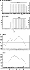
Figure 1.
Figure 1.
Estimated Membrane-Spanning Domains and Hydrophobic Plots for the Membrane Fusion Chimeras with GFP. Detailed are the most C-terminal amino acids of the chimeras (x axes). AA, amino acid. (A) Sequences from the chimeras were submitted to the TMHMM program to estimate their probability of being inside a membrane (y axes). Thick black lines represent the position of the transmembrane domain as estimated by TMHMM. The sequence at top shows the TM23 transmembrane domain and the derived TM20 and TM17. The amino acids removed from the original LAMP1 transmembrane domain from TM23 to produce TM20 are single underlined. The three amino acids removed from TM20 to produce TM17 are double underlined. The sequence at bottom shows the BP19 chimera, including the transmembrane domain of the vacuolar sorting receptor BP-80 and a corresponding derivative, BP22, with the added tripeptide LAL in the transmembrane domain. (B) Hydropathy plots of TM23 and BP19 using the program of Kyte and Doolittle (1982). The hydropathy index of each residue is represented on the y axes. The position of the transmembrane domain estimated by the TMHMM program is underlined. Two Phe residues internal to the membrane-spanning domain of BP-80 are indicated with stars.
Figure 2.
Figure 2.
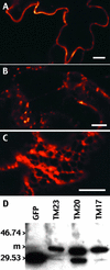
Differential Accumulation Pattern of Transiently Expressed TM23, TM20, and TM17 Reflects the Accumulation of a Membrane-Bound Reporter. (A) to (C) Tobacco lower leaf epidermis expressing the transmembrane chimeras at 72 hr after infiltration. The GFP accumulation pattern was observed with a confocal microscope from cells expressing TM23 (A), TM20 (B), or TM17 (C). The scale of colors from red to white represents the amount of GFP accumulation from the lowest to the highest GFP signal, respectively. Images were collected in a linear mode of GFP fluorescence. Bars = 10 μm. (D) Immunoblot with anti-GFP antibodies of a crude extract of leaves transiently expressing TM23, TM20, or TM17. A soluble GFP was used as a control (GFP lane). m, positions of the membrane forms of the transmembrane chimeras. Positions of molecular mass markers (in kilodaltons) are shown at left.
Figure 3.
Figure 3.
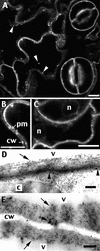
Tobacco Lower Leaf Epidermis Stably Expressing TM23 Accumulates GFP in the Plasma Membrane. Small pieces of a leaf stably expressing the GFP chimera were cut off and either observed using a confocal microscope ([A] to [C]) or fixed and immunolabeled with anti-GFP antibodies for electron microscopy ([D] and [E]). In addition to the dominant line-type of labeling, dots are visible (arrowheads, [A]) that are equivalent to structures associated with the plasma membrane (arrowhead with a star [E]). The plasma membrane-specific location of TM23 is especially visible within a 900-nm-long portion of membrane delimited by arrowheads (D). No labeling was found on the tonoplast, marked by arrows ([D] and [E]). c, chloroplast; cw, cell wall; n, nucleus; pm, plasma membrane; v, vacuole. Bars = 10 μm for (A) to (C) and 200 nm for (D) and (E).
Figure 4.
Figure 4.
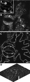
Tobacco Lower Leaf Epidermal Cells Stably Transformed by TM17 Accumulate GFP in the Endoplasmic Reticulum. (A) to (C) Small pieces of a leaf stably expressing the GFP chimera were cut off and observed using a confocal microscope. The same area of cells is presented in various confocal planes at the level of the nucleus of stomata cells (A), at the level of the cortical ER (B), or on a median plane of the epidermal cell layer (C). n, nucleus. Bars = 10 μm. (D) Three-dimensional reconstruction of labeled epidermal cells using the program NIH Image 1.62/3DV1.01. Fourteen individual confocal sections were taken from the same labeled area, each corresponding to 1 μm of thickness, and were treated to rebuild the three-dimensional view.
Figure 5.
Figure 5.
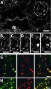
A Single Transmembrane Domain of 20 Amino Acids Leads to the Accumulation of a GFP Chimera in the Golgi. Small pieces of a leaf, stably ([A] to [F]) or transiently ([G] and [H]) expressing the GFP chimera TM20, were cut off and observed using a confocal microscope. In (G) and (H), epidermal cells were double transformed with the GFP chimera TM20 (green) and the AtERD2-YFP marker for the Golgi apparatus and the ER (red). The two images collected from the same optical section were merged to show the colocalization of the two markers (right panels, yellow). n, nucleus. Bars = 10 μm for (A) to (F) and 5 μm for (G) and (H).
Figure 6.
Figure 6.
Lengthening the Plant Transmembrane Domain of the Vacuolar Sorting Receptor BP-80 from 19 to 22 Amino Acids Affects the Localization of the Reporter from the Golgi to the Plasma Membrane. Small pieces of leaves transiently expressing the GFP chimeras BP19 and BP22 were observed using a confocal microscope. (A) Cells were double transformed with the GFP chimera BP19 (green) and the AtERD2-YFP marker for the Golgi apparatus and the ER (red). The two images collected from the same optical section were merged to show the colocalization of the two markers (right panels, yellow). (B) and (C) Epidermal cells expressing the BP22 construct were observed using the quantitative mode of the microscope. The scale of colors from red to white represents the amount of GFP accumulation from the lowest to the highest GFP signal, respectively. Blue represents the saturating emission of fluorescence (arrowheads). Plasma membrane labeling of two adjacent cells is clearly visible in some areas of the cells (arrows). (D) Cells were double transformed with the GFP chimera BP22 (green) and the AtERD2-YFP marker for the Golgi apparatus and the ER (red). The two images collected from the same optical section were merged to show that none of the BP22 bodies (arrowheads) localize together with the Golgi (arrow). Bars = 5 μm for (A) and (D) and 10 μm for (B) and (C).
Figure 7.
Figure 7.
Model for the Distribution of Membrane Proteins within the Plant Secretory Pathway. Membrane-bound chimeras to GFP (stars) are found at various locations in living tobacco cells. The membrane corresponds to the ER, the Golgi, or the plasma membrane for a transmembrane domain length of 17, 20, and 23 amino acids, respectively. This length-dependent distribution also is found when using the transmembrane domain of BP-80, a vacuolar sorting receptor (black triangles) normally localized in the trans-Golgi network (TGN) and the prevacuole. In some cases, reporter accumulated in the Golgi may be in contact with a lytic environment that is responsible for the cleavage (scissors) of the linker between the GFP and the membrane. The soluble released reporter then could reach the vacuole in a process that remains to be characterized (dotted arrows). Importantly, no tonoplast accumulation of the reporters was detected. This finding suggests that some positive information is required for membrane proteins in plants to efficiently reach the compartments located beyond the Golgi on the vacuolar route (gray compartments).

Similar articles

Cited by

References

    1. Ahmed, S.U., Bar-Peled, M., and Raikhel, N.V. (1997). Cloning and subcellular location of an Arabidopsis receptor-like protein that shares common features with protein-sorting receptors of eukaryotic cells. Plant Physiol. 114, 325–336. - PMC - PubMed
    1. Ahmed, S.U., Rojo, E., Kovaleva, V., Venkataraman, S., Dombrowski, J.E., Matsuoka, K., and Raikhel, N.V. (2000). The plant vacuolar sorting receptor AtELP is involved in transport of NH(2)-terminal propeptide-containing vacuolar proteins in Arabidopsis thaliana. J. Cell Biol. 149, 1335–1344. - PMC - PubMed
    1. Barrieu, F., and Chrispeels, M.J. (1999). Delivery of a secreted soluble protein to the vacuole via a membrane anchor. Plant Physiol. 120, 961–968. - PMC - PubMed
    1. Batoko, H., Zheng, H.-Q., Hawes, C., and Moore, I. (2000). A Rab1 GTPase is required for transport between endoplasmic reticulum and Golgi apparatus and for normal Golgi movement in plants. Plant Cell 12, 2201–2217. - PMC - PubMed
    1. Benghezal, M., Wasteneys, G.O., and Jones, D.A. (2000). The C-terminal dilysine motif confers endoplasmic reticulum localization to type I membrane proteins in plants. Plant Cell 12, 1179–1201. - PMC - PubMed

Publication types

MeSH terms