Disruption of a long-range cis-acting regulator for Shh causes preaxial polydactyly
- PMID: 12032320
- PMCID: PMC124279
- DOI: 10.1073/pnas.112212199
Disruption of a long-range cis-acting regulator for Shh causes preaxial polydactyly
Abstract
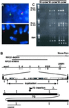
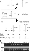
Preaxial polydactyly (PPD) is a common limb malformation in human. A number of polydactylous mouse mutants indicate that misexpression of Shh is a common requirement for generating extra digits. Here we identify a translocation breakpoint in a PPD patient and a transgenic insertion site in the polydactylous mouse mutant sasquatch (Ssq). The genetic lesions in both lie within the same respective intron of the LMBR1/Lmbr1 gene, which resides approximately 1 Mb away from Shh. Genetic analysis of Ssq reveals that the Lmbr1 gene is incidental to the phenotype and that the mutation directly interrupts a cis-acting regulator of Shh. This regulator is most likely the target for generating PPD mutations in human.
Figures


Similar articles
-
A novel candidate gene for mouse and human preaxial polydactyly with altered expression in limbs of Hemimelic extra-toes mutant mice.Genomics. 2000 Jul 1;67(1):19-27. doi: 10.1006/geno.2000.6225. Genomics. 2000. PMID: 10945466
-
Elimination of a long-range cis-regulatory module causes complete loss of limb-specific Shh expression and truncation of the mouse limb.Development. 2005 Feb;132(4):797-803. doi: 10.1242/dev.01613. Development. 2005. PMID: 15677727
-
Single base pair change in the long-range Sonic hedgehog limb-specific enhancer is a genetic basis for preaxial polydactyly.Dev Dyn. 2005 Feb;232(2):345-8. doi: 10.1002/dvdy.20254. Dev Dyn. 2005. PMID: 15637698
-
[Progress on polydactyly character of vertebrate].Yi Chuan. 2004 May;26(3):387-93. Yi Chuan. 2004. PMID: 15640026 Review. Chinese.
-
Sonic hedgehog: restricted expression and limb dysmorphologies.J Anat. 2003 Jan;202(1):13-20. doi: 10.1046/j.1469-7580.2003.00148.x. J Anat. 2003. PMID: 12587915 Free PMC article. Review.
Cited by
-
Extensive promoter-centered chromatin interactions provide a topological basis for transcription regulation.Cell. 2012 Jan 20;148(1-2):84-98. doi: 10.1016/j.cell.2011.12.014. Cell. 2012. PMID: 22265404 Free PMC article.
-
A new locus for split hand/foot malformation with long bone deficiency (SHFLD) at 2q14.2 identified from a chromosome translocation.Hum Genet. 2007 Sep;122(2):191-9. doi: 10.1007/s00439-007-0390-7. Epub 2007 Jun 14. Hum Genet. 2007. PMID: 17569090
-
Why study human limb malformations?J Anat. 2003 Jan;202(1):27-35. doi: 10.1046/j.1469-7580.2003.00130.x. J Anat. 2003. PMID: 12587917 Free PMC article.
-
A high-resolution map of the three-dimensional chromatin interactome in human cells.Nature. 2013 Nov 14;503(7475):290-4. doi: 10.1038/nature12644. Epub 2013 Oct 20. Nature. 2013. PMID: 24141950 Free PMC article.
-
Conserved noncoding elements follow power-law-like distributions in several genomes as a result of genome dynamics.PLoS One. 2014 May 2;9(5):e95437. doi: 10.1371/journal.pone.0095437. eCollection 2014. PLoS One. 2014. PMID: 24787386 Free PMC article.
References
-
- Heutink P, Zguricas J, van Oosterhout L, Breedveld G J, Testers L, Sandkuijl L A, Snijders P J, Weissenbach J, Lindhout D, Hovius S E, et al. Nat Genet. 1994;6:287–292. - PubMed
-
- Hing A V, Helms C, Slaugh R, Burgess A, Wang J C, Herman T, Dowton S B, Donis-Keller H. Am J Med Genet. 1995;58:128–135. - PubMed
-
- Heus H C, Hing A, van Baren M J, Joosse M, Breedveld G J, Wang J C, Burgess A, Donnis-Keller H, Berglund C, Zguricas J, et al. Genomics. 1999;57:342–351. - PubMed
Publication types
MeSH terms
Substances
Associated data
- Actions
LinkOut - more resources
Full Text Sources
Molecular Biology Databases
Miscellaneous

