A fragment of the HMGN2 protein homes to the nuclei of tumor cells and tumor endothelial cells in vivo
- PMID: 12032302
- PMCID: PMC124250
- DOI: 10.1073/pnas.062189599
A fragment of the HMGN2 protein homes to the nuclei of tumor cells and tumor endothelial cells in vivo
Abstract
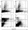
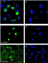
We used a screening procedure to identify protein domains from phage-displayed cDNA libraries that bind both to bone marrow endothelial progenitor cells and tumor vasculature. Screening phage for binding of progenitor cell-enriched bone marrow cells in vitro, and for homing to HL-60 human leukemia cell xenograft tumors in vivo, yielded a cDNA fragment that encodes an N-terminal fragment of human high mobility group protein 2 (HMGN2, formerly HMG-17). Upon i.v. injection, phage displaying this HMGN2 fragment homed to HL-60 and MDA-MB-435 tumors. Testing of subfragments localized the full binding activity to a 31-aa peptide (F3) in the HMGN2 sequence. Fluorescein-labeled F3 peptide bound to and was internalized by HL-60 cells and human MDA-MB-435 breast cancer cells, appearing initially in the cytoplasm and then in the nuclei of these cells. Fluorescent F3 accumulated in HL-60 and MDA-MB-435 tumors after an i.v. injection, appearing in the nuclei of tumor endothelial cells and tumor cells. Thus, F3 can carry a payload (phage, fluorescein) to a tumor and into the cell nuclei in the tumor. This peptide may be suitable for targeting cytotoxic drugs and gene therapy vectors into tumors.
Figures


Similar articles
-
High mobility group nucleosomal binding domain 2 (HMGN2) SUMOylation by the SUMO E3 ligase PIAS1 decreases the binding affinity to nucleosome core particles.J Biol Chem. 2014 Jul 18;289(29):20000-11. doi: 10.1074/jbc.M114.555425. Epub 2014 May 28. J Biol Chem. 2014. PMID: 24872413 Free PMC article.
-
A tumor-homing peptide with a targeting specificity related to lymphatic vessels.Nat Med. 2002 Jul;8(7):751-5. doi: 10.1038/nm720. Epub 2002 Jun 10. Nat Med. 2002. PMID: 12053175
-
Progesterone signals through membrane progesterone receptors (mPRs) in MDA-MB-468 and mPR-transfected MDA-MB-231 breast cancer cells which lack full-length and N-terminally truncated isoforms of the nuclear progesterone receptor.Steroids. 2011 Aug;76(9):921-8. doi: 10.1016/j.steroids.2011.01.008. Epub 2011 Feb 1. Steroids. 2011. PMID: 21291899 Free PMC article.
-
HDAC6 Deacetylates HMGN2 to Regulate Stat5a Activity and Breast Cancer Growth.Mol Cancer Res. 2016 Oct;14(10):994-1008. doi: 10.1158/1541-7786.MCR-16-0109. Epub 2016 Jun 29. Mol Cancer Res. 2016. PMID: 27358110 Free PMC article.
-
[Application of HMGN2-tag constructs to analysis of HMGN2 distribution in HeLa cells].Sheng Wu Yi Xue Gong Cheng Xue Za Zhi. 2005 Oct;22(5):1015-9. Sheng Wu Yi Xue Gong Cheng Xue Za Zhi. 2005. PMID: 16294743 Chinese.
Cited by
-
Quantum Dot-Based Screening Identifies F3 Peptide and Reveals Cell Surface Nucleolin as a Therapeutic Target for Rhabdomyosarcoma.Cancers (Basel). 2022 Oct 14;14(20):5048. doi: 10.3390/cancers14205048. Cancers (Basel). 2022. PMID: 36291832 Free PMC article.
-
Synthesis and evaluation of an 18 F-labeled derivative of F3 for targeting surface-expressed nucleolin in cancer and tumor endothelial cells.J Labelled Comp Radiopharm. 2016 Oct;59(12):492-499. doi: 10.1002/jlcr.3439. Epub 2016 Sep 4. J Labelled Comp Radiopharm. 2016. PMID: 27594091 Free PMC article.
-
Baculovirus display: a multifunctional technology for gene delivery and eukaryotic library development.Adv Virus Res. 2006;68:91-112. doi: 10.1016/S0065-3527(06)68003-2. Adv Virus Res. 2006. PMID: 16997010 Free PMC article. Review.
-
Molecular changes in the vasculature of injured tissues.Am J Pathol. 2007 Aug;171(2):702-11. doi: 10.2353/ajpath.2007.061251. Epub 2007 Jun 28. Am J Pathol. 2007. PMID: 17600129 Free PMC article.
-
Silk Fibroin as a Functional Biomaterial for Drug and Gene Delivery.Pharmaceutics. 2019 Sep 26;11(10):494. doi: 10.3390/pharmaceutics11100494. Pharmaceutics. 2019. PMID: 31561578 Free PMC article. Review.
References
-
- Hanahan D, Folkman J. Cell. 1996;86:353–364. - PubMed
-
- Jain R K. Adv Drug Delivery Rev. 1997;26:71–90. - PubMed
-
- Hanahan D, Weinberg R A. Cell. 2000;100:57–70. - PubMed
-
- Holash J, Maisonpierre P C, Compton D, Boland P, Alexander C R, Zagzag D, Yancopoulos G D, Wiegand S J. Science. 1999;284:1994–1998. - PubMed
-
- Yancopoulos G D, Davis S, Gale N W, Rudge J S, Wiegand S J, Holash J. Nature (London) 2000;407:242–248. - PubMed
Publication types
MeSH terms
Substances
Grants and funding
LinkOut - more resources
Full Text Sources
Other Literature Sources
Miscellaneous

