. 2002 Jun 3;21(11):2724-35.
doi: 10.1093/emboj/21.11.2724.

p110, a novel human U6 snRNP protein and U4/U6 snRNP recycling factor

Affiliations

p110, a novel human U6 snRNP protein and U4/U6 snRNP recycling factor

Mathias Bell et al. EMBO J. .

Abstract

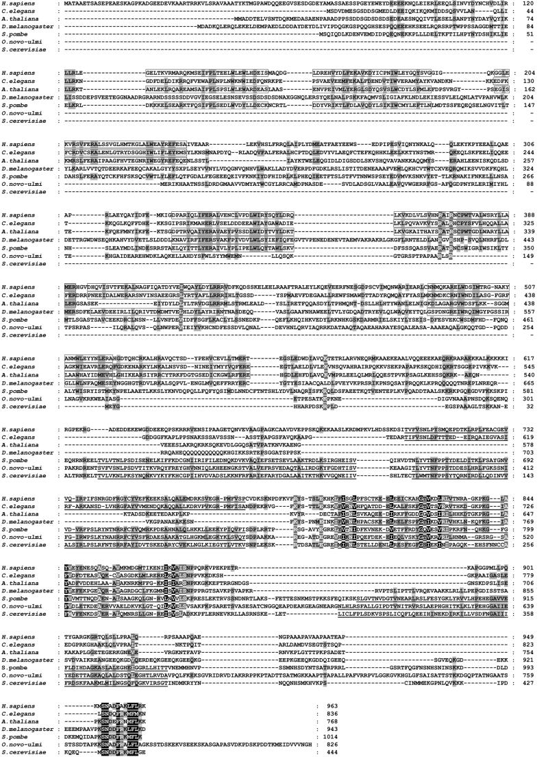
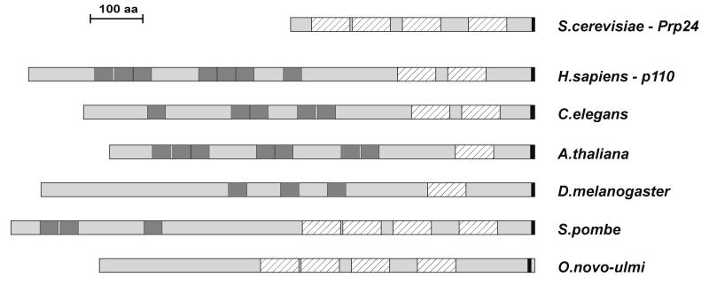
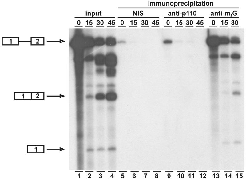
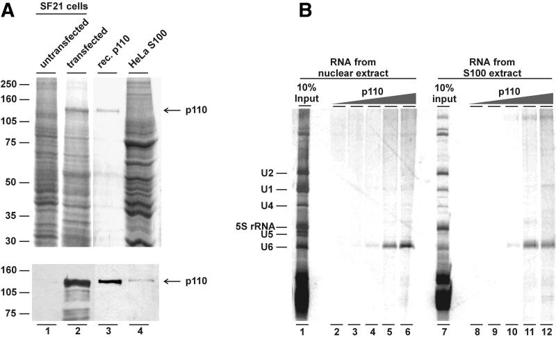
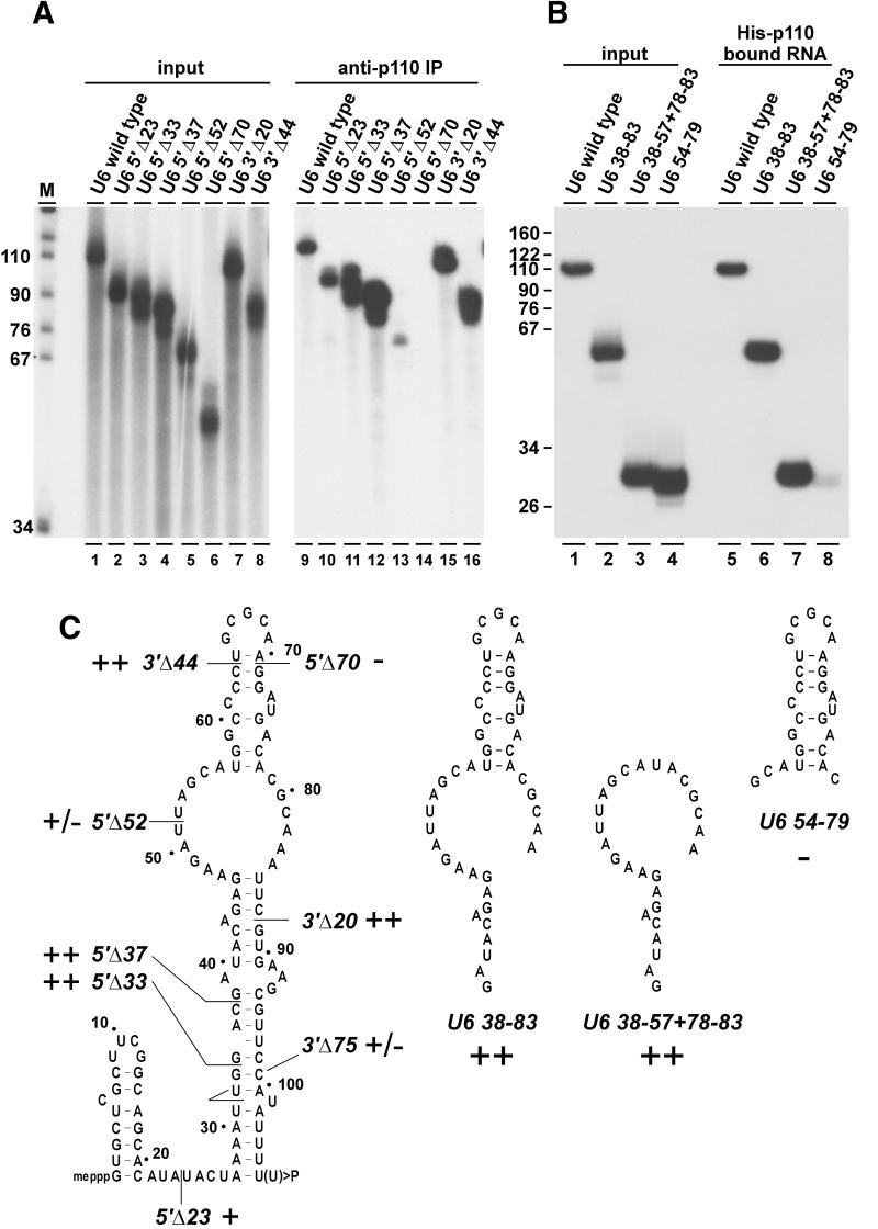
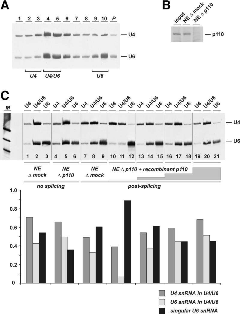
During each spliceosome cycle, the U6 snRNA undergoes extensive structural rearrangements, alternating between singular, U4-U6 and U6-U2 base-paired forms. In Saccharomyces cerevisiae, Prp24 functions as an snRNP recycling factor, reannealing U4 and U6 snRNAs. By database searching, we have identified a Prp24-related human protein previously described as p110(nrb) or SART3. p110 contains in its C-terminal region two RNA recognition motifs (RRMs). The N-terminal two-thirds of p110, for which there is no counterpart in the S.cerevisiae Prp24, carries seven tetratricopeptide repeat (TPR) domains. p110 homologs sharing the same domain structure also exist in several other eukaryotes. p110 is associated with the mammalian U6 and U4/U6 snRNPs, but not with U4/U5/U6 tri-snRNPs nor with spliceosomes. Recom binant p110 binds in vitro specifically to human U6 snRNA, requiring an internal U6 region. Using an in vitro recycling assay, we demonstrate that p110 functions in the reassembly of the U4/U6 snRNP. In summary, p110 represents the human ortholog of Prp24, and associates only transiently with U6 and U4/U6 snRNPs during the recycling phase of the spliceosome cycle.


Figures

None
Fig. 1. Sequence alignment of the human p110 protein with homologous proteins. The human p110 sequence (Homo sapiens; NP_055521) has been aligned with the following sequences: Caenorhabditis elegans (CAA97405); Arabidopsis thaliana (CAB45062); Drosophila melanogaster (CAA75535); Schizosaccharomyces pombe (CAB52740); Ophiostoma novo-ulmi (AAA76605); and Saccharomyces cerevisiae Prp24 (P49960). The total numbers of amino acids are given on the right. TPR (HAT) domains are boxed, RRM domains are underlined. Amino acid identity in the alignment is shown by the black background, shading indicates conservation of the physicochemical property of the residues.
None
Fig. 2. Conserved domain structures of the S.cerevisiae Prp24 protein, human p110 and related proteins from other species. The proteins are aligned relative to their C-terminal ends. The RRM (striped boxes) and TPR (HAT) motifs (in dark gray) as well as the short C-terminal region (in black) are indicated.
None
Fig. 3. Human p110 protein is associated with U4 and U6 snRNAs: immunoprecipitation from cell extracts and [32P]pCp labeling. Immunoprecipitations were done from HeLa nuclear extract and S100 extract, using anti-p110 antibodies (lanes α) or non-immune antiserum (lanes NIS). The reactions were carried out in parallel at different stringencies, varying from 200 to 600 mM NaCl, as indicated above the lanes. Co-precipitated RNAs were purified, [32P]pCp labeled and analyzed by denaturing gel electrophoresis. For comparison, RNA from 10% of the input is shown (lanes 10% input). The identities of the [32P]pCp-labeled RNA bands are given on the left.
None
Fig. 4. p110 protein is present in U6 and U4/U6 snRNPs, but not in U4/U5/U6 tri-snRNPs: glycerol gradient sedimentation of nuclear extract. HeLa nuclear extract was fractionated through glycerol gradient sedimentation, and RNA was prepared from each fraction (1–21, as indicated above). (A) The snRNA distribution was analyzed by denaturing gel electrophoresis and silver staining. The sedimentation profile of the 5S, 16S and 23S rRNA markers is given on the top and the identity of the snRNAs U1, U2, U4, U5 and U6 on the right. (B and C) Anti-p110 immunoprecipitations were carried out from the same gradient fractions, and co-precipitated RNA was prepared and analyzed by both northern blot hybridization with U4- and U6-specific probes (B) and [32P]pCp labeling (C). (D) The gradient fractions were analyzed for p110 protein by western blotting. The mobility of two marker proteins and p110 is marked on the right.
None
Fig. 5. p110 protein is not detectable in spliceosomes. 32P-labeled MINX pre-mRNA was spliced in vitro in HeLa cell nuclear extract for 0, 15, 30 and 45 min, followed by immunoprecipitation by non-immune control serum (NIS; lanes 5–8), anti-p110 antibodies (lanes 9–12) or anti-m3G antibodies (lanes 13–15). Aliquots of the total reactions were also analyzed (lanes 1–4). The mobilities of pre-mRNA, spliced mRNA and exon 1 intermediate are marked on the left.
None
Fig. 6. Recombinant p110 protein specifically binds U6 snRNA. (A) Expression and purification of recombinant p110 protein from baculovirus- transfected SF21 cells. Aliquots of extract prepared from untransfected SF21 cells (lane 1) or from SF21 cells transfected with the p110 expression construct (lane 2), purified His-tagged p110 protein (lane 3) and HeLa S100 extract (lane 4) were analyzed by SDS–PAGE and Coomassie Blue staining (top panel) and western blotting with anti-p110 antibodies (bottom panel). The arrow marks the position of p110 protein. (B) U6-specific RNA binding of p110 protein. Purified His-tagged p110 protein (0, 0.05, 0.25, 1.25 and 6.25 µg per assay; see lanes 2–6 and 8–12, respectively) was incubated with total RNA prepared from 125 µl of HeLa nuclear extract or S100 extract, respectively. After anti-p110 immunoprecipitation, co-precipitating RNAs were purified and analyzed by denaturing gel electrophoresis and silver staining (nuclear extract, lanes 2–6; S100 extract, lanes 8–12). The RNA composition of the total nuclear and S100 extracts is also shown (lanes 1 and 7, respectively). The positions of U1, U2, U4, U5, U6 and 5S rRNA are marked on the left.
None
Fig. 7. U6 snRNA sequence requirements for p110 binding. (A) p110 binding to 5′ and 3′ truncated mutant derivatives of U6 snRNA. 32P-labeled wild-type U6 and U6 derivatives (as indicated above the lanes; see C) were incubated with recombinant p110 protein, followed by immunoprecipi tation with anti-p110 antibodies. In lanes 1–8, 20% of the input RNAs were analyzed and in lanes 9–16 the total immunoprecipitated material (anti-p110 IP). (B) p110 binding to short internal U6 fragments. 32P-labeled wild-type U6 and three derivatives containing internal fragments of U6 (as indicated above the lanes; see C) were incubated with recombinant p110 protein, followed by binding to Ni-NTA–agarose and recovery of bound material. In lanes 1–4, 20% of the input RNAs were analyzed and in lanes 5–8 the total precipitated material. (C) U6 singular secondary structure model (Rinke and Steitz, 1985). The 5′ and 3′ truncations and the short internal fragments of U6 are represented schematically as well as their p110 binding properties (in comparison with full-length wild-type U6 snRNA: ++, >50%; +, 10–50%; +/–, <10%, but above background level; –, undetectable).
None
Fig. 8. p110 is required for U4/U6 snRNP recycling in vitro. (A) Nuclear extract was fractionated by CsCl gradient centrifugation, and RNA from fractions 1–10 (top to bottom; P, pellet fraction) was analyzed by denaturing gel electrophoresis and northern blotting with U4 and U6 probes. The distribution of U4/U6 and U4 snRNPs as well as U6 snRNA is indicated below. (B) p110 immunodepletion of nuclear extract. Aliquots of normal nuclear extract (input), and nuclear extract after mock depletion (NE Δ mock) and p110 depletion (NE Δ p110) were analyzed by western blotting with anti-p110 antibodies. (C) Analysis of the distribution of U4/U6, free U4 and free U6 snRNPs. Mock-depleted (lanes 1–3) and p110-depleted nuclear extracts (lanes 4–6) were fractionated by CsCl gradient centrifugation as shown in (A). RNA was purified from pooled fractions 2/3 (free U4), 4/5 (U4/U6) and 9/10 (free U6) (as indicated above the lanes) and was analyzed by northern blotting with U4 and U6 probes. In addition, splicing reactions were performed in mock-depleted extract (lanes 7–9), and in p110-depleted extract, without p110 protein (lanes 10–12) and after complementation with recombinant p110 protein (200 ng per 25 µl reaction, lanes 13–15; 500 ng, lanes 16–18; 1000 ng, lanes 19–21). For each assay, the three lanes derived from one CsCl gradient are separated by black lines. Below the northern blot, the U4–U6 distribution is represented quantitatively for each extract or reaction (dark gray bars, fraction of U4 in the U4/U6 snRNP; light gray bars, fraction of U6 in the U4/U6 snRNP; black bars, free U6 fraction). M, DIG molecular weight marker V (Roche; 122, 110 and 89 nucleotides).

Similar articles

Cited by

References

    1. Achsel T., Brahms,H., Kastner,B., Bachi,A., Wilm,M. and Lührmann,R. (1999) A doughnut-shaped heteromer of human Sm-like proteins binds to the 3′-end of U6 snRNA, thereby facilitating U4/U6 duplex formation in vitro. EMBO J., 18, 5789–5802. - PMC - PubMed
    1. Bell M. and Bindereif,A. (1999) Cloning and mutational analysis of the Leptomonas seymouri U5 snRNA gene: function of the Sm site as a conserved sequence element in core RNP formation and nuclear localization. Nucleic Acids Res., 27, 3986–3994. - PMC - PubMed
    1. Bindereif A. and Green,M.R. (1987) An ordered pathway of snRNP binding during mammalian pre-mRNA splicing complex assembly. EMBO J., 6, 2415–2424. - PMC - PubMed
    1. Bindereif A., Wolff,T. and Green,M.R. (1990) Discrete domains of human U6 snRNA required for the assembly of U4/U6 snRNP and splicing complexes. EMBO J., 9, 251–255. - PMC - PubMed
    1. Blatch G.L. and Lassle,M. (1999) The tetratricopeptide repeat: a structural motif mediating protein–protein interactions. BioEssays, 21, 932–939. - PubMed

Publication types

MeSH terms