. 2002 Jun 3;21(11):2591-601.
doi: 10.1093/emboj/21.11.2591.

Brain abnormalities, defective meiotic chromosome synapsis and female subfertility in HSF2 null mice

Affiliations

Brain abnormalities, defective meiotic chromosome synapsis and female subfertility in HSF2 null mice

Marko Kallio et al. EMBO J. .

Abstract

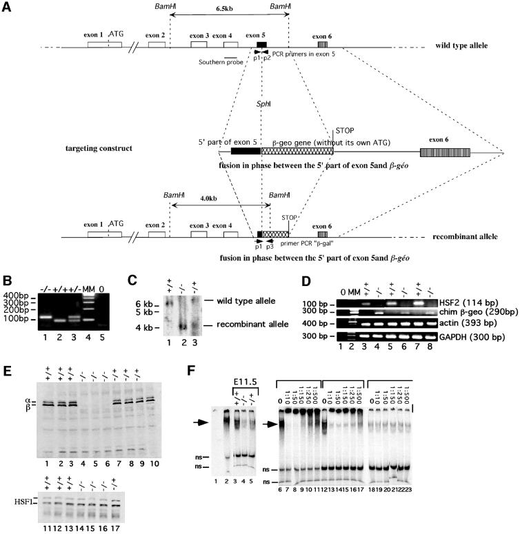
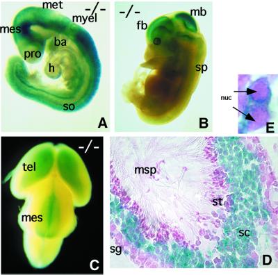
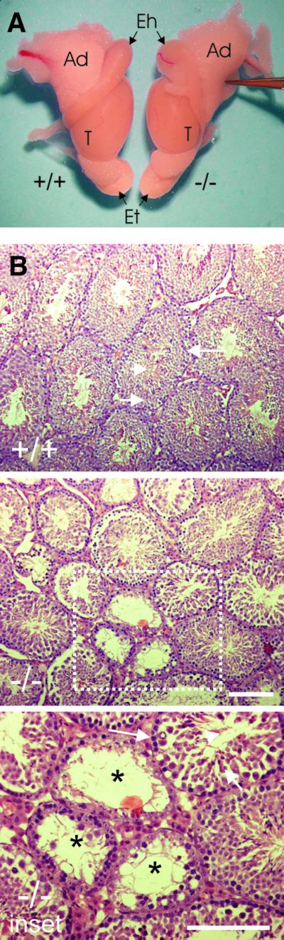
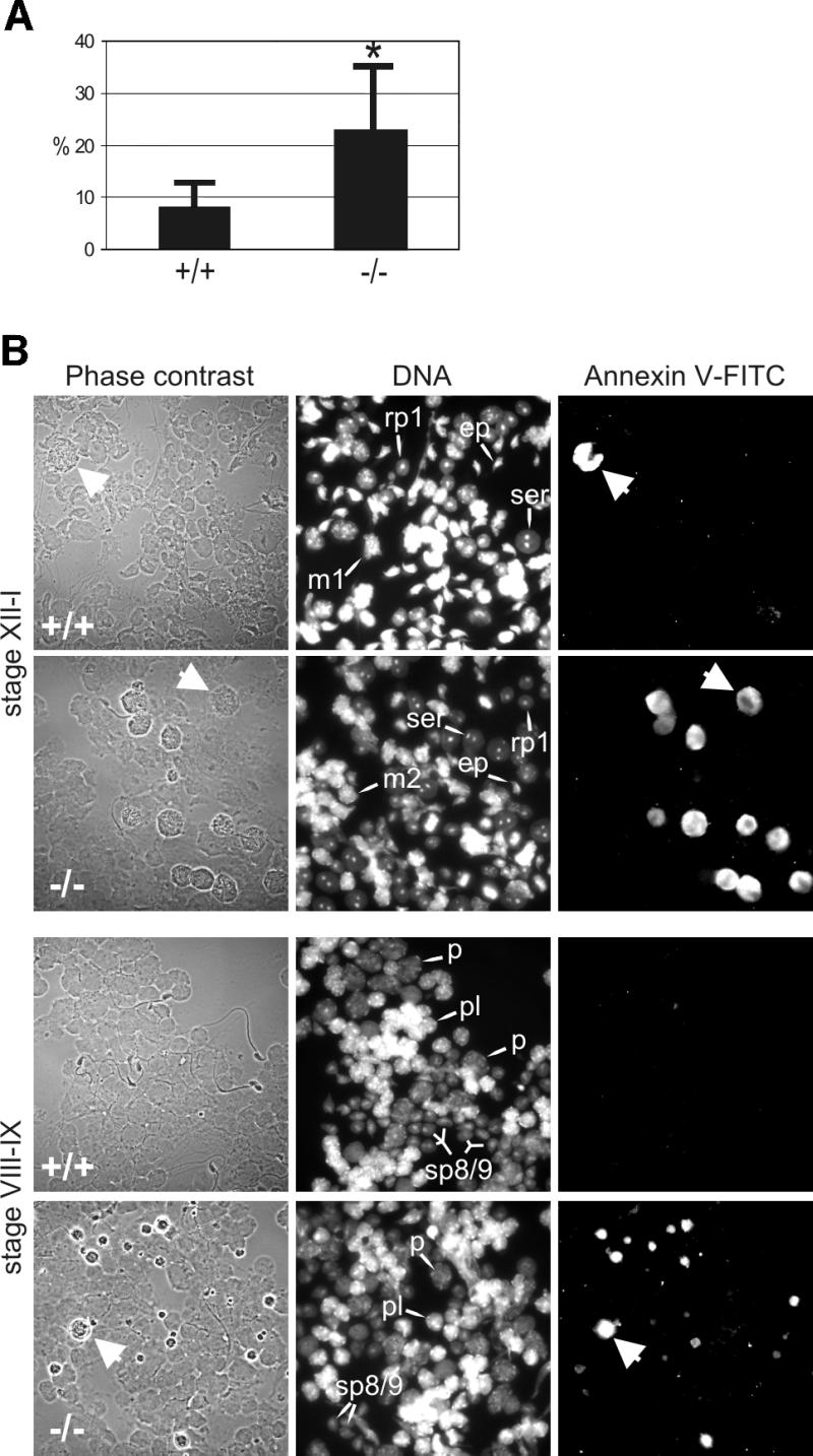
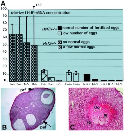
Heat shock factor 2, one of the four vertebrate HSFs, transcriptional regulators of heat shock gene expression, is active during embryogenesis and spermatogenesis, with unknown functions and targets. By disrupting the Hsf2 gene, we show that, although the lack of HSF2 is not embryonic lethal, Hsf2(-/-) mice suffer from brain abnormalities, and meiotic and gameto genesis defects in both genders. The disturbances in brain are characterized by the enlargement of lateral and third ventricles and the reduction of hippocampus and striatum, in correlation with HSF2 expression in proliferative cells of the neuroepithelium and in some ependymal cells in adults. Many developing spermatocytes are eliminated via apoptosis in a stage-specific manner in Hsf2(-/-) males, and pachytene spermatocytes also display structural defects in the synaptonemal complexes between homologous chromosomes. Hsf2(-/-) females suffer from multiple fertility defects: the production of abnormal eggs, the reduction in ovarian follicle number and the presence of hemorrhagic cystic follicles are consistent with meiotic defects. Hsf2(-/-) females also display hormone response defects, that can be rescued by superovulation treatment, and exhibit abnormal rates of luteinizing hormone receptor mRNAs.


Figures

None
Fig. 1. Targeted inactivation of the Hsf2 gene. (A) Schematic representation of the wild-type and mutated alleles. Horizontal small arrows show the location of the three primers used for PCR genotyping. (B) PCR genotyping of offspring from F1 heterozygous intercrosses. (C) Southern blot of BamHI-digested tail DNA. Lanes 1 and 3: wild-type allele 6.5 kb fragment. Lanes 2 and 3: disrupted allele 4 kb fragment. (D) RT–PCR analysis of HSF2, chimeric HSF2-βgeo, actin and GAPDH mRNA levels in Hsf2+/+, Hsf2+/– and Hsf2–/– tissues. Lane 1: no reverse transcription (0); lane 2, molecular markers (MM); lanes 3 and 4, testis; lanes 5 and 6, E9.5 embryos; lanes 7 and 8, E13.5 embryos. (E) Western blot analysis of whole E11.5 embryo extracts of littermates with polyclonal anti-mouse HSF2 (upper panel). Equal loading and transfer were assessed with a monoclonal anti-HSF1 (lower panel). Lanes 1–3 and 11–13, Hsf2+/+; lanes 4–6 and 14–17, Hsf2–/–; lanes 7–9 and 17, Hsf2+/–; lane 10, unstressed F9 EC cells. (F) EMSA analysis of E11.5 embryo extracts with an HSE-containing double-stranded oligonucleotide. Lane 1, no extracts; lanes 2 and 6–11, unstressed F9 EC cells; lanes 3–5 and 12–23, embryos. The dilutions of polyclonal anti-mouse HSF2 in supershift experiment are indicated. Arrowheads, specific HSF2–HSE complexes; ns, non-specific complexes.
None
Fig. 2. LacZ expression as a reporter of the HSF2 expression profile. (A and B) Lateral view of an E9.5 and E13.5 Hsf2–/– embryo, respectively. (C) Dorsal view of an E15.5 Hsf2–/– embryo. (D) Transverse section of a seminiferous tubule showing X-gal staining of spermatocytes. (E) Cytoplasmic localization of the chimeric recombinant protein using β-galactosidase detection by X-gal staining (blue) in mouse pachytene spermatocytes (Hsf2–/– male). nuc, nucleus; h, heart; so, somites; pro, prosencephalon; mes, mesencephalon; met, metencephalon; myel, myel encephalon; tel, telencephalon; ba, branchial arches; fb, forebrain; mb, midbrain; sp, spinal chord; msp, mature spermatozoa; sg, spermatogonia; sc, spermatocytes; st, spermatids.
None
Fig. 3. HSF2 and β-gal expression in the ventricular zone of embryonic and adult brain. (A) Parasagittal section of an E13.5 Hsf2–/– embryonic brain showing β-gal expression along the lumen of the ventricles. fb, forebrain; mb, midbrain; chp, choroid plexus. (B and C) HSF2 immunolocalization and BrdU staining, respectively, at the level of midbrain in E12.5 Hsf2+/+ embryos. vz, ventricular zone; lv, lumen of the ventricle. (DF) β-gal detection in the ependymal layer of adult Hsf2–/– brain. st, striatum; se, septum; ep, ependymal zone; lv, lateral ventricle. Scale bar: 50 µm. (E) Detail of (D), magnification: 20×. (F) Detail of a transverse section at the level of the hippocampus (h), magnification: 20×.
None
Fig. 4. Abnormal structure of the HSF2-deficient adult brain. Brain transverse sections (A–F) from Hsf2+/+ (A, C and E) or Hsf2–/– (B, D and F) 3-month-old males, illustrating the enlargment of the lateral and third ventricles all along the brain. (G and H) Parasagittal sections of the adult brain showing the reduced size of the hippocampus in Hsf2–/– (–/–) (H) compared with Hsf2+/+ (WT) (G) brain. ST, striatum; SE, septum; H, hippocampus; LV, lateral ventricle; TV, third ventricle; CE, cerebellum.
None
Fig. 5. Gross anatomy of male reproductive organs and analysis of testis cross-sections of adult Hsf2+/+ (+/+) and Hsf2–/– mice (–/–). (A) Testis and epididymis size. T, testis; Eh, head of epididymis; Et, tail of epididymis; Ad, adipose tissues. (B) Cross-sections from testes. For (+/+): arrowhead, elongating spermatids; short arrow, spermatocytes; long arrow, spermatogonia. For (–/–): reduction in the diameter of the seminiferous tubules and indications of disruption of spermatogenesis. For inset: lack of spermatocytes (short arrow) and spermatids (arrowhead), vacuolization of the tubules (asterisk). Bars = 100 µm.
None
Fig. 6. Apoptosis of developing germ cells in the testes of Hsf2–/– mice. (A) Flow cytometric analysis of annexin V–FITC-stained testicular cells. (B) Stage-specific apoptosis in the testis of Hsf2–/– mice. Translumination-assisted dissection of the seminiferous tubules followed by annexin V–FITC immunofluorescence and microscopic analysis revealed two populations of dying cells in the Hsf2–/– mice at stages VIII–IX and XII–I; late pachytene and meiotically dividing spermatocytes (arrows). Tubule segments isolated from stages XII–I containing type A3 and A4 spermatogonia, early pachytene spermatocytes, meiotically dividing spermatocytes (m1, m2), round step-1 spermatids (rp1) and elongating spermatids (ep), and from stages VIII–IX containing type A1 spermatogonia, pre-leptotene spermatocytes (pl), late pachytene spermatocytes (p) and elongating step-8/9 spermatids (sp8/9). Note the reduction in the number of post-meiotic round and elongated spermatids in the Hsf2–/– mice.
None
Fig. 7. Synapsis between the homologous chromosomes is defective in the Hsf2–/– mice. (A) Immunofluorescence labeling with anti-SCP3 antibody (Cy3 channel, red) and Crest anti-centromere sera (FITC channel, green). A part of a seminiferous tubule in developmental stage VIII (Clermont, 1972), with middle pachytene spermatocytes (mp), round spermatids (rs) and elongated spermatids (es). Loop-like configurations are present between one or more pairs of homologous chromosomes (arrow in the inset A). (B) An SC with one loop-like structure near the centromere terminus of a chromosome pair (arrow) (Z-level section). (C) Separation of the lateral elements at the centromere region (arrowheads in the inset C; stack of five Z-level sections from a confocal microscope series). Merge of anti-SCP3 (red) and Crest (green) in images (B) and (C). Bars = 10 µm.
None
Fig. 8. Ovarian defects in Hsf2–/ females. (A) Abnormally elevated levels of luteinizing hormone receptor (LH-R) mRNAs in Hsf2–/– with anovulatory problems. Seven Hsf2+/+ and seven Hsf2–/– 5-month-old females were mated with OF1 males. On the day of plug detection, eggs were harvested and counted in the ampulla. In parallel, LH-R mRNA levels were analyzed by RT–PCR. The relative mRNA concentration is given in arbitrary units. Value 1 was attributed to the ovary exhibiting the lowest expression level. Each plot corresponds to the average determined from three or four PCR experiments (except for female B that was tested only once). Error bars are indicated. Dense hatched bars, Hsf2–/– females that produced no eggs or only abnormal fragmented eggs. Hatched bars, Hsf2–/– females that produced a few (an average of two) fertilized eggs. Empty bars, Hsf2+/+ females with no ovulated fertilized eggs. Filled bars, Hsf2+/+ females with normal ovulation scores and a normal number of fertilized eggs. (B) Paraffin section of an Hsf2–/– ovary with marked ovulatory problems: low number of primary (prf) and pre-antral (paf) follicles. (C) A pre-antral hemorrhagic follicle in the section of an Hsf2–/– ovary which never gave offspring. Oo, oocyte; Gr, granulosa cells; Bl, blood.

Similar articles

Cited by

References

    1. Alastalo T.P., Lonnstrom,M., Leppa,S., Kaarniranta,K., Pelto-Huikko,M., Sistonen,L. and Parvinen,M. (1998) Stage-specific expression and cellular localization of the heat shock factor 2 isoforms in the rat seminiferous epithelium. Exp. Cell Res., 240, 16–27. - PubMed
    1. Baudat F., Manova,K., Pui Yuen,J., Jasin,M. and Keeney,S. (2000) Chromosone synapsis defects and sexually dimorphic meiotic progression in mice lacking Spo11. Mol. Cell, 6, 989–998. - PubMed
    1. Christians E., Michel,E., Adenot,P., Mezger,V., Rallu,M., Morange,M. and Renard,J.P. (1997) Evidence for the involvement of mouse heat shock factor 1 in the atypical expression of hsp70.1 heat shock gene during mouse zygotic genome activation. Mol. Cell. Biol., 17, 778–788. - PMC - PubMed
    1. Christians E., Davis,A.A., Thomas,S.D. and Benjamin,I.J. (2000) Maternal effect of Hsf1 on reproductive success. Nature, 407, 693–694. - PubMed
    1. Clermont Y. (1972) Kinetics of spermatogenesis in mammals: seminiferous epithelium cycle and spermatogonia renewal. Physiol. Rev., 52, 198–236. - PubMed

Publication types

MeSH terms