Comparative Study
. 2002 Jun;22(11):3794-802.
doi: 10.1128/MCB.22.11.3794-3802.2002.

Growth and early postimplantation defects in mice deficient for the bromodomain-containing protein Brd4

Affiliations
Comparative Study

Growth and early postimplantation defects in mice deficient for the bromodomain-containing protein Brd4

Denis Houzelstein et al. Mol Cell Biol. 2002 Jun.

Abstract

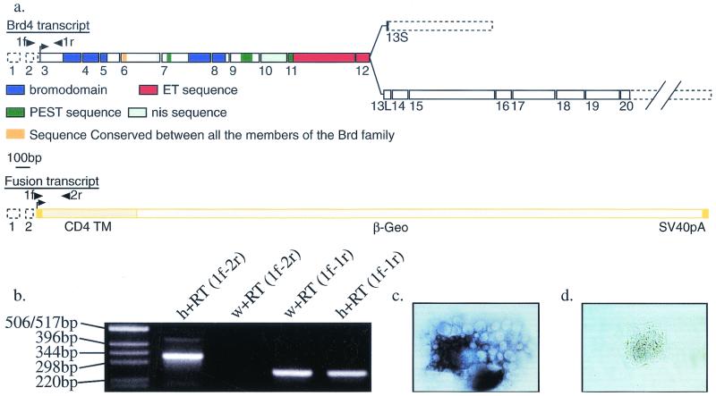
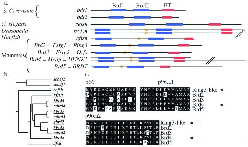
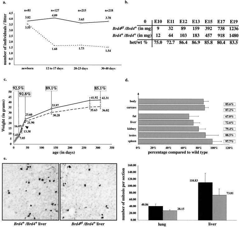
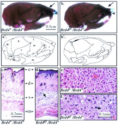
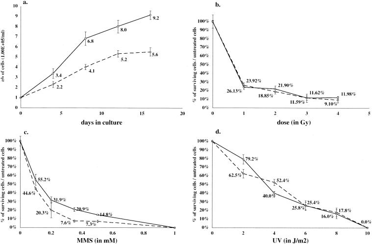
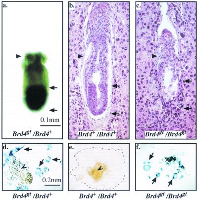
In a gene trap screen we recovered a mouse mutant line in which an insertion generated a null allele of the Brd4 gene. Brd4 belongs to the Fsh/Brd family, a group of structurally related proteins characterized by the association of two bromodomains and one extraterminal domain. Members of this family include Brd2/Ring3/Fsrg1 in mammals, fs(1)h in Drosophila, and Bdf1 in Saccharomyces cerevisiae. Brd4 heterozygotes display pre- and postnatal growth defects associated with a reduced proliferation rate. These mice also exhibit a variety of anatomical abnormalities: head malformations, absence of subcutaneous fat, cataracts, and abnormal liver cells. In primary cell cultures, heterozygous cells also display reduced proliferation rates and moderate sensitivity to methyl methanesulfonate. Embryos nullizygous for Brd4 die shortly after implantation and are compromised in their ability to maintain an inner cell mass in vitro, suggesting a role in fundamental cellular processes. Finally, sequence comparisons suggest that Brd4 is likely to correspond to the Brd-like element of the mediator of transcriptional regulation isolated by Y. W. Jiang, P. Veschambre, H. Erdjument-Bromage, P. Tempst, J. W. Conaway, R. C. Conaway, and R. D. Kornberg (Proc. Natl. Acad. Sci. USA 95:8538-8543, 1998) and the Brd4 mutant phenotype is discussed in light of this result. Together, our results provide the first genetic evidence for an in vivo role in mammals for a member of the Fsh/Brd family.


Figures

FIG. 1.
FIG. 1.
The gene trap strategy and Brd4 gene. (a) Structure of the Brd4 and Brd4gt transcripts. A long Brd4 mRNA (6 kb, encoding a 1,400-amino acid protein) is transcribed from 20 exons (1 to 20), and a short Brd4 mRNA (3.5 kb, encoding a 722-amino acid protein) is formed by the use of an alternative 13th exon (13S). The fusion transcript containing the first two noncoding exons from Brd4 fused to the gene trap sequences is transcribed from the Brd4gt allele. 1f, 1r, and 2r are the specific primers used for RT-PCR in Fig. 1b. Dotted boxes, UTR; plain boxes, coding exons; nis, nuclear localization signal; TM, transmembrane domain. (b) RT-PCR with primers 1f (5′ RACE fragment specific), 1r (Brd4-specific coding sequence), and 2r (gene trap-specific primer). Primers 1f and 2r (1f-2r) amplified the expected 338-bp product in total RNA from Brd4gt/Brd4+ (h) but not Brd4+/Brd4+(w) individuals. Primers 1f and 1r (1f-1r) amplified the expected 256-bp fragment in total RNA from both wild types and heterozygotes. (c and d) Brd4+/Brd4+ (c) and Brd4gt/Brd4gt (d) blastocyst explants cultured for 2 days and hybridized with a probe covering exons 1 to 9 and part of exon 10. No transcript could be detected in the homozygote.
FIG. 2.
FIG. 2.
Structure of the Brd4 gene and conservation of the Brd family. (a) Domain organizations of the different members of the Brd/fs(1)h family. A convenient nomenclature for the vertebrate members of this family based on the root fsrg (female sterile homeotic related) was proposed by Rhee et al. (32). However, here we use the nomenclature retained by the Mouse Gene Informatics Nomenclature Committee, who proposed the root Brd to designate the members of this bromodomain-containing family. Bromodomains are shown in blue, ET domains are shown in red, and the conserved 10-amino acid motif is shown in orange. Note that the hffsh sequence is only partial and terminates in the ET domain. (b) Similarity tree of the members of the Brd family (vertebrate members of the family are underlined). Note that Brd2/RING3 and Brd3/ORFX group together, in agreement with the important conservation of synteny observed between human chromosomes 6 and 9 (see reference for review). Brd5/BRDT, the only member of the Brd family that exhibits a nonubiquitous pattern of expression (19), is found outside the mammalian group, suggesting a possible divergent function. Bdf1 and Bdf2 group together, suggesting a duplication after the separation between yeast and metazoans. Alignments of bromodomains also confirmed this result (data not shown). (c) Sequence comparison of the Ring3-like peptides (p66, p96a1, and p96a2) (isolated in the study cited in reference 18) with those of the four mammalian members of the Brd family. Arrows point at the sequences of the peptides and Brd4.
FIG. 3.
FIG. 3.
In vivo phenotype of the heterozygotes. (a) Postnatal lethality: comparison between the average numbers of Brd4+/Brd4+ (plain line) and Brd4gt/Brd4+ (dotted line) individuals per litter from matings of Brd4gt/Brd4+ males with Brd4+/Brd4+ females, from birth to adulthood. The number indicated above each point represents the average number of individuals per litter. Note that the expected ratio of heterozygous to wild-type individuals was observed at birth. n, number of individuals counted. (b) Growth defect before birth: average weights of Brd4+/Brd4+ and Brd4gt/Brd4+ individuals from E10 to E19. At least three littermate individuals from each genotype were counted at every point. Heterozygote versus wild-type weight percentages are indicated. Note that the difference in weight between heterozygous and wild-type individuals is most pronounced between E10 and E11. (c) Growth defect after birth. Six Brd4gt/Brd4+ and five Brd4+/Brd4+ Brd4gt/Brd4+ males were weighed over a period of 320 days. Wild-type individuals (plain line) were significantly heavier than their heterozygous (dotted line) littermates. Weights of both males and females from several litters gave results quantitatively and qualitatively similar. The average weight is indicated over each point, and heterozygote versus wild-type weight percentages are indicated in the squares. (d) Growth defect of individual organs. Individual organs from Brd4+/Brd4+ and Brd4gt/Brd4+ 10-month-old females were weighed separately. Every organ was affected by the mutation. (e) Reduced numbers of mitotic cells in the Brd4gt/Brd4+ individuals. An anti-phospho-histone H3 antibody was used to reveal mitotic cells from E15 liver and lung sections. An enlargement of part of such a section is shown. The average numbers of mitotic cells per section are indicated. There were significantly fewer mitotic cells in sections from heterozygous embryos than in those from wild-type embryos, although the cell density was unaffected.
FIG. 4.
FIG. 4.
Anatomical and histological defects in the heterozygotes. (a, a′, b, and b′) Skeletal preparations of two-month-old Brd4+/Brd4+ (a) and Brd4gt/Brd4+ (b) male skulls, respectively; bone is stained in red and cartilage in blue. Note the shorter and bent nasal bone (arrow), the shorter incisor bone (arrowhead), and the shorter mandible of the heterozygote. f, frontal bone; i, incisive bone; ip, interparietal bone; lf, lacrimal foramen; m, mandible; mp, mastoid process; n, nasal bone; o, occipital bone; os, orbital surface of the frontal bone; p, parietal bone; t, temporal bone; tb, tympanic bulla; z, zygomatic bone. (c and d) Sections through the skin of two Brd4+/Brd4+ (c) and Brd4gt/Brd4+ (d) 1-month-old individuals. In the heterozygote, the epidermis (e) was thickened and the amount of subcutaneous adipose tissue (s) was reduced and contained degenerating adipose cells surrounded by phagocytic cells (black arrow). d, dermis; m, muscle. (e and f) Sections through the liver of two Brd4+/Brd4+ (e) and Brd4gt/Brd4+ (f) 2-month-old individuals. The black arrow in Fig. 4f points to degenerating hepatocytes, and the white arrowheads point at surrounding phagocytes in the heterozygote.
FIG. 5.
FIG. 5.
Effect of DNA damaging agents on survival of MEFs. (a) 1.00 × 10⁵ Brd4+/Brd4+ (plain line) or Brd4gt/Brd4+ (dotted line) MEFs were harvested on individual plates at day 0 and counted at 4-day intervals. (b to d) MEFs were plated as for Fig. 5a. After they adhered to the plate, they were submitted to increasing doses of IR (b), MMS (c), or UV (d), cultured for 8 days, and counted.
FIG. 6.
FIG. 6.
The homozygote phenotype. (a to c) E6 embryos. Black arrows demarcate the embryonic part of the conceptus, whereas the black arrowhead points at the extraembryonic part. Whole-mount β-galactosidase staining of a Brd4gt/Brd4+ embryo (a) and hematoxylin-eosin staining of sections of Brd4+/Brd4+ (b) and Brd4gt/Brd4gt (c) embryos are shown. Note that the embryonic part which normally expresses the gene strongly is missing in the homozygote. (d to f) Blastocyst explants stained with β-galactosidase after 5 days in culture. The arrows point at the giant trophoblastic cells, and the arrowhead points at the inner cell mass. In Fig. 6e, the limit of trophoblast giant cell spread is indicated by a dotted line. In Fig. 6f, the inner cell mass of the homozygote has degenerated and is missing.

Similar articles

Cited by

References

    1. Beck, S., I. Hanson, A. Kelly, D. J. Pappin, and J. Trowsdale. 1992. A homologue of the Drosophila female sterile homeotic (fsh) gene in the class II region of the human MHC. DNA Sequence 2:203-210. - PubMed
    1. Bergmann, E., and J. M. Egly. 2001. Trichothiodystrophy, a transcription syndrome. Trends Genet. 17:279-286. - PubMed
    1. Berneburg, M., and A. R. Lehmann. 2001. Xeroderma pigmentosum and related disorders: defects in DNA repair and transcription. Adv. Genet. 43:71-102. - PubMed
    1. Bullock, S. L., J. M. Fletcher, R. S. Beddington, and V. A. Wilson. 1998. Renal agenesis in mice homozygous for a gene trap mutation in the gene encoding heparan sulfate 2-sulfotransferase. Genes Dev. 12:1894-1906. - PMC - PubMed
    1. Chua, P., and G. S. Roeder. 1995. Bdf1, a yeast chromosomal protein required for sporulation. Mol. Cell. Biol. 15:3685-3696. - PMC - PubMed

Publication types

MeSH terms