. 2002 Jun;76(11):5769-83.
doi: 10.1128/jvi.76.11.5769-5783.2002.

Functional interaction between the pp71 protein of human cytomegalovirus and the PML-interacting protein human Daxx

Affiliations

Functional interaction between the pp71 protein of human cytomegalovirus and the PML-interacting protein human Daxx

Heike Hofmann et al. J Virol. 2002 Jun.

Abstract

The tegument protein pp71 (UL82) of human cytomegalovirus (HCMV) has previously been shown to transactivate the major immediate-early enhancer-promoter of HCMV. Furthermore, this protein is able to enhance the infectivity of viral DNA and to accelerate the infection cycle, suggesting an important regulatory function during viral replication. To gain insight into the underlying mechanisms that are used by pp71 to exert these pleiotropic effects, we sought for cellular factors interacting with pp71 in a yeast two-hybrid screen. Here, we report the isolation of the human Daxx (hDaxx) protein as a specific interaction partner of HCMV pp71. hDaxx, which was initially described as an adapter protein involved in apoptosis regulation, has recently been identified as a nuclear protein that interacts and colocalizes with PML in the nuclear domain ND10. In order to assess whether pp71 can also be detected in ND10 structures, a vector expressing pp71 in fusion with the green fluorescent protein was used for transfection of human fibroblasts. This revealed a colocalization of pp71 with the ND10 proteins PML and Sp100. In addition, cotransfection of a hDaxx expression vector resulted in an enhanced recruitment of pp71 to ND10. Targeting of pp71 to nuclear dots could also be observed in infected human fibroblasts in the absence of de novo viral protein synthesis. Moreover, cotransfection experiments revealed that pp71-mediated transactivation of the major immediate-early enhancer-promoter was synergistically enhanced in the presence of hDaxx. These results suggest an important role of hDaxx for pp71 protein function.


Figures
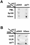
FIG. 1.
FIG. 1.
Specific interaction between HCMV pp71 and the hDaxx protein in yeast cells. Yeast cells were transformed with two separate vectors, one of which encoded a GAL4 activation domain either alone (pGAD) or as fusion with the indicated protein (AD), the second plasmid encoded a GAL4 DNA-binding domain fusion as indicated (DBD). Thereafter, yeast colonies were selected for the presence of both plasmids on dropout medium lacking tryptophane and leucine and subsequently analyzed for the expression of β-galactosidase. (A) Interaction analysis between pp71 fused to the GAL4 activation domain and the ND10-associated factors PML, Sp100, and hDaxx as fusions with the GAL4 DNA-binding domain. (B) Interaction analysis between hDaxx fused to the GAL4 activation domain and the HCMV tegument proteins UL69, UL26, and pp71 as fusions with the GAL4 DNA-binding domain.
FIG. 2.
FIG. 2.
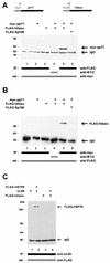
Specific interaction between pp71 and hDaxx as determined in coimmunoprecipitation experiments. 293 cells were transfected with eukaryotic expression vectors encoding myc-pp71, FLAG-hDaxx, and FLAG-hSPT6, as well as the HCMV protein ppUL69, as indicated and prepared for immunoprecipitation as described in Materials and Methods. Immunoprecipitation was performed with antibodies as indicated by bars. Precipitates were washed three times and separated by SDS-10% PAGE. (A) Immunoprecipitations were performed with the anti-FLAG monoclonal antibody or an anti-IE2 monoclonal antibody as indicated. Thereafter, coprecipitated pp71protein was detected in Western blot analysis with the anti-myc monoclonal antibody. Lanes: 1, lysates from untransfected cells; 2, transfection with plasmid myc-pp71 alone; 3, transfection with vector FLAG-hDaxx alone; 4 and 5, transfections with a combination of vectors encoding myc-pp71 and FLAG-hDaxx; 6, transfection with plasmid myc-pp71 and a plasmid encoding FLAG-Sp100. (B) Immunoprecipitations were performed with the anti-myc monoclonal antibody or the anti-IE2 monoclonal antibody. Interacting proteins were thereafter detected in a Western blot with the anti-FLAG monoclonal antibody. The lanes are analogous to those in panel A. (C) Immunoprecipitations were performed with the anti-UL69 monoclonal antibody; thereafter, coprecipitated proteins were detected by Western blot analysis with the anti-FLAG monoclonal antibody. The interaction between ppUL69 and the cellular protein hSPT6 served as a positive control (lane 2). Lanes: 1, transfection of expression vector pCB6-UL69; 2, transfection with a combination of plasmids encoding ppUL69 and FLAG-hSPT6; 3, transfection with plasmid FLAG-hDaxx; 4, transfection with constructs expressing FLAG-hDaxx and ppUL69. Molecular masses are indicated in kilodaltons. Abbreviations: IP, immunoprecipitation; wb, Western blot; IgG, immunoglobulin G.
FIG. 3.
FIG. 3.
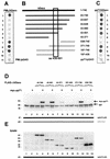
Delineation of the pp71 interaction domain within hDaxx. (A, B, and C) Yeast cells were transformed with two separate vectors, one of which encoded either PML as a positive control (A) or pp71 (C) fused to the GAL4 DNA-binding domain. The second plasmid encoded amino- or carboxy-terminal fragments of hDaxx as fusion with the GAL4 activation domain; the amino acids contained in the respective deletion mutants are indicated in panel B. Yeast colonies were selected for the presence of both plasmids on dropout medium lacking tryptophane and leucine and subsequently analyzed for the expression of β-galactosidase by filter lift assays. As negative controls, the activation domain vector pGAD424 (pGAD) was either transformed with the pp71 or the PML DNA-binding domain fusion (lanes 11, panels A and C, respectively). The pp71 interaction domain within hDaxx is depicted by the box. (D) Interaction between pp71 and hDaxx deletion mutants after coimmunoprecipitation from 293 cells. The hDaxx mutants were precipitated with the anti-FLAG monoclonal antibody; therafter, bound pp71 protein was detected in Western blot experiments employing the anti-myc antibody. Lanes: 1, lysates from untransfected cells; 2, transfection with plasmid myc-pp71 alone; 3, transfection with vector FLAG-hDaxx alone; 4, transfection with a combination of vectors encoding myc-pp71 and FLAG-hDaxx; 5, transfection with a plasmid encoding FLAG-hDaxx 43-501; 6, transfection with vectors encoding myc-pp71 and FLAG-hDaxx 43-501; 7, transfection with vector FLAG-hDaxx 43-371; 8, transfection with a combination of vectors myc-pp71 and FLAG-hDaxx 43-371; 9, transfection with a plasmid expressing FLAG-hDaxx 197-439; 10, transfection with vectors encoding myc-pp71 and FLAG-hDaxx 197-439; 11, transfection with plasmid FLAG-hDaxx 371-740; 12, transfection with a combination of vector myc-pp71 and FLAG-hDaxx 371-740; 13, transfection with a plasmid encoding FLAG-hDaxx 538-740; 14, transfection with vectors expressing myc-pp71 and FLAG-hDaxx 538-740. (E) Western blot analysis of the cell lysates used for immunoprecipitation in panel D. The expression of the hDaxx deletion mutants was investigated with the anti-FLAG monoclonal antibody. Molecular masses are indicated in kilodaltons. Abbreviations: IP, immunoprecipitation; wb, Western blot; IgG, immunoglobulin G.
FIG. 4.
FIG. 4.
Schematic overview depicting interaction domains within hDaxx required for association with previously published cellular proteins and HCMV pp71 (33, 35, 41, 54, 75). The amino acids of hDaxx required for the respective interactions are indicated.
FIG. 5.
FIG. 5.
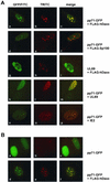
(A) Subcellular localization of pp71-GFP in HFF cells. HFF cells grown on coverslips were transfected with an expression vector encoding pp71 fused to GFP (pp71-GFP) (panels a, d, and g). In order to detect endogenous ND10 domains, indirect immunofluorescence analyses were performed with a monoclonal antibody against PML (panel b) or polyclonal antisera recognizing Sp100 (panel e) and hDaxx (panel h), followed by incubation with TRITC-conjugated anti-mouse or anti-rabbit secondary antibodies. (B) Analysis of hDaxx distribution in HFF cells. HFF cells were costained for endogenous expression of hDaxx as detected by the polyclonal antiserum (panel a) and PML with a monoclonal antibody (panel b).
FIG. 6.
FIG. 6.
Recruitment of pp71 after expression of FLAG-hDaxx. HFF cells (A) and HeLa cells (B) grown on coverslips were transfected with eukaryotic expression vectors encoding pp71-GFP, FLAG-hDaxx, and FLAG-Sp100, as well as the HCMV proteins ppUL69 and IE2, as indicated. Thereafter, indirect immunofluorescence analyses were carried out. pp71 was visible through its GFP moiety (panels Aa, d, k, and n; panels Ba and d). FLAG-hDaxx and FLAG-Sp100 were detected with the anti-FLAG monoclonal antibody (panels Ab and e; panel Be). ppUL69 was stained by using the polyclonal anti-UL69 antiserum (panels Ag and l); IE2 was detected with monoclonal antibody MAb810 (panel Ao). Staining for endogenous hDaxx was performed with a polyclonal anti-hDaxx antiserum (panel Bb). Thereafter, TRITC-conjugated anti-mouse secondary antibodies, as well as FITC-conjugated anti-rabbit secondary antibodies, were employed.
FIG. 7.
FIG. 7.
Subcellular localization of pp71 in HCMV infected cells. HFF cells were treated with 100 μg of cycloheximide/ml 30 min prior to infection (0.5 PFU/cell) and extending for 7 h after infection. Indirect immunofluorescence analyses were carried out with monoclonal antibodies directed against pp71 (a), pp65 (d) and IE1 (g). ND10 domains were visualized by staining the Sp100 protein (b, e, and h). DAPI staining of the respective cell nuclei is shown in panels c, f, and i.
FIG. 8.
FIG. 8.
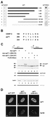
Analyses of pp71 deletion mutants. (A) Mapping of DIDs within pp71. Yeast cells were transformed with two separate vectors, one of which encoded full-length pp71 (lane 1) or amino- and carboxy-terminal deletion mutants fused to the GAL4 DNA-binding domain (lanes 2 to 7); the amino acids contained in the respective deletion mutants are indicated. The second plasmid encoded either the empty GAL4 activation domain (lefthand side) or the hDaxx fusion (righthand side). Yeast colonies were selected for the presence of both plasmids on dropout medium lacking tryptophane and leucine and subsequently analyzed for the expression of β-galactosidase by filter lift assays. (B) Specific amino acid sequence homologies between theDIDs of CENP-C and HCMV pp71 (41). Within pp71, internal deletion mutagenesis was performed, resulting in mutants pp71Δ(206-213) and pp71Δ(324-331), respectively. (C) Loss of interaction between pp71 deletion mutants and hDaxx as determined by coimmunoprecipitation experiments. 293 cells were transfected with eukaryotic expression vectors encoding myc-pp71, the internal deletion mutants myc-pp71Δ(206-213) and myc-pp71Δ(324-331), and FLAG-hDaxx as indicated and prepared for immunoprecipitation as described in Materials and Methods. Immunoprecipitation was performed with the monoclonal anti-FLAG antibody. Precipitates were washed three times and separated by SDS-10% PAGE. Thereafter, coprecipitated pp71 proteins were detected in Western blot analysis with the anti-myc monoclonal antibody. Lanes: 1, transfection with plasmid myc-pp71 alone; 2, transfection with vector FLAG-hDaxx alone; 3, transfection with a combination of vectors encoding myc-pp71 and FLAG-hDaxx; 4, transfection with plasmid myc-pp71Δ(206-213), together with the FLAG-hDaxx vector; 5, transfection of plasmid myc-pp71Δ(206-213) alone; 6, combination of plasmids myc-pp71Δ(324-331) and FLAG-hDaxx; 7, transfection of vector myc-pp71Δ(324-331) alone. Prior to immunoprecipitation, an aliquot of each sample was analyzed for expression of pp71 and the respective mutants, as determined by Western blot with the anti-myc monoclonal antibody (lysate; lower part of panel C). Molecular masses are indicated in kilodaltons. Abbreviations: IP, immunoprecipitation; wb, Western blot; IgG, immunoglobulin G. (D) Subcellular localization of the pp71 mutants in HFF cells. HFF cells grown on coverslips were transfected with an expression vector encoding pp71-GFP as control (panel a) and the mutants pp71Δ(206-213) and pp71Δ(324-331) (panels b and c, respectively). ND10 domains were visualized employing the polyclonal Sp26 serum detecting Sp100, followed by incubation with a TRITC-conjugated anti-rabbit secondary antibody (panels d, e, and f).
FIG. 9.
FIG. 9.
Analyses of pp71 point mutants. Within the two potential DIDs of pp71, several amino acid residues were substituted by alanine as indicated in the left part of the figure. These mutants were expressed as GFP fusion proteins and were tested for ND10 localization via indirect immunofluorescence staining of the PML protein (ND10/PML); subsequently, recruitment of the inividual mutants was investigated after coexpression of FLAG-hDaxx (recruitment). Additionally, all mutants were expressed as myc-fusions and tested for hDaxx binding in coimmunoprecipitation experiments (co-IP). Positive or negative results in the individual experiments are indicated by “+” or “−.” Wild-type pp71 (wt-pp71) and the internal deletion mutants [Δ(206-213) and Δ(324-331)] were tested in parallel.
FIG. 10.
FIG. 10.
Luciferase analyses after cotransfection of a reporter construct carrying the HCMV MIEP with expression vectors for wild-type pp71 or the pp71 mutants pp71Δ(206-213) and pp71Δ(324-331), FLAG-hDaxx and ppUL69 as indicated. Each experiment was performed in triplicate and repeated at least three times. The fold activation was calculated relative to the basal activity of the reporter construct after cotransfection of the empty expression vectors pCB6 or myc-pcDNA3 and FLAG-pcDNA3. Western blot analyses reveal that similiar amounts of pp71 and hDaxx were expressed (insert). (A) Lanes: 1, cotransfection was performed with the empty expression vectors pCB6 and FLAG-pcDNA3; 2, cotransfection was performed with a construct expressing pp71 (pCB6-pp71); 3, cotransfection was performed with the FLAG-hDaxx plasmid; 4, cotransfection was performed with vectors encoding pp71 and FLAG-hDaxx; 5, cotransfection was performed with a plasmid expressing ppUL69 (pCB6-UL69); 6, cotransfection was performed with plasmids encoding ppUL69 and FLAG-hDaxx. (B) Lanes: 1, cotransfection was performed with the empty expression vectors FLAG-pcDNA3 and myc-pcDNA3; 2, cotransfection was performed with a construct expressing myc-pp71; 3, cotransfection was performed with the FLAG-hDaxx plasmid; 4, cotransfection was performed with vectors encoding myc-pp71 and FLAG-hDaxx; 5, cotransfection was performed with a plasmid expressing the mutant myc-pp71Δ(206-213); 6, cotransfection was performed with vectors encoding myc-pp71Δ(206-213) and FLAG-hDaxx; 7, cotransfection was performed with a plasmid expressing the mutant myc-pp71Δ(324-331); 8, cotransfection was performed with plasmids encoding myc-pp71Δ(324-331) and FLAG-hDaxx.

Similar articles

Cited by

References

    1. Ahn, J. H., E. J. Brignole III, and G. S. Hayward. 1998. Disruption of PML subnuclear domains by the acidic IE1 protein of human cytomegalovirus is mediated through interaction with PML and may modulate a RING finger-dependent cryptic transactivator function of PML. Mol. Cell. Biol. 18:4899-4913. - PMC - PubMed
    1. Ahn, J. H., and G. S. Hayward. 2000. Disruption of PML-associated nuclear bodies by IE1 correlates with efficient early stages of viral gene expression and DNA replication in human cytomegalovirus infection. Virology 274:39-55. - PubMed
    1. Ahn, J. H., W. J. Jang, and G. S. Hayward. 1999. The human cytomegalovirus IE2 and UL112-113 proteins accumulate in viral DNA replication compartments that initiate from the periphery of promyelocytic leukemia protein-associated nuclear bodies (PODs or ND10). J. Virol. 73:10458-10471. - PMC - PubMed
    1. Alford, C. A., and W. J. Britt. 1990. Cytomegalovirus, p. 1981-2010. In B. N. Fields, D. M. Knipe, and P. M. Howley (ed.), Virology. Raven Press, Ltd., New York, N.Y.
    1. Andreoni, M., M. Faircloth, L. Vugler, and W. J. Britt. 1989. A rapid microneutralization assay for the measurement of neutralizing antibody reactive with human cytomegalovirus. J. Virol. Methods 23:157-167. - PubMed

Publication types

MeSH terms

Substances

LinkOut - more resources