

Targeting assay to study the cis functions of human telomeric proteins: evidence for inhibition of telomerase by TRF1 and for activation of telomere degradation by TRF2
- PMID: 11971978
- PMCID: PMC133804
- DOI: 10.1128/MCB.22.10.3474-3487.2002
Targeting assay to study the cis functions of human telomeric proteins: evidence for inhibition of telomerase by TRF1 and for activation of telomere degradation by TRF2
Abstract
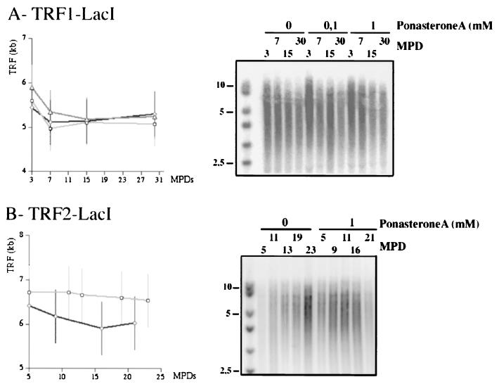
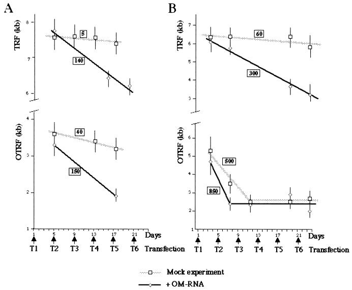
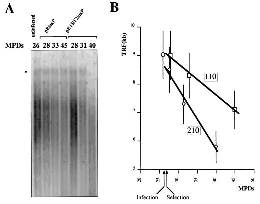
We investigated the control of telomere length by the human telomeric proteins TRF1 and TRF2. To this end, we established telomerase-positive cell lines in which the targeting of these telomeric proteins to specific telomeres could be induced. We demonstrate that their targeting leads to telomere shortening. This indicates that these proteins act in cis to repress telomere elongation. Inhibition of telomerase activity by a modified oligonucleotide did not further increase the pace of telomere erosion caused by TRF1 targeting, suggesting that telomerase itself is the target of TRF1 regulation. In contrast, TRF2 targeting and telomerase inhibition have additive effects. The possibility that TRF2 can activate a telomeric degradation pathway was directly tested in human primary cells that do not express telomerase. In these cells, overexpression of full-length TRF2 leads to an increased rate of telomere shortening.
Figures

Similar articles
-
Expression of telomeric repeat binding factor 1 and 2 and TRF1-interacting nuclear protein 2 in human gastric carcinomas.Int J Oncol. 2001 Sep;19(3):507-12. Int J Oncol. 2001. PMID: 11494028
-
Control of human telomere length by TRF1 and TRF2.Mol Cell Biol. 2000 Mar;20(5):1659-68. doi: 10.1128/MCB.20.5.1659-1668.2000. Mol Cell Biol. 2000. PMID: 10669743 Free PMC article.
-
Human telomeres contain two distinct Myb-related proteins, TRF1 and TRF2.Nat Genet. 1997 Oct;17(2):231-5. doi: 10.1038/ng1097-231. Nat Genet. 1997. PMID: 9326950
-
Regulation of telomerase by telomeric proteins.Annu Rev Biochem. 2004;73:177-208. doi: 10.1146/annurev.biochem.73.071403.160049. Annu Rev Biochem. 2004. PMID: 15189140 Review.
-
Post-translational modifications of TRF1 and TRF2 and their roles in telomere maintenance.Mech Ageing Dev. 2012 Jun;133(6):421-34. doi: 10.1016/j.mad.2012.05.002. Epub 2012 May 23. Mech Ageing Dev. 2012. PMID: 22634377 Review.
Cited by
-
The individual blood cell telomere attrition rate is telomere length dependent.PLoS Genet. 2009 Feb;5(2):e1000375. doi: 10.1371/journal.pgen.1000375. Epub 2009 Feb 13. PLoS Genet. 2009. PMID: 19214207 Free PMC article.
-
Crosstalk between telomere maintenance and radiation effects: A key player in the process of radiation-induced carcinogenesis.Mutat Res Rev Mutat Res. 2014 Jan 31:S1383-5742(14)00002-7 10.1016/j.mrrev.2014.01.001. doi: 10.1016/j.mrrev.2014.01.001. Online ahead of print. Mutat Res Rev Mutat Res. 2014. PMID: 24486376 Free PMC article. Review.
-
DNA excision repair at telomeres.DNA Repair (Amst). 2015 Dec;36:137-145. doi: 10.1016/j.dnarep.2015.09.017. Epub 2015 Sep 16. DNA Repair (Amst). 2015. PMID: 26422132 Free PMC article. Review.
-
Stem cell expansion during carcinogenesis in stem cell-depleted conditional telomeric repeat factor 2 null mutant mice.Oncogene. 2013 Oct 24;32(43):5156-66. doi: 10.1038/onc.2012.555. Epub 2012 Nov 26. Oncogene. 2013. PMID: 23178498 Free PMC article.
-
Poly(ADP-ribose) polymerase inhibitors as promising cancer therapeutics.Acta Pharmacol Sin. 2010 Sep;31(9):1172-80. doi: 10.1038/aps.2010.103. Epub 2010 Aug 2. Acta Pharmacol Sin. 2010. PMID: 20676117 Free PMC article. Review.
References
-
- Artandi, S. E., and R. A. DePinho. 2000. A critical role for telomeres in suppressing and facilitating carcinogenesis. Curr. Opin. Genet. Dev. 10:39-46. - PubMed
-
- Baumann, P., and T. R. Cech. 2001. Pot1, the putative telomere end-binding protein in fission yeast and humans. Science 292:1171-1175. - PubMed
-
- Beattie, T. L., W. Zhou, M. O. Robinson, and L. Harrington. 1998. Reconstitution of human telomerase activity in vitro. Curr. Biol. 8:177-180. - PubMed
-
- Bilaud, T., C. Brun, K. Ancelin, C. E. Koering, T. Laroche, and E. Gilson. 1997. Telomeric localization of TRF2, a novel human telobox protein. Nat. Genet. 17:236-239. - PubMed
Publication types
MeSH terms
Substances
LinkOut - more resources
Full Text Sources
Other Literature Sources
Molecular Biology Databases
Research Materials
Miscellaneous