Association of the caveola vesicular system with cellular entry by filoviruses
- PMID: 11967340
- PMCID: PMC136134
- DOI: 10.1128/jvi.76.10.5266-5270.2002
Association of the caveola vesicular system with cellular entry by filoviruses
Abstract
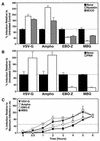
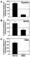
The filoviruses Ebola Zaire virus and Marburg virus are believed to infect target cells through endocytic vesicles, but the details of this pathway are unknown. We used a pseudotyping strategy to investigate the cell biology of filovirus entry. We observed that specific inhibitors of the caveola system, including cholesterol-sequestering drugs and phorbol esters, inhibited the entry of filovirus pseudotypes into human cells. We also measured slower cell entry kinetics for both filovirus pseudotypes than for pseudotypes of vesicular stomatitis virus (VSV), which has been recognized to exploit the clathrin-mediated entry pathway. Finally, visualization by immunofluorescence and confocal microscopy revealed that the filovirus pseudotypes colocalized with the caveola protein marker caveolin-1 but that VSV pseudotypes did not. Collectively, these results provide evidence suggesting that filoviruses use caveolae to gain entry into cells.
Figures


Similar articles
-
Studies of ebola virus glycoprotein-mediated entry and fusion by using pseudotyped human immunodeficiency virus type 1 virions: involvement of cytoskeletal proteins and enhancement by tumor necrosis factor alpha.J Virol. 2005 Jan;79(2):918-26. doi: 10.1128/JVI.79.2.918-926.2005. J Virol. 2005. PMID: 15613320 Free PMC article.
-
Caveola-dependent endocytic entry of amphotropic murine leukemia virus.J Virol. 2005 Aug;79(16):10776-87. doi: 10.1128/JVI.79.16.10776-10787.2005. J Virol. 2005. PMID: 16051869 Free PMC article.
-
Folate receptor alpha and caveolae are not required for Ebola virus glycoprotein-mediated viral infection.J Virol. 2003 Dec;77(24):13433-8. doi: 10.1128/jvi.77.24.13433-13438.2003. J Virol. 2003. PMID: 14645601 Free PMC article.
-
No exit: targeting the budding process to inhibit filovirus replication.Antiviral Res. 2009 Mar;81(3):189-97. doi: 10.1016/j.antiviral.2008.12.003. Epub 2008 Dec 27. Antiviral Res. 2009. PMID: 19114059 Free PMC article. Review.
-
Mechanisms of Filovirus Entry.Curr Top Microbiol Immunol. 2017;411:323-352. doi: 10.1007/82_2017_14. Curr Top Microbiol Immunol. 2017. PMID: 28601947 Review.
Cited by
-
Studies of ebola virus glycoprotein-mediated entry and fusion by using pseudotyped human immunodeficiency virus type 1 virions: involvement of cytoskeletal proteins and enhancement by tumor necrosis factor alpha.J Virol. 2005 Jan;79(2):918-26. doi: 10.1128/JVI.79.2.918-926.2005. J Virol. 2005. PMID: 15613320 Free PMC article.
-
Human papillomavirus types 16, 31, and 58 use different endocytosis pathways to enter cells.J Virol. 2003 Mar;77(6):3846-50. doi: 10.1128/jvi.77.6.3846-3850.2003. J Virol. 2003. PMID: 12610160 Free PMC article.
-
SARS coronavirus entry into host cells through a novel clathrin- and caveolae-independent endocytic pathway.Cell Res. 2008 Feb;18(2):290-301. doi: 10.1038/cr.2008.15. Cell Res. 2008. PMID: 18227861 Free PMC article.
-
Membrane Rafts: Portals for Viral Entry.Front Microbiol. 2021 Feb 4;12:631274. doi: 10.3389/fmicb.2021.631274. eCollection 2021. Front Microbiol. 2021. PMID: 33613502 Free PMC article. Review.
-
[The pathogenesis of Ebola virus disease].Zhejiang Da Xue Xue Bao Yi Xue Ban. 2015 Jan;44(1):1-8. doi: 10.3785/j.issn.1008-9292.2015.01.001. Zhejiang Da Xue Xue Bao Yi Xue Ban. 2015. PMID: 25851968 Free PMC article. Chinese.
References
-
- Anderson, R. G. 1998. The caveolae membrane system. Annu. Rev. Biochem. 67:199-225. - PubMed
-
- Antony, A. C. 1996. Folate receptors. Annu. Rev. Nutr. 16:501-521. - PubMed
-
- Bishop, N. E. 1997. An update on nonclathrin-coated endocytosis. Rev. Med. Virol. 7:199-209. - PubMed
-
- Calvo, M., F. Tebar, C. Lopez-Iglesias, and C. Enrich. 2001. Morphologic and functional characterization of caveolae in rat liver hepatocytes. Hepatology 33:1259-1269. - PubMed
Publication types
MeSH terms
Substances
Grants and funding
LinkOut - more resources
Full Text Sources
Other Literature Sources

