. 2002 May;76(10):4987-99.
doi: 10.1128/jvi.76.10.4987-4999.2002.

Genetic evidence for a structural interaction between the carboxy termini of the membrane and nucleocapsid proteins of mouse hepatitis virus

Affiliations

Genetic evidence for a structural interaction between the carboxy termini of the membrane and nucleocapsid proteins of mouse hepatitis virus

Lili Kuo et al. J Virol. 2002 May.

Abstract

The coronavirus membrane (M) protein is the most abundant virion protein and the key component in viral assembly and morphogenesis. The M protein of mouse hepatitis virus (MHV) is an integral membrane protein with a short ectodomain, three transmembrane segments, and a large carboxy-terminal endodomain facing the interior of the viral envelope. The carboxy terminus of MHV M has previously been shown to be extremely sensitive to mutation, both in a virus-like particle expression system and in the intact virion. We have constructed a mutant, M(Delta)2, containing a two-amino-acid truncation of the M protein that was previously thought to be lethal. This mutant was isolated by means of targeted RNA recombination with a powerful host range-based selection allowed by the interspecies chimeric virus fMHV (MHV containing the ectodomain of the feline infectious peritonitis virus S protein). Analysis of multiple second-site revertants of the M(Delta)2 mutant has revealed changes in regions of both the M protein and the nucleocapsid (N) protein that can compensate for the loss of the last two residues of the M protein. Our data thus provide the first genetic evidence for a structural interaction between the carboxy termini of the M and N proteins of MHV. In addition, this work demonstrates the efficacy of targeted recombination with fMHV for the systematic genetic analysis of coronavirus structural protein interactions.


Figures
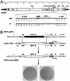
FIG. 1.
FIG. 1.
Selection of the MΔ2 mutant. (A) Construction of a transcription vector for synthesis of donor RNA. Plasmid pLK61 was derived from pMH54 (18) as detailed in Materials and Methods. The restriction sites shown are those relevant to plasmid construction (XhoI, EcoRV, BssHII, NheI, and BclI), in vitro transcription (PacI), or mutant analysis (AccI). The filled circle denotes the linker between the cDNA segments corresponding to the 5′ and 3′ ends of the MHV genome, and the arrow indicates the T7 promoter. The expanded region of sequence shows the three base changes made in pLK61 to create a stop codon at codon 227 of the M ORF and a new AccI site (underlined). The corresponding wild-type (wt) sequence from pMH54 is aligned for comparison. Also indicated is TRS7, the TRS governing synthesis of sgRNA7 (N mRNA). (B) Scheme for generation of the MΔ2 mutant by targeted RNA recombination between the interspecies chimera fMHV (18) and donor RNA transcribed from plasmid pLK61. fMHV contains the ectodomain-encoding region of the FIPV S gene (shaded rectangle) and is able to grow in feline cells but not in murine cells. A single crossover (solid line), within the HE gene, should generate a recombinant that has simultaneously reacquired the MHV S ectodomain and the ability to grow in murine cells and has also incorporated the mutant M gene (hatched rectangle). A potential second crossover (broken line), in gene 4 or the E gene, would exclude the mutant M gene from the recombinant. At the bottom is shown the mixed progeny of two independent targeted recombination experiments, forming tiny and large plaques on mouse L2 cells at 72 h postinfection.
FIG. 2.
FIG. 2.
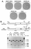
Purification and analysis of targeted recombination progeny. (A) Appearance of purified plaques on mouse L2 cells at 72 h postinfection. Alb231 and Alb233 are tiny-plaque recombinants from two independent infection-transfection experiments, and Alb243 is a large-plaque recombinant that arose in the same infection-transfection as Alb231. Alb239 is a wild-type control recombinant reconstructed from fMHV and pMH54 donor RNA. A control mock-infected L2 cell monolayer is included for comparison. (B) RT-PCR analysis. RNA was isolated from infected cells, reverse transcribed with random primer p(N)6, and amplified with primers PM145 and PM159. PCR products were analyzed by electrophoresis in 1.2% agarose with (+) or without (−) prior digestion with AccI. Lane M, DNA fragment size markers. wt, wild type.
FIG. 3.
FIG. 3.
Sequence analysis summary for the E, M, and N genes of revertants of MΔ2 mutants Alb231 and Alb233. Revertant R1 and sets 1, 2, and 3 (left column) constitute four independent groups of revertants. R1 and sets 1 and 3 were obtained from Alb231; set 2 was from Alb233. The E, M, and N proteins are represented linearly at the top. The solid rectangles indicate the membrane-bound domains of the E and M proteins (10, 39); the hatched rectangle indicates the RNA-binding domain of the N protein (20, 27). Arrows show the positions within the M and N proteins at which potential reverting mutations were found, and amino acid changes are grouped under the corresponding wild-type residue at each of these positions. Asterisks indicate stop codons. No changes were found in the E gene in any of the revertants. Those amino acid residues or positions that were chosen for further analysis are boxed. Numbers denote the following: 1, mutation of the MΔ2 stop codon from UAG to UAA; 2, mutation of the wild-type codon 228 from ACC to AUC; 3, replacement of nt 650 to 678 of the MΔ2 gene by nt 31 to 56 of the MHV leader; and 4, a deletion of nt 1309 to 1318 of the N gene resulting in a frameshift and truncation of the 15 carboxy-terminal amino acids of the N protein.
FIG. 4.
FIG. 4.
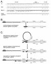
Recombination of part of the MHV leader RNA into the 3′ terminus of the M gene. (A) Nucleotide sequence of the M gene-N gene region of revertant 1I aligned with the corresponding regions of the MΔ2 sequence and the leader-N gene region of sgRNA7. The broken line indicates the locus of the apparent crossover between MΔ2 and sgRNA7. Revertants 1H and 3E, which contain mutations in both the MΔ2 stop codon and codon 228 (underlined), and revertants 1A and 1M, which contain just the codon 228 mutation, also appear to result from more-limited crossovers between MΔ2 and sgRNA7. TRS7 denotes the TRS governing synthesis of sgRNA7 (N mRNA). (B) Potential mechanism of formation of the substitution in revertant 1I via a nonhomologous recombination event between sgRNA7 and genomic RNA. (C) Proposed mechanism of formation of the substitution in revertant 1I through an aberrant shift from synthesis of negative-strand sgRNA7 to synthesis of the full-length antigenome, as detailed in Discussion. In panels B and C, note that the leader region is not drawn to scale. UTR, untranslated region.
FIG. 5.
FIG. 5.
Reconstruction of viruses containing potential M gene reverting mutations of the MΔ2 lesion. Derivative plasmids were constructed from pLK61, containing the MΔ2 stop codon at codon 227 in addition to a single candidate compensating mutation elsewhere in the M gene. Donor RNAs transcribed from these vectors were used to construct recombinants from fMHV, and the plaque sizes of the resulting double mutants were assessed at 68 h postinfection.
FIG. 6.
FIG. 6.
Reconstruction of viruses containing potential N gene reverting mutations of the MΔ2 lesion. Derivative plasmids were constructed from pLK61, containing the MΔ2 stop codon at codon 227 in addition to a single candidate compensating mutation in the N gene. Donor RNAs transcribed from these vectors were used to construct recombinants from fMHV, and the plaque sizes of the resulting double mutants were assessed at 68 h postinfection.
FIG. 7.
FIG. 7.
Summary of intramolecular interactions within the M protein and intermolecular interactions between the M and N proteins suggested by this study. The schematic at the top shows the relative topologies and interfaces of the M, N, and E proteins at or within the virion envelope. The linear representation of the M and N proteins depicts the genetic interactions revealed by the MΔ2 revertant analysis. Solid rectangles indicate the three transmembrane domains of the M protein (39); the hatched rectangle indicates the RNA-binding domain of the N protein (20, 27). The carboxy-terminal truncations of the M and N proteins introduced, respectively, by the MΔ2 mutation and the Q437MMA mutation are also indicated by solid rectangles. gRNA, genomic RNA.

Similar articles

Cited by

References

    1. Almazan, F., J. M. Gonzalez, Z. Penzes, A. Izeta, E. Calvo, J. Plana-Duran, and L. Enjuanes. 2000. Engineering the largest RNA virus genome as an infectious bacterial artificial chromosome. Proc. Natl. Acad. Sci. USA 97:5516-5521. - PMC - PubMed
    1. Baudoux, P., C. Carrat, L. Besnardeau, B. Charley, and H. Laude. 1998. Coronavirus pseudoparticles formed with recombinant M and E proteins induce alpha interferon synthesis by leukocytes. J. Virol. 72:8636-8643. - PMC - PubMed
    1. Bos, E. C. W., W. Luytjes, H. van der Meulen, H. K. Koerten, and W. J. M. Spaan. 1996. The production of recombinant infectious DI-particles of a murine coronavirus in the absence of helper virus. Virology 218:52-60. - PMC - PubMed
    1. Bos, E. C. W., W. Luytjes, and W. J. M. Spaan. 1997. The function of the spike protein of mouse hepatitis virus strain A59 can be studied on virus-like particles: cleavage is not required for infectivity. J. Virol. 71:9427-9433. - PMC - PubMed
    1. Casais, R., V. Thiel, S. G. Siddell, D. Cavanagh, and P. Britton. 2001. Reverse genetics system for the avian coronavirus infectious bronchitis virus. J. Virol. 75:12359-12369. - PMC - PubMed

Publication types

Substances

LinkOut - more resources