

p53 regulates cell survival by inhibiting PIK3CA in squamous cell carcinomas
- PMID: 11959846
- PMCID: PMC152354
- DOI: 10.1101/gad.973602
p53 regulates cell survival by inhibiting PIK3CA in squamous cell carcinomas
Abstract
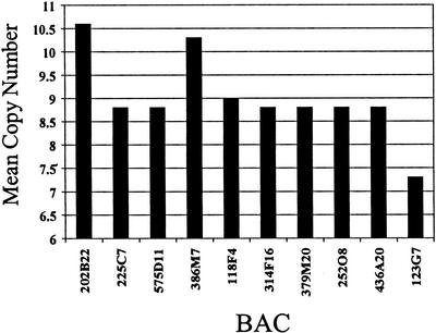
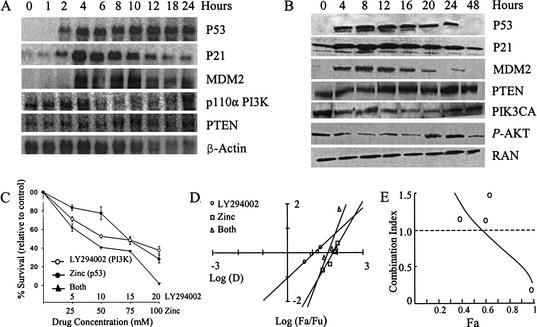
Interactions between the p53 and PI3K/AKT pathways play a significant role in the determination of cell death/survival. In benign cells these pathways are interrelated through the transcriptional regulation of PTEN by p53, which is required for p53-mediated apoptosis. PTEN exerts its effects by decreasing the phosphorylated AKT fraction, thereby diminishing prosurvival activities. However, the link between these pathways in cancer is not known. In this study, PIK3CA, encoding the p110alpha catalytic subunit of PI3K, is identified as an oncogene involved in upper aerodigestive tract (UADT) carcinomas. Simultaneous abnormalities in both pathways are rare in primary tumors, suggesting that amplification of PIK3CA and mutation of p53 are mutually exclusive events and either event is able to promote a malignant phenotype. Moreover, the negative effect of p53 induction on cell survival involves the transcriptional inhibition of PIK3CA that is independent of PTEN activity, as PTEN is not expressed in the primary tumors. Conversely, constitutive activation of PIK3CA results in resistance to p53-related apoptosis in PTEN deficient cells. Thus, p53 regulates cell survival by inhibiting the PI3K/AKT prosurvival signal independent of PTEN in epithelial tumors. This inhibition is required for p53-mediated apoptosis in malignant cells.
Figures


Similar articles
-
Frequent monoallelic deletion of PTEN and its reciprocal associatioin with PIK3CA amplification in gastric carcinoma.Int J Cancer. 2003 Apr 10;104(3):318-27. doi: 10.1002/ijc.10962. Int J Cancer. 2003. PMID: 12569555
-
Activation of PI3K/Akt pathway by PTEN reduction and PIK3CA mRNA amplification contributes to cisplatin resistance in an ovarian cancer cell line.Gynecol Oncol. 2005 Apr;97(1):26-34. doi: 10.1016/j.ygyno.2004.11.051. Gynecol Oncol. 2005. PMID: 15790433
-
PTEN reverses MDM2-mediated chemotherapy resistance by interacting with p53 in acute lymphoblastic leukemia cells.Cancer Res. 2003 Oct 1;63(19):6357-62. Cancer Res. 2003. PMID: 14559824
-
Pten signaling in gliomas.Neuro Oncol. 2002 Jul;4(3):196-211. Neuro Oncol. 2002. PMID: 12084351 Free PMC article. Review.
-
The PTEN, Mdm2, p53 tumor suppressor-oncoprotein network.Trends Biochem Sci. 2002 Sep;27(9):462-7. doi: 10.1016/s0968-0004(02)02166-7. Trends Biochem Sci. 2002. PMID: 12217521 Review.
Cited by
-
Clinical-Pathologic Analysis of Breast Cancer With PIK3CA Mutations in Chinese Women.Technol Cancer Res Treat. 2020 Jan-Dec;19:1533033820950832. doi: 10.1177/1533033820950832. Technol Cancer Res Treat. 2020. PMID: 33047659 Free PMC article.
-
New and emerging factors in tumorigenesis: an overview.Cancer Manag Res. 2015 Jul 28;7:225-39. doi: 10.2147/CMAR.S47797. eCollection 2015. Cancer Manag Res. 2015. PMID: 26251629 Free PMC article. Review.
-
HOTAIR Promotes the Hyperactivation of PI3K/Akt and Wnt/β-Catenin Signaling Pathways via PTEN Hypermethylation in Cervical Cancer.Cells. 2024 Sep 4;13(17):1484. doi: 10.3390/cells13171484. Cells. 2024. PMID: 39273054 Free PMC article.
-
Deficiency in the p110alpha subunit of PI3K results in diminished Tie2 expression and Tie2(-/-)-like vascular defects in mice.Blood. 2005 May 15;105(10):3935-8. doi: 10.1182/blood-2004-10-3955. Epub 2005 Feb 1. Blood. 2005. PMID: 15687236 Free PMC article.
-
LKB1 is necessary for Akt-mediated phosphorylation of proapoptotic proteins.Cancer Res. 2008 Sep 15;68(18):7270-7. doi: 10.1158/0008-5472.CAN-08-1484. Cancer Res. 2008. PMID: 18794113 Free PMC article.
References
-
- Alper O, De Santis ML, Stromberg K, Hacker NF, Cho-Chung YS, Salomon DS. Anti-sense suppression of epidermal growth factor receptor expression alters cellular proliferation, cell-adhesion, and tumorigenicity in ovarian cancer cells. Int J Cancer. 2000;88:566–574. - PubMed
-
- Bhuyan DK, Reddy PG, Bhuyan KC. Growth factor receptor gene and protein expressions in the human lens. Mech Aging Dev. 2000;113:205–218. - PubMed
-
- Bjorkqvist AM, Husgafvel-Pursiainen K, Anttila S, Karjalainen A, Tammilehto L, Mattson K, Vainio H, Knuutila S. DNA gains in 3q occur frequently in squamous cell carcinoma of the lung, but not in adenocarcinoma. Genes Chromosomes Cancer. 1998;22:79–82. - PubMed
-
- Chou TC, Talalay P. Quantitative analysis of dose-effect relationships: The combined effects of multiple drugs or enzyme inhibitors. Adv Enzyme Regul. 1984;22:27–55. - PubMed
MeSH terms
Substances
LinkOut - more resources
Full Text Sources
Other Literature Sources
Medical
Molecular Biology Databases
Research Materials
Miscellaneous