. 2002 Apr 1;16(7):836-45.
doi: 10.1101/gad.966402.

The plexin-B1/Rac interaction inhibits PAK activation and enhances Sema4D ligand binding

Affiliations

The plexin-B1/Rac interaction inhibits PAK activation and enhances Sema4D ligand binding

Haris G Vikis et al. Genes Dev. .

Abstract

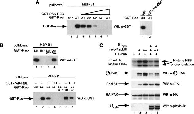
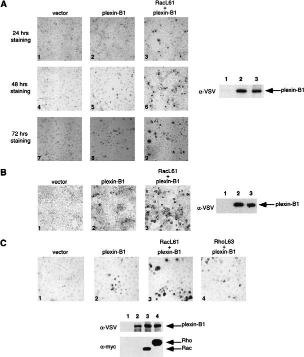
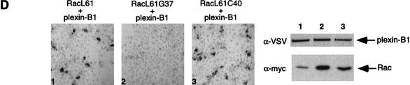
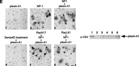
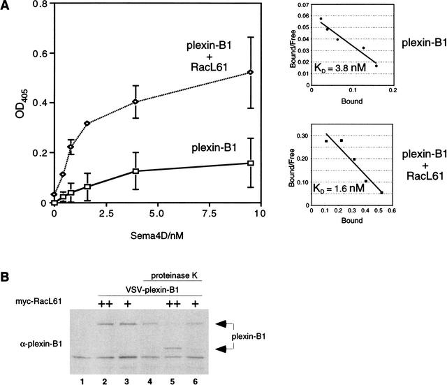
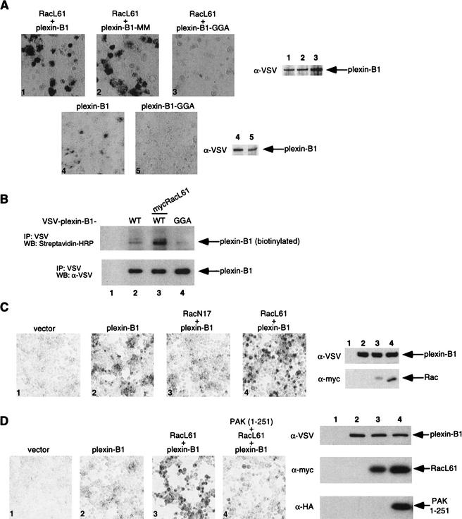
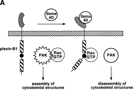
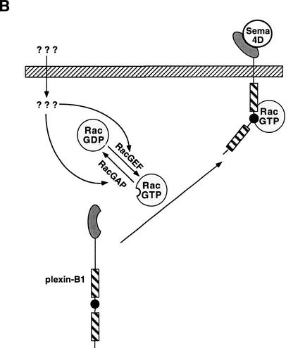
The small GTPase Rac has been implicated in growth cone guidance mediated by semaphorins and their receptors. Here we demonstrate that plexin-B1, a receptor for Semaphorin4D (Sema4D), and p21-activated kinase (PAK) can compete for the interaction with active Rac and plexin-B1 can inhibit Rac-induced PAK activation. We have also demonstrated that expression of active Rac enhances the ability of plexin-B1 to interact with Sema4D. Active Rac stimulates the localization of plexin-B1 to the cell surface. The enhancement in Sema4D binding depends on the ability of Rac to bind plexin-B1. These observations support a model where signaling between Rac and plexin-B1 is bidirectional; Rac modulates plexin-B1 activity and plexin-B1 modulates Rac function.


Figures

Figure 1
Figure 1
(A) The Rac-binding domain (RBD) of p21-activated kinase (PAK) competes with plexin-B1 for RacL61. In vitro binding was performed using purified glutathione S-transferase (GST)–RacN17 (5 μg, lane 1) or GST–RacL61 (lane 2) and incubated with MBP–plexin-B1 (10 μg) prebound to amylose resin in the absence or presence of increasing amounts of GST–PAK–RBD (lane 3, 0.5 μg; lane 4, 1 μg; lane 5, 5 μg; lane 6, 15 μg; lane 7, 25 μg, left panel). Similarly, 15 μg of GST or GST–PAK–RBD were used (right panel). Associated GST–Rac was isolated and subject to Western blot with α-GST antibody (Zymed). (B) The RBD of PAK can compete with plexin-B1 for RacL61G37, but not RacL61C40. MBP-B1 (10 μg) prebound to amylose resin was subjected to in vitro binding with GST–RacL61/N17/L61G37/L61C40 (5 μg) (upper panel). Bound GST–Rac was determined by α-GST Western blot. GST–RacN17/L61 (5 μg) was incubated with MBP–plexin-B1 (10 μg) prebound to amylose resin in the presence or absence of GST–PAK–RBD (lane 3, 1 μg; lanes 4 and 7, 15 μg) (lower panel). (C) Plexin B1 inhibits RacL61-induced PAK activation. HEK 293 cells were transfected with 200 ng of HA-PAK, 500 ng plexin-B1cyto, and 25 ng or 50 ng myc–RacL61. HA-PAK was immunoprecipitated with α-HA (BAbCO). Kinase activity was determined by in vitro kinase assay using histone H2B as a substrate or by α-phospho PAK antibody.
Figure 2
Figure 2
(A) Expression of RacL61 enhances the binding of Sema4D to plexin-B1 in COS-7 cells. COS-7 cells were transfected with expression vectors for VSV–plexin-B1, mycRacL61, or pcDNA3. Binding of AP-Sema4D was performed. Pictures were taken at 100× magnification and show staining over the course of 24 h (panels 1–3), 48 h (panels 4–6), and 72 h (panels 7–9). The expression level of plexin-B1 from a duplicate plate was determined by α-VSV Western blot. Lanes 1–3 correspond to panels 1–3, respectively. Differences in background color are attributable to differences in shutter speed of the digital camera. (B) Expression of RacL61 enhances the binding of Sema4D to plexin-B1 in HEK293 cells. Cells were transfected as in A, however staining is shown after 5 h. (C) RhoL63 does not enhance the binding of Sema4D to plexin-B1. COS-7 cells were transfected with the indicated expression vectors and subject to the same staining procedures as in A. (D) Mutations in RacL61 ablate the ability to stimulate binding of plexin-B1 to Sema4D. Transfections and staining were carried out as in A. (E) Rac does not affect the interaction of Sema3A with NP-1/Plexin-A1. COS-7 cells were transfected with the indicated expression vectors. Cells were treated with Sema3A for 1 h prior to fixation and staining as described in A. Sema4D ligand was used in panel 4 and 400× magnification was used to take the photos.
Figure 2
Figure 2
(A) Expression of RacL61 enhances the binding of Sema4D to plexin-B1 in COS-7 cells. COS-7 cells were transfected with expression vectors for VSV–plexin-B1, mycRacL61, or pcDNA3. Binding of AP-Sema4D was performed. Pictures were taken at 100× magnification and show staining over the course of 24 h (panels 1–3), 48 h (panels 4–6), and 72 h (panels 7–9). The expression level of plexin-B1 from a duplicate plate was determined by α-VSV Western blot. Lanes 1–3 correspond to panels 1–3, respectively. Differences in background color are attributable to differences in shutter speed of the digital camera. (B) Expression of RacL61 enhances the binding of Sema4D to plexin-B1 in HEK293 cells. Cells were transfected as in A, however staining is shown after 5 h. (C) RhoL63 does not enhance the binding of Sema4D to plexin-B1. COS-7 cells were transfected with the indicated expression vectors and subject to the same staining procedures as in A. (D) Mutations in RacL61 ablate the ability to stimulate binding of plexin-B1 to Sema4D. Transfections and staining were carried out as in A. (E) Rac does not affect the interaction of Sema3A with NP-1/Plexin-A1. COS-7 cells were transfected with the indicated expression vectors. Cells were treated with Sema3A for 1 h prior to fixation and staining as described in A. Sema4D ligand was used in panel 4 and 400× magnification was used to take the photos.
Figure 3
Figure 3
(A) RacL61 enhances the affinity and number of plexin-B1 receptors at the cell surface. COS-7cells were transfected as described in Materials and Methods. AP-Sema4D binding was assayed as described in Materials and Methods. Background activity of vector-transfected cells was subtracted. Scatchard plots are shown and linear regression analysis was used to determine the line of best fit. The data shown is an average of three replicates with error bars indicating the calculated standard deviation. (B) RacL61 recruits plexin-B1 to the cell surface. HEK293 cells were transfected with VSV–plexin-B1 (1 μg) and myc–RacL61 (25 ng or 100 ng) in the combinations indicated. Cells were treated with 0.2 mg/mL proteinase K and lysates were analyzed by Western blot with anti-plexin-B1 antibody.
Figure 4
Figure 4
(A) The Rac binding deficient mutant plexin-B1–GGA is deficient in Sema4D binding and cannot be stimulated by RacL61. COS-7 cells were transfected with the indicated expression plasmids and treated with Sema4D as described in Figure 2A. Plexin-B1–GGA showed lower basal and RacL61-stimulated Sema-4D binding (panel 1 vs. 3 and 4 vs. 5). (B) Cell surface expression of plexin-B1–GGA is diminished. HEK293 cells were transfected with the indicated expression plasmids. Cells were biotinylated with Sulfo-NHS-LC-Biotin as described in Materials and Methods. Plexin-B1 and plexin-B1–GGA were immunoprecipitated with α-VSV and detected by Western blot with streptavidin-HRP and α-VSV Western blots. (C) RacN17 inhibits the Sema4D/plexin-B1 interaction. HEK293 cells were transfected as indicated and subject to the same Sema4D treatment and detection as described in Figure 2A. (D) The N-terminal domain of p21-activated kinase (PAK) inhibits RacL61-induced Sema4D/plexin-B1 binding. HEK 293 cells were transfected as indicated and subject to the same Sema4D treatment and detection as described in Figure 2A.
Figure 5
Figure 5
(A) Sema4D inhibits p21-activated kinase (PAK) activity and promotes growth cone collapse. Treatment of plexin-B1 expressing neurons with Sema4D results in the recruitment and sequestration of active Rac. This inhibits the ability of Rac to activate PAK and thus results in the disassembly of cytoskeletal structures associated with growth cone collapse/turning. (B) Active Rac enhances the affinity of plexin-B1 for Sema4D and its localization at the cell surface. Expression of plexin-B1 at the cell surface requires endogenous GTP–Rac levels. Stimulation of Rac by signals acting on RacGEFs and/or RacGAPs result in enhanced localization of plexin-B1 at the membrane in addition to an enhanced affinity for Sema4D.
Figure 5
Figure 5
(A) Sema4D inhibits p21-activated kinase (PAK) activity and promotes growth cone collapse. Treatment of plexin-B1 expressing neurons with Sema4D results in the recruitment and sequestration of active Rac. This inhibits the ability of Rac to activate PAK and thus results in the disassembly of cytoskeletal structures associated with growth cone collapse/turning. (B) Active Rac enhances the affinity of plexin-B1 for Sema4D and its localization at the cell surface. Expression of plexin-B1 at the cell surface requires endogenous GTP–Rac levels. Stimulation of Rac by signals acting on RacGEFs and/or RacGAPs result in enhanced localization of plexin-B1 at the membrane in addition to an enhanced affinity for Sema4D.

Similar articles

Cited by

References

    1. Bentley D, O'Connor TP. Cytoskeletal events in growth cone steering. Curr Opin Neurobiol. 1994;4:43–48. - PubMed
    1. Buchwald G, Hostinova E, Rudolph MG, Kraemer A, Sickmann A, Meyer HE, Scheffzek K, Wittinghofer A. Conformational switch and role of phosphorylation in PAK activation. Mol Cell Biol. 2001;21:5179–5189. - PMC - PubMed
    1. Culotti JG, Kolodkin AL. Functions of netrins and semaphorins in axon guidance. Curr Opin Neurobiol. 1996;6:81–88. - PubMed
    1. Dharmawardhane S, Sanders LC, Martin SS, Daniels RH, Bokoch GM. Localization of p21-activated kinase 1 (PAK1) to pinocytic vesicles and cortical actin structures in stimulated cells. J Cell Biol. 1997;138:1265–1278. - PMC - PubMed
    1. Driessens MH, Hu H, Nobes CD, Self A, Jordens I, Goodman CS, Hall A. Plexin-B semaphorin receptors interact directly with active Rac and regulate the actin cytoskeleton by activating Rho. Curr Biol. 2001;11:339–344. - PubMed

Publication types

MeSH terms

LinkOut - more resources