. 2002 Mar;13(3):1071-82.
doi: 10.1091/mbc.01-08-0409.

Role of adaptor complex AP-3 in targeting wild-type and mutated CD63 to lysosomes

Affiliations

Role of adaptor complex AP-3 in targeting wild-type and mutated CD63 to lysosomes

Brian A Rous et al. Mol Biol Cell. 2002 Mar.

Abstract

CD63 is a lysosomal membrane protein that belongs to the tetraspanin family. Its carboxyterminal cytoplasmic tail sequence contains the lysosomal targeting motif GYEVM. Strong, tyrosine-dependent interaction of the wild-type carboxyterminal tail of CD63 with the AP-3 adaptor subunit mu 3 was observed using a yeast two-hybrid system. The strength of interaction of mutated tail sequences with mu 3 correlated with the degree of lysosomal localization of similarly mutated human CD63 molecules in stably transfected normal rat kidney cells. Mutated CD63 containing the cytosolic tail sequence GYEVI, which interacted strongly with mu 3 but not at all with mu 2 in the yeast two-hybrid system, localized to lysosomes in transfected normal rat kidney and NIH-3T3 cells. In contrast, it localized to the cell surface in transfected cells of pearl and mocha mice, which have genetic defects in genes encoding subunits of AP-3, but to lysosomes in functionally rescued mocha cells expressing the delta subunit of AP-3. Thus, AP-3 is absolutely required for the delivery of this mutated CD63 to lysosomes. Using this AP-3-dependent mutant of CD63, we have shown that AP-3 functions in membrane traffic from the trans-Golgi network to lysosomes via an intracellular route that appears to bypass early endosomes.


Figures
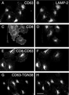
Figure 1
Figure 1
Indirect immunofluorescence steady-state localization of wild-type human CD63, CD8, and chimeric proteins in stably transfected NRK cells. NRK cells were transfected with ΔpMEP4 constructs containing cDNA encoding either human CD63 (A and B), CD8 (C and D), CD8-CD63 (E and F), or CD63-TGN38 (G and H). Stably transfected cell lines were treated with 3 μM CdCl2 to induce protein expression and then double labeled with a mouse mAb to either CD63 (A and G) or CD8 (C and E) and a rabbit polyclonal antibody against endogenous LAMP-2 (B, D, F, and H). Bar, 20 μm.
Figure 2
Figure 2
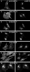
Indirect immunofluorescence steady-state localization of mutated human CD63-LIMP-1 in stably transfected NRK cells. NRK cells were transfected with ΔpMEP4 constructs containing cDNA encoding human CD63 with point mutations in the cytosolic tail, either AYEVM (A and B), GAEVM (C and D), GYAVM (E and F), GYEAM (G and H), GYEVA (I and J), GYEVI (K and L), or GFEVM (M and N). Stably transfected cell lines were treated with 3 μM CdCl2 to induce protein expression and then double labeled with a mouse mAb to CD63 (A, C, E, G, I, K, and M) and a rabbit polyclonal antibody against endogenous LAMP-2 (B, D, F, H, J, L, and N). Bar, 20 μm.
Figure 3
Figure 3
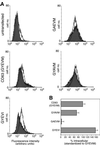
Fluorescence-activated cell sorting analysis of human CD63-LIMP-I and mutated proteins in stably transfected NRK cells. NRK cells were transfected with ΔpMEP4 constructs containing cDNA encoding human CD63 and constructs with point mutations in the cytosolic tail. After induction of protein expression the distribution of the wild-type and mutated human CD63 proteins between the cell surface and intracellular structures was determined by FACS analysis. (A) Fluorescence intensity for untransfected NRK cells (top left) and cells transfected with wild-type CD63 (GYEVM), and mutated constructs (GYEVI, GAEVM, and GYAVM) are shown. The filled traces represent intact cells and the unfilled traces the permeabilized cells. A larger shift to the right of the unfilled trace compared with the filled trace represents a larger proportion of the CD63 at intracellular sites. (B) Histogram showing the amount of intracellular mutated CD63 in each cell line relative to the amount of intracellular wild-type CD63 in the cell line expressing this construct.
Figure 4
Figure 4
Interaction of the cytosolic domains of CD63 and mutated CD63 with μ2 and μ3A in the yeast two-hybrid system. Yeast cells were transformed with bait constructs (LexA fusion) containing the C-terminal cytosolic domains of CD63, and the mutant forms AYEVM, GAEVM, GYAVM, GYEVA, GYAVM, GYEAM, GYEVA, and GYEVI and a prey construct (either VP16-μ2, VP16-μ3A). Transformed yeast were grown on agar plates in the absence of histidine for 3 d (μ3A) or 10 d (μ2). All transformed yeast grew in the presence of histidine and no transformed yeast containing “empty” VP16 grew in the absence of histidine.
Figure 5
Figure 5
Characterization of the interaction of the cytosolic domains of CD63 and mutated CD63 with μ3A in the yeast two-hybrid system. Yeast cells were transformed as described in Figure 4. (A and B) Cultures were set up containing 0.05 OD600 units of transfected cells into 2 ml of selective medium lacking histidine and grown at 30°C with shaking. OD600 measurements were made up to 180 h (B shows an expanded time scale). The control interaction between the C-terminal cytosolic domain of wild-type CD63 and empty VP16 (filled circles) is shown in each figure. Vector controls for other tails also showed no significant growth. Each point represents the mean ± SEM of the activity of three separate cultures. (C) Cultures of transformed yeast were established in selective medium lacking histidine, but containing 0–1 M 3-AT, and grown at 30°C with shaking. After incubation for 48 h, OD600 was measured and the ratio in the presence versus absence of 3-AT recorded. Each point represents the mean ± SEM of the activity of three separate cultures. (D) Cultures of transformed yeast were grown in medium containing histidine to an OD600 of ∼1.2. The β-galactosidase activity of the cultures was then measured. Each bar in the histogram represents the mean ± SEM of the activity of five separate cultures.
Figure 6
Figure 6
Uptake of anti-CD63 antibody by stably transfected NRK lines expressing either wild-type human CD63 (A and B) or the GYEVI construct (C and D). Protein expression was induced in stably transfected NRK cell lines with 3 μM CdCl2 for 16 h. The cells were then incubated at 37°C for 30 min in the presence of anti-CD63 mAb and then incubated for a further 30 min without antibody (i.e., chased). The cells were then fixed and immunofluorescence localization of the anti-CD63 mAb (A and C) and of endogenous LAMP-2 (B and D) carried out. Bar, 20 μm.
Figure 7
Figure 7
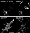
Steady-state localization of wild-type human CD63 and the GYEVI construct in transiently transfected pearl cells. Pearl cells were transiently transfected with ΔpMEP containing cDNA encoding either wild-type CD63 (A and B) or the GYEVI construct (C and D). The transfected cells were treated with 3 μM CdCl2 to induce protein expression and then double labeled, for indirect immunofluorescence localization, with a mouse mAb to CD63 (A and D) and a rat mAb to endogenous mouse LAMP-1 (B and E). Bar, 20 μm.
Figure 8
Figure 8
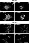
Steady-state localization of CD63 and GYEVI constructs in stably transfected mocha and 3T3 cell lines. Mocha cells (A–D) and 3T3 cells (E–H) were transfected with ΔpMEP constructs containing cDNA encoding wild-type human CD63 (A, B, E, and F) or the GYEVI construct (C, D, G, and H). Stably transfected cell lines were treated with 3 μM CdCl2 to induce protein expression and then double labeled, for indirect immunofluorescence localization, with a mouse mAb to CD63 (A, C, E, and G) and a rat mAb to endogenous mouse LAMP-1 (B, D, F, and H). Bar, 20 μm.
Figure 9
Figure 9
Localization of the GYEVI construct of human CD63 in rescued mocha cells expressing the δ subunit of AP-3. A stably transfected mocha cell line expressing the δ subunit of AP-3 was transiently transfected with a ΔpMEP construct containing cDNA encoding the human CD63/LIMP-I, GYEVI construct. After treating the transfected cells with 3 μM CdCl2 to induce protein expression they were double labeled, for indirect immunofluorescence localization with a mouse mAb to human CD63 (A) and with a rabbit polyclonal antibody to the δ subunit of AP-3 (B). Bar, 20 μm. (C) Cells that were expressing the GYEVI construct were then counted and grouped according to the presence of intracellular human CD63 or only cell surface human CD63 and the presence or absence of detectable δ subunit of AP-3. The data shown in the histogram represent the mean of two experiments. In the two experiments the counted numbers of human CD63-positive cells showing only surface expression were 208 and 349. The counted numbers of CD63-positive cells also showing intracellular localization of human CD63 were 91 and 44.
Figure 10
Figure 10
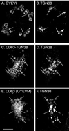
Indirect immunofluorescence steady-state localization of wild-type human CD63, GYEVI, and CD63-TGN38 constructs in stable lines of transfected NRK cells treated with chloroquine. Stably transfected NRK cell lines expressing either the GYEVI construct (A and B), CD63-TGN38 (C and D), or wild-type CD63 (E and F) were incubated for 2 h in the presence of both 3 μM CdCl2 and 100 μM chloroquine before fixation. The cells were then double labeled, for indirect immunofluorescence localization, with a mouse mAb to human CD63 (A, C, and E) and a rabbit polyclonal antibody to the lumenal domain of rat TGN38 (B, D, and F). Structures positive for TGN38, but which do not label with the anti-CD63 antibody, are indicated by single-headed arrows. Structures that label with both the TGN38 antibody and the CD63 antibody are indicated by the double-headed arrows. Bar, 10 μm.

Similar articles

Cited by

References

    1. Akasaki K, Michihara A, Mibuka K, Fujiwara Y, Tsuji H. Biosynthetic transport of a major lysosomal membrane glycoprotein, lamp-1: Convergence of biosynthetic and endocytic pathways occurs at three distinctive points. Exp Cell Res. 1995;220:464–473. - PubMed
    1. Akasaki K, Michihara A, Fujiwara Y, Mibuka K, Tsuji H. Biosynthetic transport of a major lysosome-associated membrane glycoprotein 2, lamp-2: a significant fraction of newly synthesized lamp-2 is delivered to lysosomes by way of early endosomes. J Biochem. 1996;120:1088–1094. - PubMed
    1. Andrejewski N, Punnonen E-L, Guhde G, Tanaka Y, Lüllmann-Rauch R, Hartmann D, von Figura K, Saftig P. Normal lysosomal morphology and function in LAMP-1-deficient mice. J Biol Chem. 1999;274:12692–12701. - PubMed
    1. Banting G, Maile R, Roquemore EP. The steady state distribution of humTGN46 is not significantly altered in cells defective in clathrin-mediated endocytosis. J Cell Sci. 1998;111:3451–3458. - PubMed
    1. Barriocanal JG, Bonifacino JS, Yuan L, Sandoval IV. Biosynthesis, glycosylation, movement through the Golgi system, and transport to lysosomes by an N-linked carbohydrate-independent mechanism of three lysosomal integral membrane proteins. J Biol Chem. 1986;261:16755–16763. - PubMed

Publication types

MeSH terms

LinkOut - more resources