. 2002 Mar;13(3):767-81.
doi: 10.1091/mbc.01-05-0275.

Palmitoylation of tetraspanin proteins: modulation of CD151 lateral interactions, subcellular distribution, and integrin-dependent cell morphology

Affiliations

Palmitoylation of tetraspanin proteins: modulation of CD151 lateral interactions, subcellular distribution, and integrin-dependent cell morphology

Xiuwei Yang et al. Mol Biol Cell. 2002 Mar.

Abstract

Here we demonstrate that multiple tetraspanin (transmembrane 4 superfamily) proteins are palmitoylated, in either the Golgi or a post-Golgi compartment. Using CD151 as a model tetraspanin, we identified and mutated intracellular N-terminal and C-terminal cysteine palmitoylation sites. Simultaneous mutations of C11, C15, C242, and C243 (each to serine) eliminated >90% of CD151 palmitoylation. Notably, palmitoylation had minimal influence on the density of tetraspanin protein complexes, did not promote tetraspanin localization into detergent-resistant microdomains, and was not required for CD151-alpha 3 beta 1 integrin association. However, the CD151 tetra mutant showed markedly diminished associations with other cell surface proteins, including other transmembrane 4 superfamily proteins (CD9, CD63). Thus, palmitoylation may be critical for assembly of the large network of cell surface tetraspanin-protein interactions, sometimes called the "tetraspanin web." Also, compared with wild-type CD151, the tetra mutant was much more diffusely distributed and showed markedly diminished stability during biosynthesis. Finally, expression of the tetra-CD151 mutant profoundly altered alpha 3 integrin-deficient kidney epithelial cells, such that they converted from a dispersed, elongated morphology to an epithelium-like cobblestone clustering. These results point to novel biochemical and biological functions for tetraspanin palmitoylation.


Figures

Figure 1
Figure 1
Palmitoylation of tetraspanin proteins. (A) A431 cells were pulsed with [3H]palmitate and lysed in RIPA buffer, and then CD9, CD147, CD151, fyn, and caveolin were immunoprecipitated using mAbs Du-Al, 8G6, 5C11, FYN3, and C13630, respectively. (B) A15, CD82, CD81, CD63, and CD9 were immunoprecipitated using mAbs AZM30.4, M104, M38, 6H1, and Du-All-1, respectively. The A15 and CD82 samples were obtained from transiently transfected 293 cells. CD81, CD63, and CD9 samples were from A431 cells. Molecular masses are given in kilodaltons.
Figure 2
Figure 2
Tetraspanin protein complexes. A431 cells were labeled with [3H]palmitate and then lysed in the buffers containing either 1% Brij-96 (lanes a–g) or 1% Triton X-100 (lanes h–n). Equal amounts of lysate were used for each immunoprecipitation. Antibodies for individual immunoprecipitations were A2-IIE10, A3-X8, A6-ELE, Du-All-1, 5C11, M104, and 6H1 for α2, α3, α6, CD9, CD151, CD82, and CD63, respectively. Unknown proteins of ∼60 and 50 kDa (distinct from CD63) are indicated by arrowheads. At least part of the 50-kDa protein is likely to be a CD151 dimer, as confirmed by immunoblotting for CD151 (Yang, Claas, Kraeft, Chen, Wang, Kreidberg, and Hemler, unpublished results). Note: Compared with Brij-96 extraction (lane d), Triton X-100 extraction (lane k) yielded less CD9, because of destabilization of the Du-All-1 epitope. Also, more CD151 appears in the anti-α3 lane (lane b) compared with the anti-CD151 lane (lane e), because in Brij-96 lysate, the 5C11 epitope on CD151 is likely obscured by associated proteins.
Figure 3
Figure 3
Palmitoylation sensitivity to brefeldin A (BFA). A431 cells were incubated with 10 μg/ml brefeldin A, 10 μg/ml nocodazole, or 20 μg/ml monensin for 1 h in DMEM in the absence of serum. Medium was then removed, and drugs were added again (same dose) in the presence of [3H]palmitate and 5% dialyzed fetal calf serum for 1 h. From Brij-96 lysates, either CD9 (left) or CD151 (right) was immunoprecipitated. Precipitated samples (IP) were analyzed for [3H]palmitate incorporation (top) or by immunoblotting (Ib) for CD9 or α3 integrin (bottom). Because CD151 tightly associates with α3 integrin, α3 was used as a marker to assess CD151 loading. SDS-PAGE was carried out on a 4–12% acrylamide gradient.
Figure 4
Figure 4
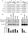
CD151 palmitoylation sites. (A) Schematic diagram of candidate CD151 palmitoylation sites. Shown are the four transmembrane domains (TM1–4), short extracellular loop (SEL), inner loop (IL), large extracellular loop (LEL), and membrane-proximal cysteine residues. Various C→S (CYS→SER) mutants are also indicated. Unless otherwise indicated, all mutants contained a GFP domain fused to the carboxy terminus. Wt, wild type. (B and C) In separate experiments, 293 cells were transiently transfected with various CD151 constructs and pulsed with [3H]palmitate (palm.). After transfection (24 h), cells were lysed in RIPA buffer and immunoprecipitated using mAb 5C11. Samples were then divided equally, resolved by SDS-PAGE, and then either dried for the detection of [3H]palmitate or transferred to nitrocellulose for blotting (Ib) with GFP polyclonal antibody.
Figure 5
Figure 5
Sucrose density gradient analysis of CD151. Stably transfected MDA-231 cells containing either wild-type CD151 (Wt) or CD151-tetra mutant (Te) were lysed in 1% Brij-96. Lysates were then subjected to 5–35-45% discontinuous sucrose density gradient centrifugation and 12 fractions (Frac.) of 400 μl each were collected from the top of the gradient. (A) From each fraction, 50 μl was immunoprecipitated with anti-GFP polyclonal antibody, and samples were immunoblotted (Ib) using a monoclonal anti-GFP antibody to detect CD151-GFP. (B) From each fraction, 50 μl of lysate was resolved by SDS-PAGE, transferred to membrane, and then immunoblotted for CD71, a cell surface transmembrane protein typically found in dense fractions.
Figure 6
Figure 6
CD151 detergent extractability. (A) Equal numbers of semiconfluent stably transfected MDA-231 cells (mock, wt-CD151, tetra-CD151) were seeded and grown for 24 h. Cells were then washed, cooled on ice, and rocked gently in the presence of 1% Brij-96 for 5–10 min at 4°C. After removal of the soluble fraction (S), the remaining insoluble material was further extracted in RIPA for an additional 1 h at 4°C (to yield fraction I). Samples were resolved by SDS-PAGE and blotted (Ib) for CD151-GFP or GFP (using polyclonal anti-GFP) or for CD9 (using mAb C9BB). Note: The greater insolubility of CD151 compared with CD9 is possibly due to much stronger CD151 association with α3 and α6 integrins. Results shown are representative of multiple experiments. (B) MDA-231 cells were lysed in 1.0% Brij-96 for 10 min, and then soluble (S) and insoluble (I) fractions were obtained as in A. In this experiment, anti-CD151 mAb 1A5 was used to blot both endogenous CD151 and CD151-GFP proteins. Note: Expression of endogenous CD151 (lanes c–f) is greatly reduced because of the presence of exogenous CD151-GFP.
Figure 7
Figure 7
Integrity of CD151 complexes. (A) Mock or stable CD151 transfectants (MDA-231 cells) were surface labeled with biotin for 1 h at 4°C, washed, and lysed in 1.0% Brij-96 buffer. Equal amounts of lysates were subsequently immunoprecipitated (IP) using anti-GFP polyclonal antibody or anti-CD81 mAb M38. Samples were resolved by SDS-PAGE and blotted using avidin-horseradish peroxidase. The small panel to the right (bottom) represents a longer exposure (exp) of a portion of the large panel to the left. SDS-PAGE was carried out on a 5–15% acrylamide gradient. (B) Samples prepared as in A were immunoblotted for either CD63 or CD9. (C) CD151 transfectants were labeled with 0.3 mCi/ml [35S] methionine for 1 h and lysed in 1% Triton X-100, and then wt-CD151 and tetra-CD151 were immunoprecipitated (IP) using anti-GFP polyclonal antibody. After SDS-PAGE, samples were transferred to a PVDF membrane, exposed to film to detect associated α3 integrin (top), and subsequently blotted using anti-GFP mAb (bottom).
Figure 8
Figure 8
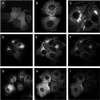
Distribution of CD151-GFP. MCF-7 cells stably transfected with various GFP-tagged CD151 proteins were grown on glass coverslips overnight, fixed in 1.0% paraformaldehyde for 20 min at 4°C, and mounted onto glass slides using the ProLong Antifade Kit from Molecular Probes (Eugene, OR). Slides were analyzed using a confocal model LSM4 scanning microscope (Zeiss, Thornwood, NY) equipped with an external argon-krypton laser. (A) Cells expressing GFP only; (B) cells expressing C15S-CD151-GFP; (C) cells expressing C241/242S-CD151-GFP; (D–F) cells expressing wild-type CD151-GFP; (G–I) cells expressing tetra-CD151; (D and G) highest optical plane; (E and H) lower plane; (F and I) lowest plane. Expression levels for mutant and wild-type CD151 were comparable, as seen by flow cytometry (GFP fluorescence) and by immunoblotting (anti-GFP antibody).
Figure 9
Figure 9
Internalization and recycling of CD151. (A) NIH3T3 cells were stably transfected with Myc-tagged wild-type CD151 (Wt) or tetra-CD151 (Te). Stable transfectants were surface labeled with reducible NHS-SS-biotin at 4°C. Some labeled cells were then lysed immediately in RIPA (lanes a and b). Other cells (lanes c–j) were incubated at 37°C for various times (to allow CD151 internalization) and then treated with reducing agent to remove all biotin remaining on the cell surface. (B) NHS-SS-biotin-labeled cells were incubated at 37°C for 60 min to allow accumulation of internalized CD151 proteins and then treated with reducing agent to remove the noninternalized surface proteins. Some cells were then lysed immediately (lanes k and n). Other cells were then incubated for an additional 30 min at 37°C to allow recycling of labeled-internalized CD151 back to the cell surface. These cells were then treated either without (lanes l and o) or with (lanes m and p) reducing agent before lysis in RIPA. From all lysate samples (A and B), CD151 was immunoprecipitated (using mAb 5C11), and biotin labeling was detected by blotting with avidin-horseradish peroxidase (HRP). Each sample was also blotted (Ib) for tubulin (bottom).
Figure 10
Figure 10
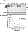
Biosynthesis and degradation of CD151. (A) MDA-231 cells stably transfected with GFP-tagged CD151 were pulsed with 0.5 mCi/ml [35S]methionine for 1 h and then either lysed (time 0) or chased in medium containing 5% dialyzed serum and 25× excess cold methionine for the indicated times (2, 6, 24, 48, and 77 h) before lysis in RIPA detergent. CD151was then immunoprecipitated using anti-GFP polyclonal antibody and samples were resolved on 4–12% SDS-PAGE and transferred to a PVDF membrane. To detect [35S]methionine, the membrane was exposed to BioMax MS film using a BioMax Transcreen LE system (top). The membrane was also blotted using anti-GFP mAb to estimate CD151 loading (bottom). (B) Levels of radioactivity (for mature CD151 only) were estimated by densitometry, and the relative levels were calculated as a percentage of the labeled protein at time 0.
Figure 11
Figure 11
Mutant CD151 alters cell morphology. (A) Stably transfected semiconfluent B12 kidney epithelial cells were lightly trypsinized, incubated at 37°C for 20 min, then stained on ice with either control immunoglobulin G or anti-CD151 mAb 5C11, and visualized using phycoerythrin-conjugated secondary antibody (Biosource, Camarillo, CA). Flow cytometry was carried out using a FACSCalibur (Becton-Dickinson), measuring GFP fluorescence (left) and phycoerythrin fluorescence (right), for tetra (Te) and wild-type (Wt) CD151-transfected cells. Background staining is indicated as “IgG.” Mean fluorescence intensity values for Te and Wt CD151, respectively, are 1017 and 740 (for GFP fluorescence) and 660 and 616 (for phycoerythrin fluorescence). (B) B12 cells expressing GFP alone, wild-type CD151, or tetra-CD151 were plated on tissue culture plastic for 24 h and then analyzed using a Zeiss Axiovert 135 microscope and photographed as described previously (Stipp and Hemler, 2000).
Figure 12
Figure 12
Potential tetraspanin palmitoylation sites. Cysteine residues in CD151 (C11, C15, C241, C242) used as palmitoylation sites and proximal to TM1 and TM4 are compared with cysteine alignments in other tetraspanin proteins. Positions of additional membrane proximal cysteines that could be palmitoylated are marked with asterisks. Only portions of TM1, TM2, TM3, and TM4 are shown. IL, inner loop.

Similar articles

Cited by

References

    1. Azorsa DO, Moog S, Cazenave JP, Lanza F. A general approach to the generation of monoclonal antibodies against members of the tetraspanin superfamily using recombinant GST fusion proteins. J Immunol Methods. 1999;229:35–48. - PubMed
    1. Banerjee SA, Hadjiargyrou M, Patterson PH. An antibody to the tetraspan membrane protein CD9 promotes neurite formation in a partially α3β1 integrin-dependent manner. J Neurosci. 1997;17:2756–2765. - PMC - PubMed
    1. Barylko B, Gerber SH, Binns DD, Grichine N, Khvotchev M, Sudhof TC, Albanesi JP. A novel family of phos-phatidylinositol 4-kinases conserved from yeast to humans. J Biol Chem. 2001;276:7705–7708. - PubMed
    1. Berditchevski F, Bazzoni G, Hemler ME. Specific association of CD63 with the VLA-3 and VLA-6 integrins. J Biol Chem. 1995;270:17784–17790. - PubMed
    1. Berditchevski F, Chang S, Bodorova J, Hemler ME. Generation of monoclonal antibodies to integrin-associated proteins: evidence that α3β1 complexes with EMMPRIN/basigin/OX47/M6. J Biol Chem. 1997;272:29174–29180. - PubMed

Publication types

MeSH terms

LinkOut - more resources