. 2002 Apr;76(8):3892-904.
doi: 10.1128/jvi.76.8.3892-3904.2002.

Minute virus of mice NS1 interacts with the SMN protein, and they colocalize in novel nuclear bodies induced by parvovirus infection

Affiliations

Minute virus of mice NS1 interacts with the SMN protein, and they colocalize in novel nuclear bodies induced by parvovirus infection

Philip J Young et al. J Virol. 2002 Apr.

Abstract

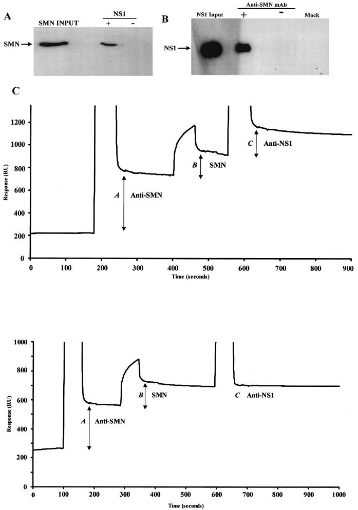
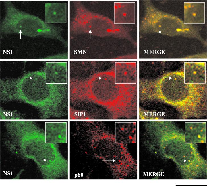
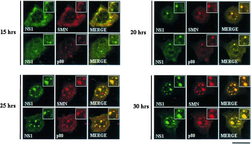
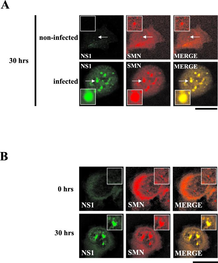
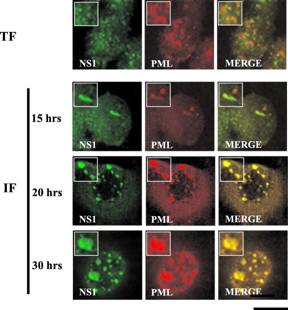
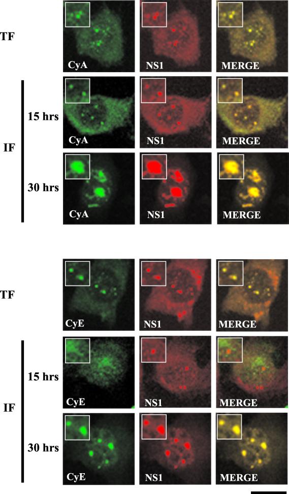
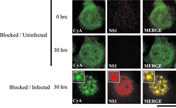
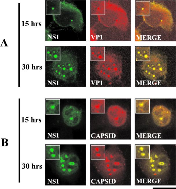
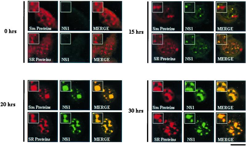
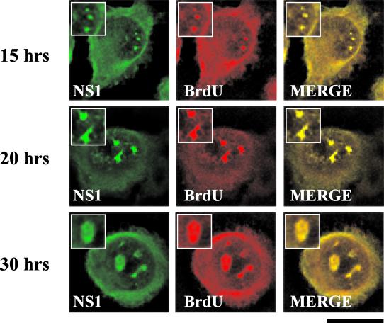
The human survival motor neuron (SMN) gene is the spinal muscular atrophy-determining gene, and a knockout of the murine Smn gene results in preembryonic lethality. Here we show that SMN can directly interact in vitro and in vivo with the large nonstructural protein NS1 of the autonomous parvovirus minute virus of mice (MVM), a protein essential for viral replication and a potent transcriptional activator. Typically, SMN localizes within nuclear Cajal bodies and diffusely in the cytoplasm. Following transient NS1expression, SMN and NS1 colocalize within Cajal bodies. At early time points following parvovirus infection, NS1 fails to colocalize with SMN within Cajal bodies; however, during the course of MVM infection, dramatic nuclear alterations occur. Formerly distinct nuclear bodies such as Cajal bodies, promyelocytic leukemia gene product (PML) oncogenic domains (PODs), speckles, and autonomous parvovirus-associated replication (APAR) bodies are seen aggregating at later points in infection. These newly formed large nuclear bodies (termed SMN-associated APAR bodies) are active sites of viral replication and viral capsid assembly. These results highlight the transient nature of nuclear bodies and their contents and identify a novel nuclear body formed during infection. Furthermore, simple transient expression of the viral nonstructural proteins is insufficient to induce this nuclear reorganization, suggesting that this event is induced specifically by a step in the viral infection process.


Figures

FIG. 1.
FIG. 1.
SMN and NS1 interact directly both in vitro and in vivo. (A) Western blot analysis of recombinant polyhistidine (six-His-)-tagged human SMN captured by GST-tagged NS1 immobilized on GST resin. The blot was developed with an anti-SMN monoclonal antibody. (B) Western blot analysis of NS1 coimmunoprecipitated with endogenous SMN from transfected A92L cells. Anti-SMN coprecipitated endogenous SMN, and anti-NS1 rabbit polyclonal sera detected NS1 complexed with SMN. NS1-SMN binding was not detected when the immunoprecipitating antibody (−) was omitted or when cells were not transfected with NS1 (mock). mAb, monoclonal antibody. (C) (Top) BIA of NS1 coimmunoprecipitated with endogenous SMN from MVM-infected double-blocked A92L cells. Total protein extracts from A92L cells 30 h postinfection were used. Anti-SMN (MANSMA3) coprecipitated SMN, and anti-NS1 rabbit polyclonal sera detected NS1 complexed with SMN. Levels of the captured anti-SMN antibody (A), SMN-NS1 complex (B), and anti-NS1 rabbit polyclonal antibody (C) are indicated. (Bottom) NS1 is not coprecipitated with SMN from double-blocked noninfected A92L cells. Total protein extracts from A92L cells 30 h after being released from the blocking process were used. Anti-SMN (MANSMA3) precipitated SMN, and anti-NS1 rabbit polyclonal sera detected no NS1 complexed with SMN. Levels the captured anti-SMN antibody (A), SMN (B), and anti-NS1 rabbit polyclonal antibody (C) are indicated.
FIG. 2.
FIG. 2.
Colocalization of SMN, p80 coilin, SIP1, and NS1 in transfected A92L cells. SMN (primary antibody: MANSMA1; secondary antibody: TRITC [red]), SIP1 (primary antibody: MANSIP1A; secondary antibody: anti-mouse TRITC conjugate [red]), p80 coilin (primary antibody: 5P10 monoclonal antibody; secondary: anti-mouse TRITC conjugate [red]), and NS1 (primary antibody: rabbit anti-NS1 polyclonal; secondary antibody: anti-rabbit FITC conjugate [green]) are indicated. Arrows, Cajal bodies containing NS1. Insets show nuclear regions of interest magnified by an additional factor of 2. Bar ≈ 30 μm.
FIG. 3.
FIG. 3.
SMN, p80 coilin, and NS1 colocalize in SAABs in MVM-infected A92L cells. SMN (primary antibody: anti-SMN monoclonal antibody MANSMA1; secondary antibody: anti-mouse TRITC conjugate [red]), p80 coilin (primary antibody: 5P10 monoclonal antibody; secondary antibody: anti-mouse TRITC conjugate [red]), and NS1 (primary antibody: rabbit anti-NS1 polyclonal; secondary antibody: anti-rabbit FITC conjugate [green]) are indicated. Synchronized infections were obtained by performing an isoleucine-aphidicolin double block on A92L cells prior to infection. Immunofluorescence experiments were performed on infected cells 15, 20, 25, and 30 h after entry into S phase. Insets show nuclear regions of interest magnified by an additional factor of 2. Bar ≈ 30 μm.
FIG. 4.
FIG. 4.
SAAB formation is neither a consequence of the blocking process nor restricted to murine cell lines. (A) Double-label experiments were performed on infected and uninfected blocked A92L cells at 30 h postrelease. SMN (primary antibody: anti-SMN monoclonal antibody MANSMA1; secondary antibody: anti-mouse TRITC conjugate) and NS1 (primary antibody: rabbit anti-NS1 polyclonal; secondary antibody: anti-rabbit FITC conjugate) are indicated. Arrows indicate SMN-positive nuclear bodies. Insets show nuclear regions of interest magnified by an additional factor of 2. Bar ≈ 30 μm. (B) SMN (primary antibody: anti-SMN monoclonal antibody MANSMA1; secondary antibody: TRITC) and NS1 (primary antibody: rabbit anti-NS1 polyclonal; secondary antibody: FITC) are indicated. Experiments were performed on NB324K cells 0 and 30 h postinfection. Insets show nuclear regions of interest magnified by an additional factor of 2. Bar ≈ 30 μm.
FIG. 5.
FIG. 5.
PML colocalizes with NS1 in SAABs at 20 h postinfection but not in A92L cells transiently expressing HA-tagged NS1. Double-label experiments were performed on A92L cells transfected with HA-tagged NS1 (TF) and blocked A92L cells 15, 20, and 30 h after infection with MVM (IF). PML (primary antibody: anti-PML monoclonal antibody; secondary antibody: anti-mouse TRITC conjugate) and NS1 (primary antibody: rabbit anti-NS1 polyclonal; secondary antibody: anti-rabbit FITC conjugate) are indicated. Insets show nuclear regions of interest magnified by an additional factor of 2. Bar ≈ 30 μm.
FIG. 6.
FIG. 6.
Cyclin A and cyclin E colocalize in SAABs. Double-label experiments were performed on A92L cells transfected with HA-tagged NS1 (TF) and blocked infected A92L cells 15 and 30 h after entry into S phase (IF). (Top) Cyclin A (CyA) colocalizes with NS1 in transfected and infected cells. Cyclin A (primary antibody: goat anti-cyclin A polyclonal; secondary antibody: anti-goat FITC conjugate) and NS1 (primary antibody: rabbit anti-NS1 polyclonal; secondary antibody: anti-rabbit TRITC conjugate) are indicated. (Bottom) Cyclin E (CyE) accumulates in SAABs at around 20 h postinfection. Cyclin E (primary antibody: goat anti-cyclin E polyclonal; secondary antibody: anti-goat FITC conjugate) and NS1 (primary antibody: rabbit anti-NS1 polyclonal; secondary antibody: anti-rabbit TRITC conjugate) are indicated. Insets show nuclear regions of interest magnified by an additional factor of 2. Bar ≈ 30 μm.
FIG. 7.
FIG. 7.
Cyclin A nuclear distribution is not altered by the blocking process. Double-label experiments were performed on noninfected A92L cells 0 and 30 h after release from the blocking process. Cyclin A (CyA; primary antibody: goat anti-cyclin A polyclonal; secondary antibody: anti-goat FITC conjugate) and NS1 (primary antibody: rabbit anti-NS1 polyclonal; secondary antibody: anti-rabbit TRITC conjugate) are indicated. In comparison, double-label experiments performed on blocked A92L cells 30 h postinfection are shown. Insets show nuclear regions of interest magnified by an additional factor of 2. Bar ≈ 30 μm.
FIG. 8.
FIG. 8.
Viral capsid components (VP1) and the assembled MVM capsid accumulate within SAABs. Double-label experiments were performed on blocked infected A9 cells 15 and 30 h postrelease. (A) NS1 and VP1 colocalize at the 15- and 30-h time points. NS1 (primary antibody: anti-NS1 polyclonal; secondary antibody: anti-rabbit FITC conjugate) and VP1 (primary antibody: anti-VP1 polyclonal peptide antibody; secondary antibody: anti-mouse TRITC conjugate) are indicated. (B) NS1 and the assembled MVM capsid colocalize at the 15- and 30-h time points. NS1 (primary antibody: anti-NS1 polyclonal; secondary antibody: anti-rabbit FITC conjugate) and the capsid (primary antibody: anticapsid monoclonal antibody [30]; secondary antibody: anti-mouse TRITC conjugate) are indicated. Insets show nuclear regions of interest magnified by an additional factor of 2. Bar ≈ 30 μm.
FIG. 9.
FIG. 9.
RNA splicing factors accumulate within SAABs. Double-label analysis was performed on blocked infected A92L cells 0, 15, 20, and 30 h after entry into S phase. Sm proteins (primary antibody: Y12; secondary antibody: anti-mouse TRITC conjugate), SR proteins (primary antibody: anti-SR monoclonal antibody; secondary antibody: anti-mouse TRITC conjugate), and NS1 (primary antibody: rabbit anti-NS1 polyclonal; secondary antibody: anti-rabbit FITC conjugate) are indicated. Insets show nuclear regions of interest magnified by an additional factor of 2. Bar ≈ 30 μm.
FIG. 10.
FIG. 10.
SAABs are the sites of viral DNA replication. Double-label experiments were performed on BrdU-treated blocked infected A92L cells 15, 20, and 30 h after entry into S phase. Cells were labeled for 20 min with BrdU at a concentration of 10 μM. BrdU (primary antibody: anti-BrdU monoclonal antibody; secondary antibody: anti-mouse TRITC conjugate) and NS1 (primary antibody: rabbit anti-NS1 polyclonal; secondary antibody: anti-rabbit FITC conjugate) are indicated. Insets show nuclear regions of interest magnified by an additional factor of 2. Bar ≈ 30 μm.

Similar articles

Cited by

References

    1. Ahn, J. H., and G. S. Hayward. 2000. Disruption of PML-associated nuclear bodies by IE1 correlates with efficient early stages of viral gene expression and DNA replication in human cytomegalovirus infection. Virology 274:39-55. - PubMed
    1. Ahn, J. K., Z. W. Pitluk, and D. C. Ward. 1992. The GC box and TATA transcription control elements in the P38 promoter of the minute virus of mice are necessary and sufficient for transactivation by the nonstructural protein NS1. J. Virol. 66:3776-3783. - PMC - PubMed
    1. Anouja, F., R. Wattiez, S. Mousset, and P. Caillet-Fauquet. 1997. The cytotoxicity of the parvovirus minute virus of mice nonstructural protein NS1 is related to changes in the synthesis and phosphorylation of cell proteins. J. Virol. 71:4671-4678. - PMC - PubMed
    1. Bashir, T., R. Horlein, J. Rommelaere, and K. Willwand. 2000. Cyclin A activates the DNA polymerase delta-dependent elongation machinery in vitro: a parvovirus DNA replication model. Proc. Natl. Acad. Sci. USA 97:5522-5527. - PMC - PubMed
    1. Bashir, T., J. Rommelaere, and C. Cziepluch. 2001. In vivo accumulation of cyclin A and cellular replication factors in autonomous parvovirus minute virus of mice-associated replication bodies. J. Virol. 75:4394-4398. - PMC - PubMed

Publication types

Substances