RNA replication of mouse hepatitis virus takes place at double-membrane vesicles
- PMID: 11907209
- PMCID: PMC136101
- DOI: 10.1128/jvi.76.8.3697-3708.2002
RNA replication of mouse hepatitis virus takes place at double-membrane vesicles
Abstract
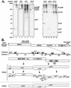
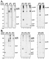
The replication complexes (RCs) of positive-stranded RNA viruses are intimately associated with cellular membranes. To investigate membrane alterations and to characterize the RC of mouse hepatitis virus (MHV), we performed biochemical and ultrastructural studies using MHV-infected cells. Biochemical fractionation showed that all 10 of the MHV gene 1 polyprotein products examined pelleted with the membrane fraction, consistent with membrane association of the RC. Furthermore, MHV gene 1 products p290, p210, and p150 and the p150 cleavage product membrane protein 1 (MP1, also called p44) were resistant to extraction with Triton X-114, indicating that they are integral membrane proteins. The ultrastructural analysis revealed double-membrane vesicles (DMVs) in the cytoplasm of MHV-infected cells. The DMVs were found either as separate entities or as small clusters of vesicles. To determine whether MHV proteins and viral RNA were associated with the DMVs, we performed immunocytochemistry electron microscopy (IEM). We found that the DMVs were labeled using an antiserum directed against proteins derived from open reading frame 1a of MHV. By electron microscopy in situ hybridization (ISH) using MHV-specific RNA probes, DMVs were highly labeled for both gene 1 and gene 7 sequences. By combined ISH and IEM, positive-stranded RNA and viral proteins localized to the same DMVs. Finally, viral RNA synthesis was detected by labeling with 5-bromouridine 5'-triphosphate. Newly synthesized viral RNA was found to be associated with the DMVs. We conclude from these data that the DMVs carry the MHV RNA replication complex and are the site of MHV RNA synthesis.
Figures


Similar articles
-
Expression and Cleavage of Middle East Respiratory Syndrome Coronavirus nsp3-4 Polyprotein Induce the Formation of Double-Membrane Vesicles That Mimic Those Associated with Coronaviral RNA Replication.mBio. 2017 Nov 21;8(6):e01658-17. doi: 10.1128/mBio.01658-17. mBio. 2017. PMID: 29162711 Free PMC article.
-
Colocalization and membrane association of murine hepatitis virus gene 1 products and De novo-synthesized viral RNA in infected cells.J Virol. 1999 Jul;73(7):5957-69. doi: 10.1128/JVI.73.7.5957-5969.1999. J Virol. 1999. PMID: 10364348 Free PMC article.
-
The putative helicase of the coronavirus mouse hepatitis virus is processed from the replicase gene polyprotein and localizes in complexes that are active in viral RNA synthesis.J Virol. 1999 Aug;73(8):6862-71. doi: 10.1128/JVI.73.8.6862-6871.1999. J Virol. 1999. PMID: 10400784 Free PMC article.
-
Biogenesis and architecture of arterivirus replication organelles.Virus Res. 2016 Jul 15;220:70-90. doi: 10.1016/j.virusres.2016.04.001. Epub 2016 Apr 9. Virus Res. 2016. PMID: 27071852 Free PMC article. Review.
-
Double-Membrane Vesicles as Platforms for Viral Replication.Trends Microbiol. 2020 Dec;28(12):1022-1033. doi: 10.1016/j.tim.2020.05.009. Epub 2020 Jun 11. Trends Microbiol. 2020. PMID: 32536523 Free PMC article. Review.
Cited by
-
The glycoprotein quality control factor Malectin promotes coronavirus replication and viral protein biogenesis.bioRxiv [Preprint]. 2024 Jul 16:2024.06.02.597051. doi: 10.1101/2024.06.02.597051. bioRxiv. 2024. PMID: 38895409 Free PMC article. Preprint.
-
Nanoscale cellular organization of viral RNA and proteins in SARS-CoV-2 replication organelles.Nat Commun. 2024 May 31;15(1):4644. doi: 10.1038/s41467-024-48991-x. Nat Commun. 2024. PMID: 38821943 Free PMC article.
-
Picornavirus VP3 protein induces autophagy through the TP53-BAD-BAX axis to promote viral replication.Autophagy. 2024 Sep;20(9):1928-1947. doi: 10.1080/15548627.2024.2350270. Epub 2024 May 16. Autophagy. 2024. PMID: 38752369
-
Identification of a membrane-associated element (MAE) in the C-terminal region of SARS-CoV-2 nsp6 that is essential for viral replication.J Virol. 2024 May 14;98(5):e0034924. doi: 10.1128/jvi.00349-24. Epub 2024 Apr 19. J Virol. 2024. PMID: 38639488 Free PMC article.
-
Classification, replication, and transcription of Nidovirales.Front Microbiol. 2024 Jan 24;14:1291761. doi: 10.3389/fmicb.2023.1291761. eCollection 2023. Front Microbiol. 2024. PMID: 38328580 Free PMC article. Review.
References
-
- Bi, W., J. D. Pinon, S. Hughes, P. J. Bonilla, K. V. Holmes, S. R. Weiss, and J. L. Leibowitz. 1998. Localization of mouse hepatitis virus open reading frame 1a derived proteins. J. Neurovirol. 4:594-605. - PubMed
-
- Bienz, K., and D. Egger. 1995. Immunocytochemistry and in situ hybridization in the electron microscope: combined application in the study of virus-infected cells. Histochem. Cell Biol. 103:325-338. - PubMed
-
- Bienz, K., D. Egger, and L. Pasamontes. 1987. Association of polioviral proteins of the P2 genomic region with the viral replication complex and virus-induced membrane synthesis as visualized by electron microscopic immunocytochemistry and autoradiography. Virology 160:220-226. - PubMed
Publication types
MeSH terms
Substances
Grants and funding
LinkOut - more resources
Full Text Sources
Other Literature Sources
Research Materials

