. 2002 Mar 18;156(6):1089-98.
doi: 10.1083/jcb.200111107. Epub 2002 Mar 18.

The role of cytochrome c in caspase activation in Drosophila melanogaster cells

Affiliations

The role of cytochrome c in caspase activation in Drosophila melanogaster cells

Loretta Dorstyn et al. J Cell Biol. .

Abstract

The release of cytochrome c from mitochondria is necessary for the formation of the Apaf-1 apoptosome and subsequent activation of caspase-9 in mammalian cells. However, the role of cytochrome c in caspase activation in Drosophila cells is not well understood. We demonstrate here that cytochrome c remains associated with mitochondria during apoptosis of Drosophila cells and that the initiator caspase DRONC and effector caspase DRICE are activated after various death stimuli without any significant release of cytochrome c in the cytosol. Ectopic expression of the proapoptotic Bcl-2 protein, DEBCL, also fails to show any cytochrome c release from mitochondria. A significant proportion of cellular DRONC and DRICE appears to localize near mitochondria, suggesting that an apoptosome may form in the vicinity of mitochondria in the absence of cytochrome c release. In vitro, DRONC was recruited to a >700-kD complex, similar to the mammalian apoptosome in cell extracts supplemented with cytochrome c and dATP. These results suggest that caspase activation in insects follows a more primitive mechanism that may be the precursor to the caspase activation pathways in mammals.


Figures

Figure 1.
Figure 1.
Caspase activation during apoptosis of BG2 cells. BG2 cells were treated with cycloheximide (A and B) or deprived of serum and insulin (C and D) over 24 h. (A and C) At various times after apoptotic stimuli, cells were assayed for viability by trypan blue exclusion (♦) and for caspase activity on either DEVD-amc (□) or VDVAD-amc (○). To determine caspase processing, lysates from cycloheximide-treated (B) or serum- and/or insulin-starved (D) BG2 cells were immunoblotted with anti-DRONC polyclonal antibody (top) or with an anti-DRICE polyclonal antibody (bottom) and proteins were visualized by enhanced chemiluminescence.
Figure 2.
Figure 2.
DARK is required for caspase activation in BG2 cells during apoptosis. BG2 cells were exposed to 37 nM of double stranded RNA for dronc, dark, or a negative control. Cells were either left untreated or treated with cycloheximide for 6 h. Cell lysates were immunoblotted using anti-DRONC and anti-DRICE antibodies.
Figure 3.
Figure 3.
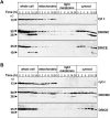
DRONC activation does not require cytochrome c release from mitochondria. BG2 cells treated with cycloheximide (A) or deprived of serum/insulin (B) for various lengths of time were gently lysed and fractionated by differential centrifugation to separate heavy membrane (P10, containing mitochondria), light membrane (P100), and cytosol (S100). Aliquots from each fraction were electrophoresed through 15% polyacrylamide gels and immunoblotted using an anti–cytochrome c monoclonal antibody (top), anti-DRONC polyclonal antibody (middle), or anti-DRICE polyclonal antibody (bottom). Signals were detected by ECL. The 50- and 36-kD DRONC bands represent the precursor and processed forms of DRONC, respectively.
Figure 4.
Figure 4.
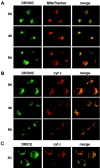
Cytochrome c is not released from mitochondria during BG2 cell apoptosis. BG2 cells treated with cycloheximide for various durations were fixed and stained with the mitochondrial marker MitoTracker red, or immunostained with either anti–cytochrome c monoclonal antibody or polyclonal antibodies for DRONC or DRICE. After incubation with appropriate FITC-coupled secondary antibodies, cells were visualized by fluorescence microscopy. Note that the DRICE antibody used in these experiments only recognizes the processed active form of DRICE, and thus cells undergoing apoptosis show a more intense staining for DRICE than the control at 0 h.
Figure 5.
Figure 5.
DRONC and active DRICE partially colocalize with mitochondria in BG2 cells. BG2 cells treated with cycloheximide for various lengths of time were costained with anti-DRONC polyclonal antibody and either MitoTracker red (A) or anti–cytochrome c monoclonal antibody (B). After incubation with FITC- (DRONC) or rhodamine- (cytochrome c) coupled secondary antibodies, cells were visualized by fluorescence microscopy using appropriate filters. The last column depicts merged images of the DRONC and MitoTracker costaining (A) or DRONC and cytochrome c costaining (B). (C) BG2 cells treated with cycloheximide for 8 h were double stained for active DRICE (FITC) and cytochrome c (rhodamine).
Figure 6.
Figure 6.
Cytochrome c localization in BG2 cells is not altered after serum deprivation. (A) Untreated BG2 cells or those deprived of serum and insulin for 24 h were fixed and costained with anti-DRONC polyclonal antibody and anti–cytochrome c monoclonal antibody. After incubation with FITC- (DRONC) or rhodamine- (cytochrome c) coupled secondary antibodies, cells were visualized by fluorescence microscopy using appropriate filters. The last column depicts merged images of the DRONC and cytochrome c costaining. (B) BG2 cells deprived of serum and insulin for various lengths of time were fixed and immunostained with anti-DRICE polyclonal antibody. As the anti-DRICE antibody only detects the active processed form of DRICE, immunofluorescence is seen in apoptotic cells only.
Figure 7.
Figure 7.
Lack of cytochrome c release during ecdysone-induced caspase activation in l(2)mbn cells. Drosophila l(2)mbn cells were treated with ecdysone for 24 h. After this, cells were either immunostained for active DRICE and cytochrome c (A) or fractionated (B) as in Fig. 3. After 24 h of ecdysone treatment, >80% of the cells were morphologically apoptotic and the majority of DRONC was processed into p36 (unpublished data). (A) Immunostaining with the active DRICE antibody clearly showed that cells contained processed DRICE. Please note that in B, the small amount of cytochrome c present in all cytosolic lanes is unlikely to represent cytochrome c released from mitochondria in cells undergoing apoptosis, as it does not show an increase over time. Furthermore, there was no reduction in the mitochondrial cytochrome c seen over the course of ecdysone treatment of cells.
Figure 8.
Figure 8.
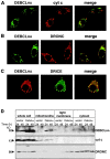
Ectopic expression of DEBCL does not induce cytochrome c release. BG2 cells were transfected with a DEBCL–HA expression construct. 24 h after transfection, cells were immunostained with an anti-HA monoclonal antibody and with either cytochrome c monoclonal antibody (A), the DRONC polyclonal antibody (B), or the active DRICE antibody (C). After incubation with FITC- (DEBCL–HA) or rhodamine- (cytochrome c and DRONC) coupled secondary antibodies, cells were visualized by fluorescence microscopy. The column on the right shows merged images of the DEBCL and cytochrome c costaining (A), DEBCL and DRONC costaining (B), or DEBCL and active DRICE costaining (C). In C, note that the DEBCL-expressing cell is positive for active DRICE. (D) Extracts prepared from DEBCL–HA-transfected BG2 cells were fractionated by centrifugation into heavy membranes (containing mitochondria), light membranes, and cytosol. Proteins were separated through 15% polyacrylamide gels and immunoblotted with an anti-HA monoclonal antibody (to detect DEBCL expression; top), anti–cytochrome c monoclonal antibody (middle), and anti-DRONC polyclonal antibody (bottom).
Figure 9.
Figure 9.
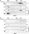
Formation of large molecular complexes containing DRONC and DRICE. BG2 cell extracts were supplemented with 2 μg cytochrome c, 2 mM dATP, and 1 mM MgCl2 and incubated for 1 h at either 4°C, 27°C, or 37°C. Extracts were then subjected to gel filtration chromatography using a Superdex 200 column. Individual fractions were collected and subjected to electrophoresis through 15% polyacrylamide gels. Fractions were immunoblotted with anti-DRONC polyclonal antibody (A) and anti-DRICE polyclonal antibody (B), and proteins were detected by ECL. Relative positions of molecular mass markers are shown. Note that the incubation of cell extracts at 27°C or 37°C causes monomeric DRONC (50 kD) to form complexes of >700 kD. Also, recruitment of DRONC in the large complex results in its processing, as demonstrated by the presence of the 36-kD band. A fraction of DRICE, most of which is processed in extracts incubated at 37°C, is also present in the complex.
Figure 10.
Figure 10.
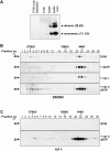
Cytochrome c enhances formation of the >670-kD complex. BG2 S100 fraction were immunodepleted with cytochrome c, as shown in A, and then incubated for 1 h at 27°C alone or supplemented with dATP, cytochrome c, or both dATP and cytochrome c (B and C). Cellular proteins were then subjected to gel filtration chromatography using a Superdex 200 column. Individual fractions were collected and subjected to electrophoresis through 15% polyacrylamide gels. Fractions were immunoblotted with anti-DRONC antibody (B) and anti–cytochrome c antibody, (C) and proteins were detected by ECL. Note that the immunodepleted S100 fraction lacks any detectable cytochrome c. Results in C show that only the dimeric (26 kD) form of cytochrome c is recruited to the >700-kD complex. Interestingly, a significant proportion of dimeric cytochrome c was detected in fractions 20–23, which corresponds to a molecular mass of ∼50 kD. This species may represent the tetrameric form of cytochrome c, which is composed of two dimers. The monomeric form (13 kD) of cytochrome c, eluted in fractions 33–39 (unpublished data), was not detected in high molecular mass fractions.

Similar articles

Cited by

References

    1. Baehrecke, E.H. 2000. Steroid regulation of programmed cell death during Drosophila development. Cell Death Differ. 7:1057–1062. - PubMed
    1. Brachmann, C.B., O.W. Jassim, B.D. Wachsmuth, and R.L. Cagan. 2000. The Drosophila bcl-2 family member dBorg-1 functions in the apoptotic response to UV-irradiation. Curr. Biol. 10:547–550. - PubMed
    1. Bratton, S.B., G. Walker, S.M. Srinivasula, X.M. Sun, M. Butterworth, E.S. Alnemri, and G.M. Cohen. 2001. Recruitment, activation and retention of caspases-9 and -3 by Apaf-1 apoptosome and associated XIAP complexes. EMBO J. 20:998–1009. - PMC - PubMed
    1. Cain, K., D.G. Brown, C. Langlais, and G.M. Cohen. 1999. Caspase activation involves the formation of the aposome, a large (∼700kDa) caspase-activating complex. J. Biol. Chem. 274:22686–22692. - PubMed
    1. Cain, K., S.B. Bratton, C. Langlais, G. Walker, D.G. Brown, X.-M. Sun, and G.M. Cohen. 2000. Apaf-1 oligomerises into biologically active ∼700-kDa and inactive ∼1.4MDa apoptosome complexes. J. Biol. Chem. 275:6067–6070. - PubMed

Publication types

MeSH terms

LinkOut - more resources