Comparative Study
. 2002 Mar;160(3):985-1000.
doi: 10.1016/S0002-9440(10)64920-6.

Abnormalities in pericytes on blood vessels and endothelial sprouts in tumors

Affiliations
Comparative Study

Abnormalities in pericytes on blood vessels and endothelial sprouts in tumors

Shunichi Morikawa et al. Am J Pathol. 2002 Mar.

Abstract

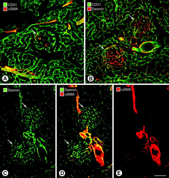
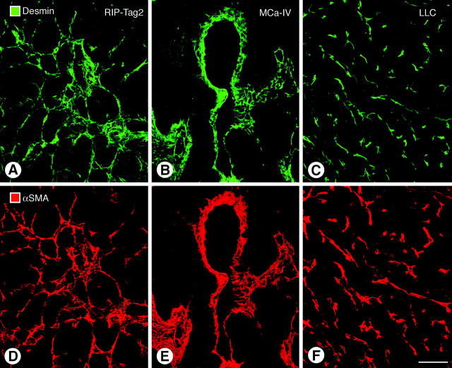
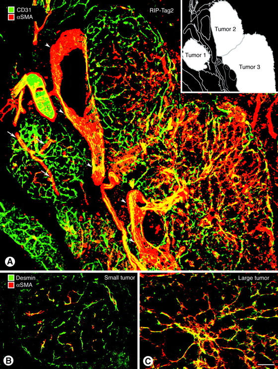
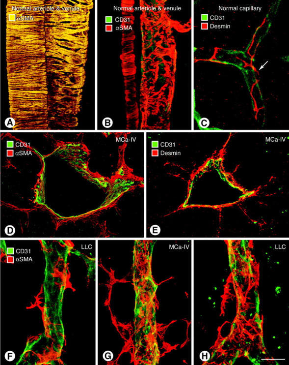
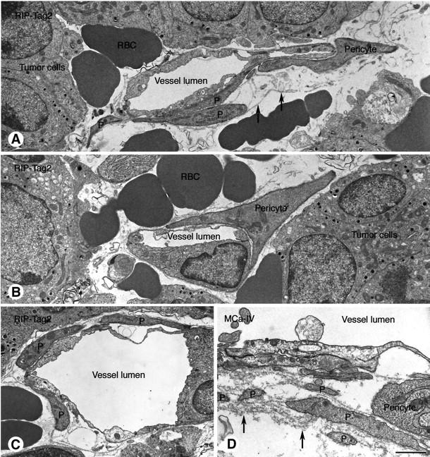
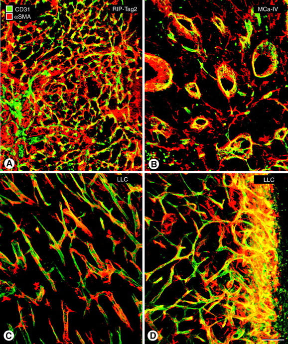
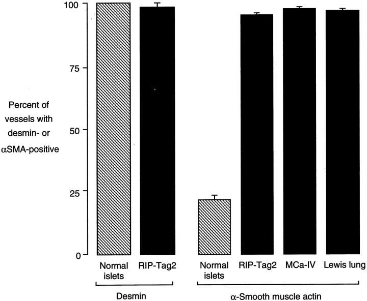
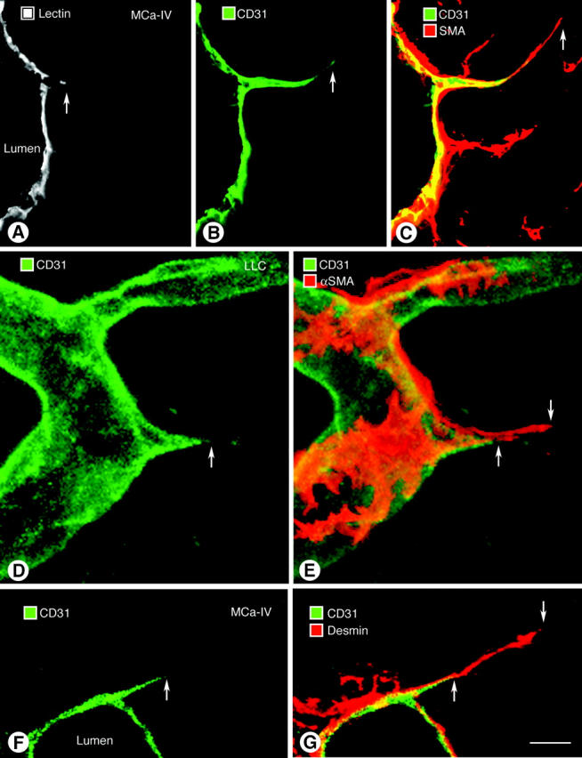
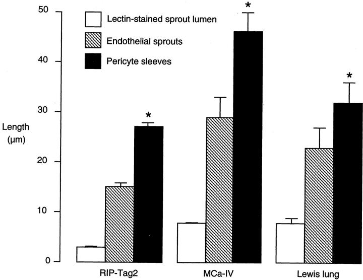
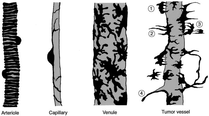
Endothelial cells of tumor vessels have well-documented alterations, but it is less clear whether pericytes on these vessels are abnormal or even absent. Here we report that alpha-smooth muscle actin (alpha-SMA) and desmin-immunoreactive pericytes were present on >97% of blood vessels viewed by confocal microscopy in 100-microm-thick sections of three different spontaneous or implanted tumors in mice. However, the cells had multiple abnormalities. Unlike pericytes on capillaries in normal pancreatic islets, which had desmin but not alpha-SMA immunoreactivity, pericytes on capillary-size vessels in insulinomas in RIP-Tag2 transgenic mice expressed both desmin and alpha-SMA. Furthermore, pericytes in RIP-Tag2 tumors, as well as those in MCa-IV breast carcinomas and Lewis lung carcinomas, had an abnormally loose association with endothelial cells and extended cytoplasmic processes deep into the tumor tissue. alpha-SMA-positive pericytes also covered 73% of endothelial sprouts in RIP-Tag2 tumors and 92% of sprouts in the other tumors. Indeed, pericyte sleeves were significantly longer than the CD31-immunoreactive endothelial cell sprouts themselves in all three types of tumors. All three tumors also contained alpha-SMA-positive myofibroblasts that resembled pericytes but were not associated with blood vessels. We conclude that pericytes are present on most tumor vessels but have multiple abnormalities, including altered expression of marker proteins. In contrast to some previous studies, the almost ubiquitous presence of pericytes on tumor vessels found in the present study may be attributed to our use of both desmin and alpha-SMA as markers and 100-microm-thick tissue sections. The association of pericytes with endothelial sprouts raises the possibility of an involvement in sprout growth or retraction in tumors.


Figures

Figure 1.
Figure 1.
Comparison of α-SMA and desmin immunoreactivities of blood vessels in normal pancreas. A and B: Vessels in pancreatic islets (arrows) and acini stained for CD31 and α-SMA or desmin immunoreactivity. Arterioles and venules but not capillaries have α-SMA-positive adventitial cells (A), whereas desmin immunoreactive cells are present on most vessels in islets (B). C–E: Desmin-immunoreactive cells are associated with most vessels including capillaries in islets (C and D, arrows), but α-SMA-positive cells are restricted to arterioles and venules (D and E). Scale bar in E applies to all figures. Bar lengths: 100 μm (A and B); 120 μm (C–E).
Figure 2.
Figure 2.
Double staining for desmin (A–C) and α-SMA (D–F) immunoreactivity of vessels in 100-μm sections of three types of tumors. A and D: Large pancreatic islet cell tumor in RIP-Tag2 transgenic mouse. B and E: Implanted MCa-IV mammary carcinoma. C and F: Implanted Lewis lung carcinoma (LLC). Unlike their different distributions on normal vessels, desmin and α-SMA are mostly co-localized in all three tumors. Scale bar in F applies to all figures. Bar length, 100 μm.
Figure 3.
Figure 3.
Increasing α-SMA expression in pericytes during tumor progression in RIP-Tag2 mice. A: In small tumor (left center and tumor 1 in inset) α-SMA-positive cells are present on arterioles (arrows) but are sparse on capillary-size vessels. In larger tumors (top center and tumor 2 in inset; bottom right and tumor 3 in inset) α-SMA-positive cells are more abundant on vessels of all size. CD31-immunoreactive endothelial cells (green). B and C: Double staining for desmin and α-SMA immunoreactivity. Pericytes on most vessels in small RIP-Tag2 tumor (B), which is a hyperplastic islet, resemble those in normal islet by expressing desmin but not α-SMA, but corresponding cells in a larger tumor (C) have both desmin and α-SMA immunoreactivities. Arrowheads mark large vein between the tumors. Scale bar in C applies to all figures. Bar lengths: 85 μm (A); 60 μm (B and C).
Figure 4.
Figure 4.
Morphology of adventitial cells on blood vessels in normal pancreas and in tumors. A: α-SMA-immunoreactive smooth muscle cells are circumferentially oriented and closely spaced on a larger arteriole (left) and venule (right) in normal pancreas, but are more uniformly shaped and closely packed on the arteriole. B: Smooth muscle cells on smaller arteriole (left) and pericytes on smaller venule (right) in normal pancreas stained for CD31 and α-SMA. The arteriole has regularly and circumferentially arranged smooth muscle cells, whereas the venule has irregularly arranged pericytes with multiple cytoplasmic processes that incompletely cover the vessel wall. C: Pericyte (arrow) on normal capillary, stained for CD31 and desmin, is positioned along the vessel axis. Most of the pericyte processes are oriented longitudinally. D–H: Pericytes on tumor vessels stained for CD31 (green) and α-SMA (red) are irregularly arranged, loosely associated with the endothelium, have cytoplasmic processes projecting in multiple directions, and have some similarities to venular pericytes. D and E: Unlike normal pericytes, some pericytes in MCa-IV carcinoma project away from the endothelium and into the tumor parenchyma. This feature is evident both with α-SMA and desmin immunoreactivity. F: Lewis lung carcinoma showing the loose association of pericytes with the endothelium of tumor vessels. G: Pericytes contacting one another near a vessel in a MCa-IV carcinoma. Pericytes also contact α-SMA-positive stromal cells that are apparently not associated with this vessel. H: Some pericytes in this Lewis lung tumor overlap other pericytes. Scale bar in H applies to all figures. Bar lengths: 35 μm (A, F–H); 30 μm (B); 15 μm (C); 80 μm (D and E).
Figure 5.
Figure 5.
Transmission electron micrographs showing abnormal pericytes on blood vessels in RIP-Tag2 tumors (A–C) and MCa-IV carcinoma (D). RBC, extravasated erythrocytes. A and B: Irregularly shaped pericyte processes (P) near a tumor vessel with a loose basement membrane (arrows). B: Pericyte process directed away from a capillary-size tumor vessel. C: Pericyte processes (P) loosely associated with the endothelium of tumor vessel. D: Pericyte processes (P) within loose, multilayered basement membrane (arrows) of tumor vessel. Scale bar in D applies to all figures. Bar lengths: 2 μm (A–C); 1 μm (D).
Figure 6.
Figure 6.
Distribution of α-SMA-immunoreactive cells (red) in three types of tumors. Some α-SMA-positive cells are pericytes on blood vessels marked by CD31 immunoreactivity (green), whereas others have no such association and are presumed to be myofibroblasts. A: RIP-Tag2 tumor: most vessels are the size of capillaries. Almost all are accompanied by α-SMA-positive cells. B: MCa-IV mammary carcinoma: vessels vary in size over a broad range and contain the largest vessels of the three tumors. Some α-SMA-positive cells surround blood vessels; others are located in the stroma between vessels. C: Lewis lung carcinoma (LLC): nearly all vessels are at least partially covered by α-SMA-positive cells. D: Lewis lung carcinoma: vessels and α-SMA-positive cells are especially densely packed at the periphery of the tumor. Some α-SMA-positive cells surround blood vessels; others do not. Scale bar in D applies to all figures. Bar length, 100 μm.
Figure 7.
Figure 7.
Bar graph comparing the percentage of blood vessels covered by desmin in normal pancreatic islets and islet cell tumors in RIP-Tag2 mice and by α-SMA-immunoreactive pericytes in normal islets, islet cell tumors in RIP-Tag2 mice, MCa-IV breast carcinomas, and Lewis lung carcinomas. Frequency of tumor vessels with pericytes was analyzed on 50 blood vessels in 100-μm sections double-stained for CD31 and desmin or α-SMA immunoreactivities of a tumor in each mouse (n = 4 mice for each type of tumor). One hundred percent of blood vessels in normal islets were covered by desmin-positive pericytes.
Figure 8.
Figure 8.
Endothelial sprouts on tumor vessels. A–C: Sprout on vessel in MCa-IV carcinoma stained by intravenous injection of FITC-L. esculentum lectin followed by staining of 100-μm sections for CD31 and α-SMA immunoreactivities. A: Only the proximal portion of the sprout is made visible by the fluorescent lectin (arrow), suggesting that the distal part of the sprout has no lumen. B: Same region showing the slender endothelial sprout with a tapered, blind ending (arrow) after CD31 staining. C: Same region showing a pericyte sleeve around and beyond (arrow) the end of the sprout. D and E: Blood vessel in Lewis lung carcinoma showing CD31-stained endothelial cells (D) and α-SMA-positive pericytes (E) with cytoplasmic processes that extend beyond the end of an endothelial sprout (arrows). F and G: Endothelial sprout in MCa-IV carcinoma accompanied by desmin-positive pericyte continuing beyond the sprout itself (arrows). Scale bar in G applies to all figures. Bar length: 25 μm (A–C); 10 μm (D–G).
Figure 9.
Figure 9.
Bar graph comparing the lengths of three components of endothelial sprouts in tumors: sprout lumen visualized by lectin staining, endothelial sprouts visualized by CD31 immunoreactivity, and pericyte sleeves on the sprouts visualized by α-SMA immunoreactivity. The sprout lumen was only 20 to 35% of the length of the sprouts, whereas the pericyte sleeves covering the sprouts were significantly longer than the sprouts in all three tumors (*, P < 0.05).
Figure 10.
Figure 10.
Schematic drawing comparing normal adventitial (periendothelial or mural) cells on an arteriole, capillary, and venule in the pancreas with abnormal pericytes on a tumor vessel representative of those found in RIP-Tag2 tumors, MCa-IV breast carcinomas, and Lewis lung carcinomas. Smooth muscle cells on arterioles were uniformly shaped, circumferentially arranged, closely packed, and tightly associated with the endothelium. Pericytes on capillaries were oriented longitudinally along the vessel, had long thin processes, were tightly positioned next to the endothelium, and covered only a small proportion of the vessel surface. Pericytes on venules had an irregular shape, close association with endothelial cells, and covered much of the vessel surface. By comparison, pericytes in the tumors we examined (1) were loosely associated with the endothelium, (2) had processes that extended away from the vessel wall, (3) in some cases overlaid other pericytes, and (4) accompanied endothelial sprouts, and even extended beyond the ends of the sprouts.

Similar articles

Cited by

References

    1. Jain RK: The next frontier of molecular medicine: delivery of therapeutics. Nat Med 1998, 4:655-657 - PubMed
    1. Arap W, Pasqualini R, Ruoslahti E: Cancer treatment by targeted drug delivery to tumor vasculature in a mouse model. Science 1998, 279:377-380 - PubMed
    1. Pasqualini R: Vascular targeting with phage peptide libraries. Q J Nucl Med 1999, 43:159-162 - PubMed
    1. Folkman J: Tumor angiogenesis. Adv Cancer Res 1985, 43:175-203 - PubMed
    1. Gilead A, Neeman M: Dynamic remodeling of the vascular bed precedes tumor growth: MLS ovarian carcinoma spheroids implanted in nude mice. Neoplasia 1999, 1:226-230 - PMC - PubMed

Publication types