Comparative Study
. 2002 Mar 1;21(5):954-65.
doi: 10.1093/emboj/21.5.954.

Epstein-Barr virus RNA confers resistance to interferon-alpha-induced apoptosis in Burkitt's lymphoma

Affiliations
Comparative Study

Epstein-Barr virus RNA confers resistance to interferon-alpha-induced apoptosis in Burkitt's lymphoma

Asuka Nanbo et al. EMBO J. .

Abstract

We investigated whether Epstein--Barr virus (EBV) infection could counteract the antitumor effect of interferon (IFN)-alpha. EBV-negative subclones isolated from EBV-positive Burkitt's lymphoma (BL) cell lines Akata, Daudi and Mutu were found to fall into apoptosis after IFN-alpha treatment. On the other hand, EBV-positive counterparts exhibited striking resistance against IFN-alpha-induced apoptosis. Transfection of an individual EBV latent gene into EBV-negative BL cells revealed that EBV-encoded poly(A)(-) RNAs (EBERs) were responsible for IFN resistance. EBERs bound double-stranded (ds) RNA-activated protein kinase (PKR), a key mediator of the antiviral effect of IFN-alpha, and inhibited its phosphorylation. Transfection of dominant-negative PKR, which was catalytically inactive and could block phosphorylation of endogenous PKR, made EBV-negative BL cells resistant to IFN-alpha-induced apoptosis. Furthermore, EBERs did not bind mutant PKR, which was catalytically active but lacked dsRNA-binding activity, nor did they inhibit its phosphorylation. These results indicate that EBERs confer resistance to IFN-alpha-induced apoptosis via binding to PKR and inhibition of its phosphorylation. This is the first report that the virus counteracts IFN-induced apoptosis in virus-associated tumors.


Figures
None
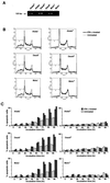
Fig. 1. IFN-α-induced apoptosis in BL-derived Akata, Daudi and Mutu cells. These cell lines were originally 100% EBV-positive, and EBV-negative subclones were isolated by limiting dilution from the parental cultures. (A) PCR analysis of EBV genomes in EBV-positive and -negative cell clones. The EBNA2 region was amplified by 30 cycles of PCR as described previously (Takeda et al., 2000). (B) DNA fluorescence histograms of propidium iodide-stained cells. EBV-positive and -negative cell clones (5 × 104/ml) were incubated in the presence or absence of human IFN-α (500 U/ml) for 60 h, and the frequency of apoptotic cells was determined by flow cytometry. A, apoptotic cells with hypodiploid DNA content. The vertical axis denotes the number of cells counted and the horizontal axis denotes fluorescence intensity. (C) The frequency of apoptotic cells determined by flow cytometry. EBV-positive and -negative cell clones (two clones each; 5 × 104/ml) were incubated in the presence or absence of human IFN-α (500 U/ml) for various times, and the frequency of apoptotic cells was determined. Results are expressed as the means of triplicate wells. (D) DNA laddering. EBV-positive and -negative cell clones (5 × 104/ml) were incubated in the presence or absence of human IFN-α (500 U/ml) for 72 h. DNA from 1 × 106 cells was subjected to 2% agarose gel electrophoresis.
None
Fig. 2. Dose response of apoptosis induction by IFN-α in EBV-positive and -negative Akata cell clones. Cells (5 × 104) were suspended in 1 ml of fresh medium containing various concentrations of IFN-α. After 84 h of incubation, cells were harvested for flow cytometry analysis.
None
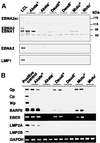
Fig. 3. EBV expression in EBV-positive and -negative Akata, Daudi and Mutu cell clones. (A) Immunoblot analysis for detection of EBNAs and LMP1. The blots were probed with EBNA-positive human serum (upper blot), an anti-EBNA2 monoclonal antibody (middle blot) and an anti-LMP1 monoclonal antibody (lower blot). Protein samples extracted from 105 cells were loaded per slot. (B) RT–PCR analysis of EBNA promoter usage and EBV latent gene expression. Akata cells were used as a positive control for detection of Qp-initiated EBNA mRNA, and a lymphoblastoid cell line immortalized by Akata EBV (LCL) was used as a positive control for detection of Cp- or Wp-initiated EBNA mRNAs, and EBER, BARF0, LMP2A and LMP2B mRNAs.
None
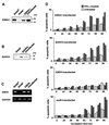
Fig. 4. IFN-α-induced apoptosis in EBV-negative Akata cell clones transfected with an individual EBV latent gene expressed in BL. An EBV-negative Akata cell clone was transfected with an individual EBV gene, and cell clones (two clones each) that stably expressed similar levels to EBV-positive Akata cells were selected and subjected to apoptosis assay. (A) EBNA1 expression. EBNA1 was detected by immunoblotting using EBNA1-positive human serum. (B) BARF0 expression in EBV-negative Akata cell clones transfected with the FLAG epitope-tagged BARF0 gene. BARF0 was detected by immunoblotting with anti-FLAG antibody. (C) EBER expression. EBER and GAPDH expression were determined by RT–PCR. (D) Apoptosis assay. Cells (5 × 104/ml) were incubated in the presence or absence of IFN-α (500 U/ml) for various times. The frequency of apoptotic cells was determined by flow cytometry. Results are expressed as the means of triplicate wells.
None
Fig. 5. IFN-α-induced apoptosis in EBV-negative Daudi and Mutu cell clones transfected with the EBER gene. EBV-negative Daudi and Mutu cell clones were transfected with the EBER plasmid, and cell clones (two clones each) that stably expressed similar levels to EBV-positive clones were selected and subjected to apoptosis assay. (A) EBER expression. EBER and GAPDH expression were determined by RT–PCR. (B) Apoptosis assay. Cells (5 × 104/ml) were incubated in the presence or absence of IFN-α (500 U/ml) for various times. The frequency of apoptotic cells was determined by flow cytometry. Results are expressed as the means of triplicate wells.
None
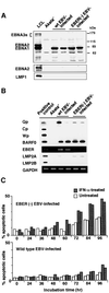
Fig. 6. IFN-α-induced apoptosis in EBER-knockout, EBV-infected Akata cells. An EBV-negative Akata cell clone was infected with EBER-positive or -negative EBV, and 100% EBV-positive cell clones (two clones each) were isolated and subjected to analysis. (A) Immunoblot analysis for detection of EBNAs and LMP1. The blots were probed with EBNA-positive human serum (upper blot), an anti-EBNA2 monoclonal antibody (middle blot) and an anti-LMP1 monoclonal antibody (lower blot). Protein samples extracted from 105 cells were loaded per slot. (B) RT–PCR analysis of EBNA promoter usage and EBV latent gene expression. EBV-positive Akata cells were used as a positive control for detection of Qp-initiated EBNA mRNA, and a lymphoblastoid cell line immortalized by Akata EBV (LCL) was used as a positive control for detection of Cp- or Wp-initiated EBNA mRNAs, and BARF0, EBER, LMP2A and LMP2B mRNAs. (C) Apoptosis assay. Cells (5 × 104/ml) were incubated in the presence or absence of IFN-α (500 U/ml) for various times. The frequency of apoptotic cells was determined by flow cytometry. Results are expressed as the means of triplicate wells.
None
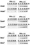
Fig. 7. Induction of PKR expression by IFN-α in EBV-positive and -negative Akata, Daudi and Mutu cell clones. Cells (5 × 104/ml) were incubated in the presence or absence of human IFN-α (500 U/ml) for various times. PKR expression was detected by immunoblotting using a polyclonal antibody to human PKR. Protein samples extracted from 2 × 105 cells were loaded per slot. IFN-α (+), IFN-α treated; IFN-α (–), IFN-α untreated.
None
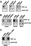
Fig. 8. Effects of EBV infection and EBER expression on phosphorylation of PKR. Cells (5 × 106) were transfected with the FLAG epitope-tagged PKR plasmid by the electroporation method. After 48 h of incubation, FLAG-PKR was immunoprecipitated with anti-FLAG antibody and subjected to in vitro kinase assay. Immunoprecipitated FLAG-PKR was detected by immunoblotting using anti-FLAG antibody (lower panel), and its phosphorylation was visualized by autoradiography (upper panel). (A) Phosphorylation of PKR in EBV-positive and -negative Akata, Daudi and Mutu cell clones. (B) Phosphorylation of PKR in EBV-negative Akata cell clones transfected with an individual EBV latent gene expressed in BL, and in Daudi and Mutu cell clones transfected with the EBER gene. (C) Phosphorylation of PKR in EBV-negative Akata cell clones that were infected with EBER-positive or -negative EBV.
None
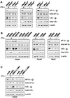
Fig. 9. Effects of EBV infection and EBER expression on phosphorylation of PKR substrates after IFN-α treatment. Cells (5 × 104/ml) were incubated in the presence of IFN-α (500 U/ml) for 12 h and subjected to immunoblotting for detection of phosphorylated eIF-2α or IκBα. Protein samples extracted from 1 × 105 cells were loaded per slot. (A) Phosphorylation of eIF-2α and IκBα in EBV-positive and -negative Akata, Daudi and Mutu cell clones. (B) Phosphorylation of eIF-2α and IκBα in EBV-negative Akata cell clones transfected with an individual EBV latent gene expressed in BL, and in Daudi and Mutu cell clones transfected with the EBER gene. (C) Phosphorylation of eIF-2α and IκBα in EBV-negative Akata cell clones that were infected with EBER-positive or -negative EBV.
None
Fig. 10. Effect of transient expression of EBER1 or EBER2 on phosphorylation of PKR in EBV-negative Akata and Daudi cell clones. Cells (5 × 106) were co-transfected with a FLAG epitope-tagged PKR plasmid (20 µg) and EBER plasmid (20 µg) by the electroporation method, incubated for 48 h, and subjected to in vitro kinase assay. EBER expression was determined by RT–PCR.
None
Fig. 11. Binding assay for association of EBER1 and EBER2 with PKR. Cells were transfected with a FLAG-tagged PKR plasmid. As a control, an EBV-positive Akata cell clone was transfected with a FLAG-tagged mutant PKR plasmid (Wu and Kaufman, 1997), which lacked the sequence coding for the dsRNA-binding domain (indicated as Akata+*). After 48 h of transfection, cells were treated with UV irradiation, digested with RNases to remove unbound RNA sequences and subjected to immunoprecipitation with anti-FLAG antibody. RNA was isolated from the immunoprecipitate, and EBER1 and EBER2 were measured by RT–PCR.
None
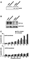
Fig. 12. Effect of dominant-negative PKR (mPKR) on phosphorylation of PKR and IFN-α-induced apoptosis in EBV-negative Daudi cells. HA-tagged mPKR plasmid was transfected into an EBV-negative Daudi cell clone, and cell clones that stably expressed mPKR were selected in the medium containing G418. (A) Expression of transfected HA-tagged mPKR that was detected by immunoblotting using an anti-HA polyclonal antibody. (B) Phosphorylation of wild-type PKR in EBV-negative Daudi cell clones expressing mPKR. Cells (5 × 106) were transfected with the FLAG epitope-tagged wild-type PKR plasmid by the electroporation method. After 48 h of incubation, FLAG-PKR was immunoprecipitated with anti-FLAG antibody and subjected to in vitro kinase assay. Immunoprecipitated FLAG-PKR was detected by immunoblotting using anti-FLAG antibody (lower panel), and its phosphorylation was visualized by autoradiography (upper panel). (C) Apoptosis assay of EBV-negative Daudi cell clones expressing mPKR. Cells (5 × 104/ml) were incubated in the presence or absence of IFN-α (500 U/ml) for various times. The frequency of apoptotic cells was determined by flow cytometry. Results are expressed as the means of triplicate wells.
None
Fig. 13. Effect of EBER expression on kinase activity of mPKR that lacked dsRNA-binding activity. EBV-positive Akata cells were transfected with FLAG-tagged wild-type PKR plasmid or with FLAG-tagged mPKR plasmid (K150A or A158D). For binding assay for association of EBER1 and EBER2 with PKR, after 48 h of transfection cells were treated with UV irradiation, digested with RNases to remove unbound RNA sequences, and subjected to immunoprecipitation with anti-FLAG antibody. RNA was isolated from the immunoprecipitate, and EBER1 and EBER2 were measured by RT–PCR. For in vitro kinase assay, after 48 h of transfection FLAG-PKR was immunoprecipitated from the cells with anti-FLAG antibody and subjected to in vitro kinase assay. Immunoprecipitated FLAG-PKR was detected by immunoblotting using anti-FLAG antibody (lower panel), and its phosphorylation was visualized by autoradiography (upper panel).
None
Fig. 14. Role of IL-10 in resistance to IFN-α-induced apoptosis. (A) EBV-negative Akata cells (5 × 104/ml) were incubated in RPMI 1640 medium containing human IFN-α (500 U/ml) in the presence or absence of recombinant IL-10 (100 pg/ml; Endogen) for 4 days. (B) EBV-positive Akata cells (5 × 104/ml) were incubated in RPMI 1640 medium containing human IFN-α (500 U/ml) in the presence or absence of purified rat anti-human IL-10 antibody (50 ng/ml; PharMingen) for 4 days. The frequency of apoptotic cells was determined by flow cytometry. Results are expressed as the means of triplicate wells.

Similar articles

Cited by

References

    1. Alas S., Emmanouilides,C. and Bonavida,B. (2001) Inhibition of interleukin 10 by rituximab results in down-regulation of bcl-2 and sensitization of B-cell non-Hodgkin’s lymphoma to apoptosis. Clin. Cancer Res., 7, 709–723. - PubMed
    1. Aman P. and von Gabain,A. (1990) An Epstein–Barr virus immortalization associated gene segment interferes specifically with the IFN-induced anti-proliferative response in human B-lymphoid cell lines. EMBO J., 9, 147–152. - PMC - PubMed
    1. Baer R. et al. (1984) DNA sequence and expression of the B95–8 Epstein–Barr virus genome. Nature, 310, 207–211. - PubMed
    1. Barber G.N. (2000) The interferons and cell death: guardians of the cell or accomplices of apoptosis. Semin. Cancer Biol., 10, 103–111. - PubMed
    1. Borden E.C., Lindner,D., Dreicer,R., Hussein,M. and Peereboom,D. (2000) Second-generation interferons for cancer: clinical targets. Semin. Cancer Biol., 10, 125–144. - PubMed

Publication types

MeSH terms