. 2002 Mar;22(5):1402-11.
doi: 10.1128/MCB.22.5.1402-1411.2002.

Mammalian selenoprotein in which selenocysteine (Sec) incorporation is supported by a new form of Sec insertion sequence element

Affiliations

Mammalian selenoprotein in which selenocysteine (Sec) incorporation is supported by a new form of Sec insertion sequence element

Konstantin V Korotkov et al. Mol Cell Biol. 2002 Mar.

Abstract

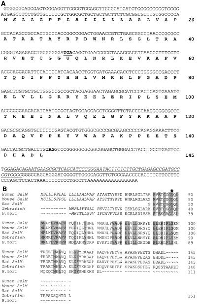
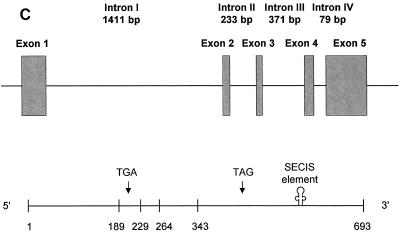
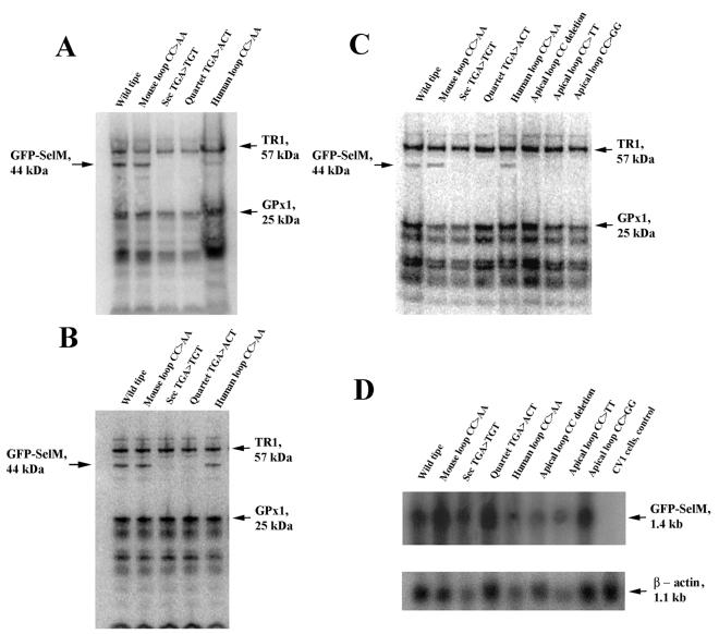
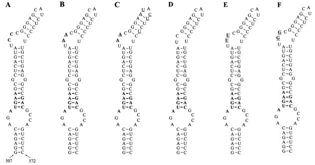
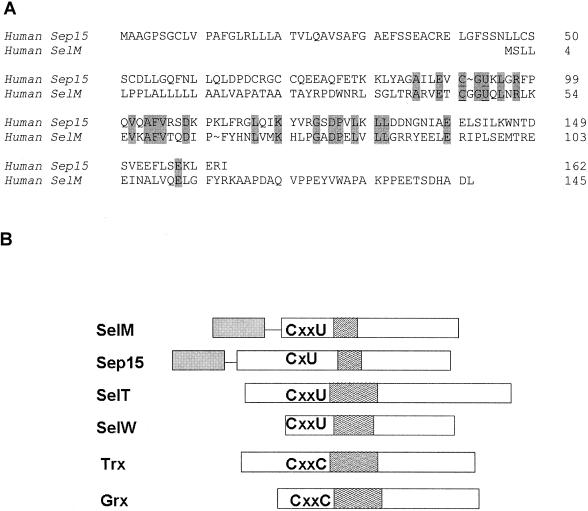
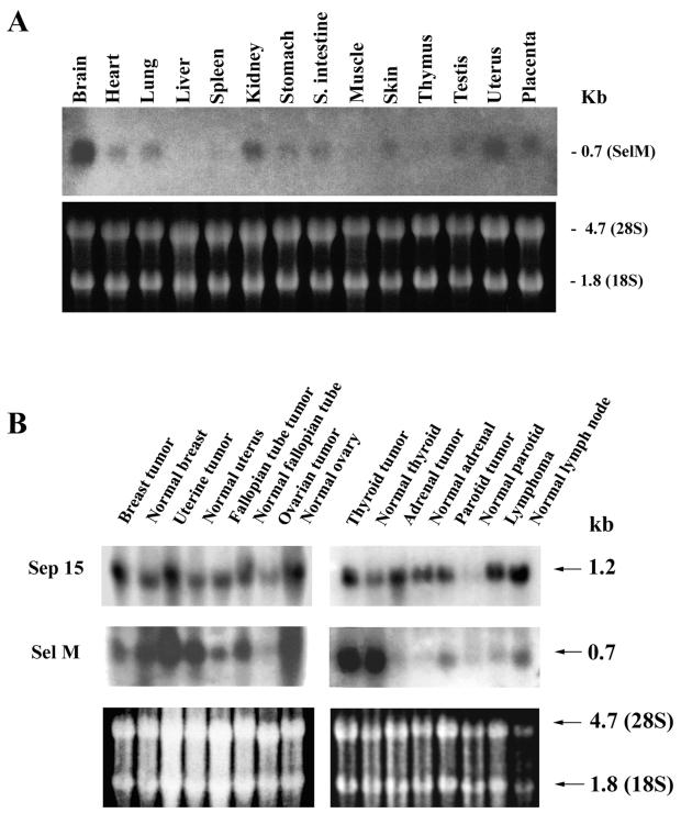
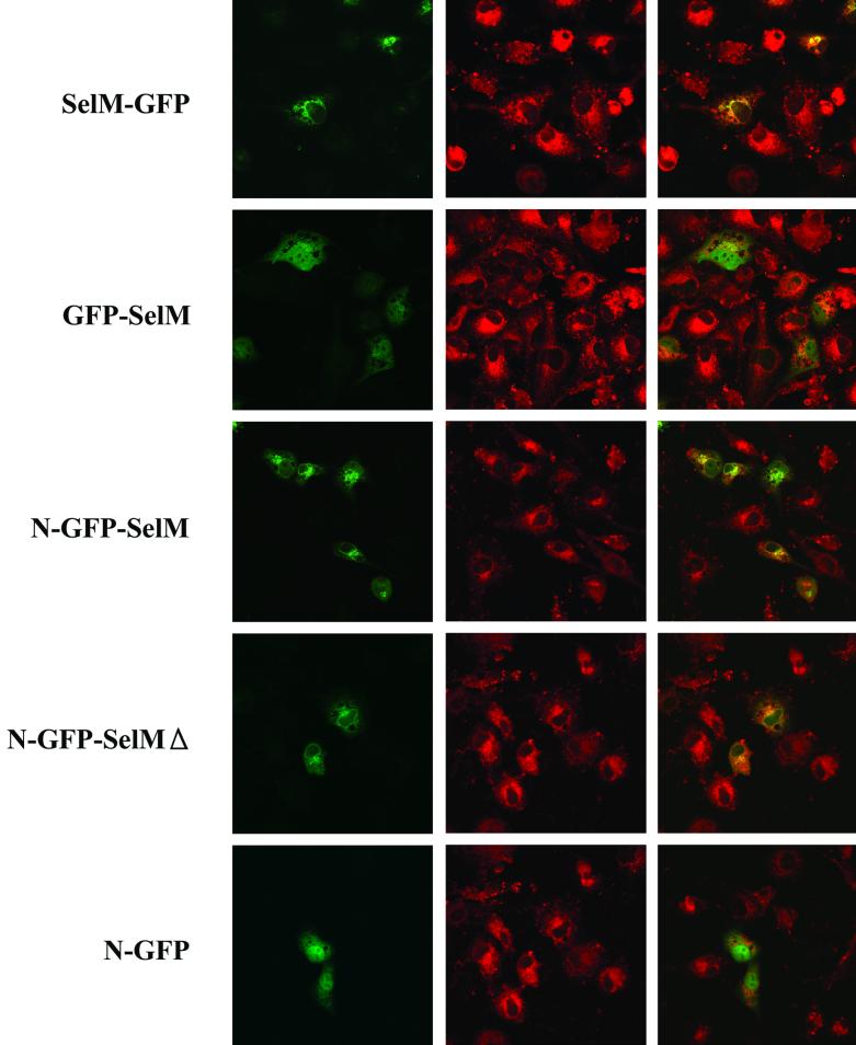
Selenocysteine (Sec), the 21st amino acid in protein, is encoded by UGA. The Sec insertion sequence (SECIS) element, which is the stem-loop structure present in 3' untranslated regions (UTRs) of eukaryotic selenoprotein-encoding genes, is essential for recognition of UGA as a codon for Sec rather than as a stop signal. We now report the identification of a new eukaryotic selenoprotein, designated selenoprotein M (SelM). The 3-kb human SelM-encoding gene has five exons and is located on chromosome 22 but has not been correctly identified by either Celera or the public Human Genome Project. We characterized human and mouse SelM cDNA sequences and expressed the selenoprotein in various mammalian cell lines. The 3" UTR of the human, mouse, and rat SelM-encoding genes lacks a canonical SECIS element. Instead, Sec is incorporated in response to a conserved mRNA structure, in which cytidines are present in place of the adenosines previously considered invariant. Substitution of adenosines for cytidines did not alter Sec incorporation; however, other mutant structures did not support selenoprotein synthesis, demonstrating that this new form of SECIS element is functional. SelM is expressed in a variety of tissues, with increased levels in the brain. It is localized to the perinuclear structures, and its N-terminal signal peptide is necessary for protein translocation.


Figures

FIG. 1.
FIG. 1.
SelM sequences. (A) Human SelM cDNA and protein sequences. The Sec-encoding TGA codon and the TAG stop signal are in bold, and TGA is underlined. U represents Sec. The numbers on the right indicate amino acid residues in the SelM sequence. The SECIS element in the 3" UTR is underlined and in italics. The predicted ER signal peptide is also in italics. (B) Alignment of multiple eukaryotic SelM sequences. Human and mouse sequences were determined in the present study. The GenBank accession numbers for the rat, zebra fish, and B. mori ESTs are AW433967, BE606173, and AU005812, respectively. U indicates Sec. This amino acid residue and the two conserved Cys amino acids upstream of Sec are underlined. In addition, a star above the sequences indicates the position of Sec. Residues conserved in all sequences are highlighted. The N-terminal signal peptide sequences are in italics. The numbers on the right indicate amino acid residues within corresponding sequences. The alignment was generated with the ClustalW program. (C) Organization of the human SelM-encoding gene. The upper portion of the panel shows the locations of exons (boxes) and introns (horizontal lines) in the human SelM-encoding gene. The lower portion of the panel shows the locations of exon-exon junctions and other functional features in the human SelM cDNA. The numbers under the boxes indicate nucleotide numbers within the human SelM cDNA that correspond to exon-exon junctions. The locations of the Sec-encoding TGA codon, the TAG stop codon, and the SECIS element in the 3" UTR are indicated.
FIG. 1.
FIG. 1.
SelM sequences. (A) Human SelM cDNA and protein sequences. The Sec-encoding TGA codon and the TAG stop signal are in bold, and TGA is underlined. U represents Sec. The numbers on the right indicate amino acid residues in the SelM sequence. The SECIS element in the 3" UTR is underlined and in italics. The predicted ER signal peptide is also in italics. (B) Alignment of multiple eukaryotic SelM sequences. Human and mouse sequences were determined in the present study. The GenBank accession numbers for the rat, zebra fish, and B. mori ESTs are AW433967, BE606173, and AU005812, respectively. U indicates Sec. This amino acid residue and the two conserved Cys amino acids upstream of Sec are underlined. In addition, a star above the sequences indicates the position of Sec. Residues conserved in all sequences are highlighted. The N-terminal signal peptide sequences are in italics. The numbers on the right indicate amino acid residues within corresponding sequences. The alignment was generated with the ClustalW program. (C) Organization of the human SelM-encoding gene. The upper portion of the panel shows the locations of exons (boxes) and introns (horizontal lines) in the human SelM-encoding gene. The lower portion of the panel shows the locations of exon-exon junctions and other functional features in the human SelM cDNA. The numbers under the boxes indicate nucleotide numbers within the human SelM cDNA that correspond to exon-exon junctions. The locations of the Sec-encoding TGA codon, the TAG stop codon, and the SECIS element in the 3" UTR are indicated.
FIG. 2.
FIG. 2.
Characterization of 75Se-labeled GFP-SelM fusion proteins. Transfected cells were grown in the presence of 75Se-selenite, and 75Se-labeled proteins were resolved by SDS-polyacrylamide gel electrophoresis and visualized with a PhosphorImager. The locations and molecular masses of the major selenoproteins TR1 and glutathione peroxidase 1 (GPx1) are indicated on the right. The location of the GFP-SelM fusion selenoprotein is on the left. (A) GFP-SelM expressed in mouse NIH 3T3 cells. NIH 3T3 cells were transfected with the plasmids encoding GFP-SelM (Wild type), GFP-SelM(CC>AA) (Mouse loop CC>AA), GFP-SelM(U48C) (Sec TGA>TGT), GFP-SelM(TGA) (Quartet TGA>ACT), and GFP-SelM(mAL>hAL) (Human loop CC>AA). (B) GFP-SelM expressed in human HEK 293 cells. HEK 293 cells were transfected with the same plasmids as in panel A. (C) GFP-SelM expressed in monkey CV-1 cells. CV-1 cells were transfected with the plasmids encoding GFP-SelM (Wild type), GFP-SelM(CC>AA) (Mouse loop CC>AA), GFP-SelM(U48C) (Sec TGA>TGT), GFP-SelM(TGA) (Quartet TGA>ACT), GFP-SelM(mAL>hAL) (Human loop CC>AA), GFP-SelM(CCdel) (Apical loop CC deletion), GFP-SelM(CC>TT) (Apical loop CC>TT), and GFP-SelM(CC>GG) (Apical loop CC>GG). (D) Northern blot analyses of GFP-SelM mRNAs. Cells were transfected with the constructs shown in panel C, and mRNAs were isolated and probed in Northern blot assays with the probe corresponding to SelM cDNA (top). The blot was reprobed to determine actin mRNA levels (bottom).
FIG. 3.
FIG. 3.
SECIS elements in vertebrate selenoprotein mRNAs. (A) Previously proposed eukaryotic SECIS element structure. The locations of structural features in the stem-loop (helix I, internal loop, quartet, helix II, and apical loop, or bulge) are indicated. N indicates any base. The quartet has non-Watson-Crick interactions. Previously invariant sequences in the SECIS element are in bold. (B) SECIS element in human SelM mRNA. Conserved sequences in the apical bulge (CC replaces the AA motif) and the quartet are in bold. The numbers under the structure indicate the locations of this SECIS element in human SelM mRNA. (C) Eukaryotic SECIS element consensus structure proposed in the present study. Characteristics of different features in the consensus SECIS element are indicated. See the text for further details. (D) Alignment of the SECIS elements in the human, mouse, rat, and zebra fish SelM-encoding genes. Locations of structural features in SECIS elements are indicated. The quartet is boxed. The CC motif in the apical loop of mammalian SelM SECIS elements and the AA motif in the zebra fish structure are in bold. nt, nucleotide.
FIG. 4.
FIG. 4.
Mouse SelM SECIS structures. The quartet, the nucleotide preceding the quartet, and the CC motif are in bold. (A) Wild-type SelM SECIS element. (B) Mouse SelM SECIS element in which CC was replaced with AA. Nucleotides that differ from the wild-type structure are underlined. (C) Mouse SelM SECIS element in which CC was replaced with AA and, in addition, the minihelix downstream of this motif and the apical loop were replaced with human sequences. Nucleotides that are different from the wild-type structure are underlined. (D) Mouse SelM SECIS element in which CC was deleted. (E) Mouse SelM SECIS element in which CC was replaced with TT. Nucleotides that differ from the wild-type structure are underlined. (F) Mouse SelM SECIS element in which CC was replaced with GG. Nucleotides that differ from the wild-type structure are underlined.
FIG. 5.
FIG. 5.
Homology analyses involving SelM. (A) Homology between human SelM and Sep15 selenoproteins. The numbers on the right indicate amino acids in selenoprotein sequences. Conserved residues are highlighted. U is Sec. (B) Comparison of SelM; selenoproteins Sep15, SelT, and SelW; and the thiol/disulfide oxidoreductases thioredoxin (Trx) and glutaredoxin (Grx). Sequences are aligned according to CxxU, CxU, and CxxC motifs. Partially filled boxes downstream of these motifs indicate α-helices. Signal peptides in SelM and Sep15 are shown by filled boxes linked by horizontal lines to other sequences. See the text for other details.
FIG. 6.
FIG. 6.
Expression of SelM mRNA. (A) Expression of mouse SelM mRNA. The upper portion shows expression of SelM mRNA in various mouse tissues. The lower portion shows expression of rRNA. (B) Expression of human SelM and Sep15 mRNAs in tumor and matched normal tissues. The upper portion shows expression of Sep15 mRNA. The middle portion shows expression of SelM mRNA. The lower portion shows expression of rRNA. The Northern Territory blot was first probed with a human Sep15 probe, stripped, and then reprobed with a human SelM probe.
FIG. 7.
FIG. 7.
Expression of GFP-SelM fusion proteins. Confocal images of CV-1 cells expressing various GFP-tagged SelM and control proteins are shown. A set of three images is shown for each construct. Each left panel shows green fluorescence corresponding to transiently expressed fusion proteins, each center panel shows fluorescence of the ER/Golgi marker, and each right panel is an image obtained by merging the left and center panels. Bar, 100 μm. The GFP fusion constructs used in this experiment are shown on the left.

Similar articles

Cited by

References

    1. Atkins, J. F., and R. F. Gesteland. 2000. The twenty-first amino acid. Nature 407:465.. - PubMed
    1. Berry, M. J., L. Banu, J. W. Harney, and P. R. Larsen. 1993. Functional characterization of the eukaryotic SECIS elements which direct selenocysteine insertion at UGA codons. EMBO J. 12:3315-3322. - PMC - PubMed
    1. Berry, M. J., G. W. Martin, 3rd, and S. C. Low. 1997. RNA and protein requirements for eukaryotic selenoprotein synthesis. Biomed. Environ. Sci. 10:182-189. - PubMed
    1. Buettner, C., J. W. Harney, and M. J. Berry. 1999. The Caenorhabditis elegans homologue of thioredoxin reductase contains a selenocysteine insertion sequence (SECIS) element that differs from mammalian SECIS elements but directs selenocysteine incorporation. J. Biol. Chem. 274:21598-21602. - PubMed
    1. Copeland, P. R., J. E. Fletcher, B. A. Carlson, D. L. Hatfield, and D. M. Driscoll. 2000. A novel RNA binding protein, SBP2, is required for the translation of mammalian selenoprotein mRNAs. EMBO J. 19:306-314. - PMC - PubMed

Publication types

MeSH terms

Associated data