. 2002 Feb 1;22(3):803-14.
doi: 10.1523/JNEUROSCI.22-03-00803.2002.

Delphilin: a novel PDZ and formin homology domain-containing protein that synaptically colocalizes and interacts with glutamate receptor delta 2 subunit

Affiliations

Delphilin: a novel PDZ and formin homology domain-containing protein that synaptically colocalizes and interacts with glutamate receptor delta 2 subunit

Yohei Miyagi et al. J Neurosci. .

Abstract

The glutamate receptor delta2 (GluRdelta2) subunit is selectively expressed in cerebellar Purkinje cells and plays an important role in cerebellar long-term depression, motor learning, motor coordination, and synapse development. We identified a novel GluRdelta2-interacting protein, named Delphilin, that contains a single PDZ domain and formin homology (FH) domains FH1 and FH2 plus coiled-coil structure. As far as we know, this is the first reported protein that contains both PDZ and FH domains. Yeast two-hybrid and surface plasmon resonance (SPR) analyses indicated that Delphilin interacts with the GluRdelta2 C terminus via its PDZ domain. This was also supported by coimmunoprecipitation experiments using a heterologous expression system in mammalian cells. Yeast cell and SPR analyses also demonstrated the possibility that the FH1 proline-rich region of Delphilin interacts with profilin, an actin-binding protein, and with the Src homology 3 domain of neuronal Src protein tyrosine kinase. In situ hybridization demonstrated the highest expression of Delphilin mRNA in Purkinje cells. Delphilin polypeptide was highly enriched in the synaptosomal membrane fraction of the cerebellum and coimmunoprecipitated with the GluRdelta2 subunit. The post-embedding immunogold technique demonstrated that Delphilin is selectively localized at the postsynaptic junction site of the parallel fiber-Purkinje cell synapse and colocalized with GluRdelta2. Thus, Delphilin is a postsynaptic scaffolding protein at the parallel fiber-Purkinje cell synapse, where it may serve to link GluRdelta2 with actin cytoskeleton and various signaling molecules.


Figures

Fig. 1.
Fig. 1.
Schematic presentations of the cDNA cloning, deduced amino acid sequence, and domain structure of Delphilin.A, The Delphilin cDNA is presented at thetop with a putative protein coding region as aheavy line associated with start (ATG) and termination (TGA) codons. The open box below designates the location of the clone MB2 [corresponding to nucleotide (nt) 76–1125], the original Delphilin clone obtained by yeast two-hybrid library screening. The locations of the clone 10-1 (nt 1–2526) and clone 9-2 (nt 426–3495), which were cloned from conventional screening of a phage cDNA library, are also presented as horizontal bars, respectively. B, Deduced amino acid sequence of Delphilin. The putative PDZ domain is marked bydouble underlining. Amino acid residues of the putative FH1 domain are in boldface, and the putative FH2 domain is underlined. Hydrophobic residues involved in the coiled-coil structure after the FH2 domain are boldfacewith underlining. C, A schematic presentation of the Delphilin domain structure. Each domain is shown as an open box with amino acid positions of the N and C terminals of the domain. The coiled-coil structure is presented as ablack box after the FH2 domain.
Fig. 2.
Fig. 2.
The PDZ and FH domains of Delphilin.A, Alignment of the PDZ domain of Delphilin with the known PDZ domains. The secondary structure elements of six β sheets and two α helices are boxed and indicated as βA–F and αA and αB, respectively, according to Doyle et al. (1996). The sequences used for the alignment are as follows: PDZ domain ofDelphilin (residues 88–165), RGS12(residues 20–97 from rat regulator of G-protein signaling 12),93PDZ-1, -2, and -3(residues 97–182, 192–297, and 420–501 from rat PSD-93 corresponding to the PDZ-1, -2, and -3 domains, respectively), and95PDZ-1 and -3 (residues 64–151 and 312–393 from mouse PSD-95 corresponding to the PDZ-1 and PDZ-3, respectively). Histidine residues at the first position of the α-helix B are boldface. B, Alignment of selected FH2 domains from Diaphanous (Castrillon and Wasserman, 1994), Bni1p (Sen-Gupta et al., 1996), mouseForminVI (Jackson-Grusby et al., 1992), cell division control protein 12 (CCP12) (Chang et al., 1997), and cell fusion protein (FUS1) (Petersen et al., 1995) with the putative FH2 domain of Delphilin. Amino acid residues conserved in more than three proteins are shown inboldface. Dots at the topof the alignment demonstrate invariant residues. C, FH1 domain of Delphilin. Prolines are shown in boldface, and two PPLP motifs are boxed.
Fig. 3.
Fig. 3.
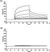
Surface plasmon resonance analyses of the interaction between the Delphilin PDZ domain and C-terminal eight-residue peptides of the GluRδ family subunits. The binding of the Delphilin PDZ domain fused with GST, at different concentrations (0.025–3.2 μm), to the GluRδ2 C-terminal peptide (A) or to the GluRδ1 corresponding peptide (B) immobilized on the surface of flow cells on a sensor chip was detected by changes in RUs over time. The sensorgrams shown were obtained after the blank sensorgram of the control nonimmobilized flow cell was subtracted from the sensorgrams of the peptide-immobilized cells.
Fig. 4.
Fig. 4.
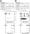
Interaction of Delphilin with GluRδ2 in HEK293T cells. HA-tagged Delphilin (Delph-HA) together with FLAG-tagged GluRδ2 (FLAG-δ2) or FLAG-tagged GluRδ2Δ1 (FLAG2Δ1) was expressed in HEK293T cells by transfection of respective expression vectors. A, Lysates of the transfected cells were immunoprecipitated with anti-FLAG antibody (anti-FLAG) or nonimmune control IgG (Ig). The immunoprecipitates were electrophoresed and immunoblotted with anti-FLAG (top panel) or anti-HA (bottom panel) antibodies. B, Lysates of the transfected cells were immunoprecipitated with anti-HA antibody or nonimmune control IgG. The immunoprecipitates were electrophoresed and immunoblotted with anti-HA (top panel) or anti-FLAG (bottom panel) antibodies. The input lanes (right two lanes of each panel) represent ∼1% of the cell lysates used for the immunoprecipitation experiments. The positions of precipitated HA-tagged Delphilin [bottom (A) and top (B)panels] and FLAG-tagged GluRδ2 [top(A) and bottom(B) panels] proteins are indicated by arrowheads. Positions of molecular weight markers are also indicated in kilodaltons on the right side of the top panel of A. In other panels, only corresponding bars are shown.IP, Immunoprecipitation.
Fig. 5.
Fig. 5.
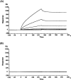
Surface plasmon resonance analyses of the interaction between the Delphilin FH1 domain and profilin II. The binding of the GST fusion protein of mouse profilin II (A), or the control GST protein itself (B) at different concentrations (0.001–1.0 μm), to the Delphilin FH1 domain peptide immobilized on the surface of a flow cell on a sensor chip was detected by changes in RUs over time. The sensorgrams shown were obtained after the blank sensorgram of the control cystein-immobilized flow cell was subtracted from the sensorgrams of the peptide-immobilized cells.
Fig. 6.
Fig. 6.
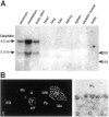
mRNA expression of Delphilin. A, Northern blot analysis of Delphilin gene transcripts. Each 20 μg of total RNA, extracted from the various mouse tissues designated above the panel, was subjected to Northern analysis. Staining of the gel with ethidium bromide after electrophoresis revealed the integrity and an equally applied amount of each RNA (data not shown). The locations of 28S and 18S ribosomal RNAs were indicated on the right side of the panel and used as size markers.B, In situ hybridization of Delphilin mRNA in the adult mouse brain. B1 demonstrates a negative image of an x-ray film autoradiogram. B2 is a bright-field micrograph of emulsion-dipped cerebellar cortex. Note that Delphilin mRNA is concentrated in the cell body of cerebellar Purkinje cells (asterisks). Cb, Cerebellum;Cx, cerebral cortex; GL, granular layer;H, hippocampus; HT, hypothalamus;MB, midbrain; Me, medulla oblongata;ML, molecular layer; OB, olfactory bulb;Po, pons; T, thalamus. Scale bars:B1, 1 mm; B2, 10 μm.
Fig. 7.
Fig. 7.
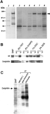
Identification and characterization of the Delphilin protein by Western blots. All blots were probed with guinea pig anti-Delphilin antibody or rabbit anti-GluRδ2 antibody.A, Delphilin expression in COS-7 cells and brain tissues. Five micrograms of each protein from transfected COS-7 cells with pZeoSV2 empty vector (lane 1) or pZeoSV2-Delphilin (lane 2) were analyzed on the left panel. Note the single 135 kDa band specific for the pZeoSV2-Delphilin transfectant. Ten micrograms of each cerebral (lanes 3,4, 5) or cerebellar (lanes 6, 7, 8) total homogenate, soluble fraction, or synaptosomal membrane fraction were also analyzed. Immunoreactive bands of the same size as the pZeoSV2-Delphilin transfectant were also identified in brain specimens except for the soluble fraction of cerebrum. B, Detergent solubility of Delphilin and GluRδ2. Ten micrograms of each cerebellar synaptosomal membrane were solubilized in the detergent presented at thetop of the panel, separated into soluble and insoluble fractions, and subjected to Western analyses.C, Immunoprecipitation of Delphilin from cerebellar synaptosomal membrane by anti-GluRδ2 antibody. Fifteen micrograms of synaptosomal membrane and each immunoprecipitate from 300 μg of membrane without serum [serum()] and with preimmune serum (pre-immune) or anti-GluRδ2 immune serum (immune) were analyzed by Western blotting with anti-Delphilin antibody. SMF, Synaptosomal membrane fraction; TRIT, Triton X-100; DOC, deoxycholate; S, supernatant; P, precipitate; IP, immunoprecipitation.
Fig. 8.
Fig. 8.
Immunohistochemistry of Delphilin on brain sections. A, B, Immunoperoxidase for Delphilin (A) and GluRδ2 (B). CE, Double immunofluorescence for Delphilin (C) and GluRδ2 (D). E is a merged view ofC and D.Asterisks mark the cell bodies of Purkinje cells. Cb, Cerebellum;Cx, cerebral cortex; GL, granular layer;H, hippocampus; HT, hypothalamus;MB, midbrain; Me, medulla oblongata;ML, molecular layer; OB, olfactory bulb;Po, pons; T, thalamus. Scale bars:B, 1 mm; E, 50 μm.
Fig. 9.
Fig. 9.
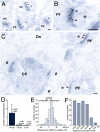
Ultrastructural localization of Delphilin (AC, arrowheads) at Purkinje cell synapses. A, Delphilin labeling at parallel fiber synapses. Purkinje cell spines forming synaptic contact with parallel fiber terminals (PF) are indicated by asterisks. B, Colocalization of Delphilin (arrowheads) and GluRδ2 (arrows) at a PF synapse. C, Lack of Delphilin labeling at climbing fiber synapses. Purkinje cell spines forming synaptic contact with climbing fiber terminals (CF) are indicated by #. Dn, Dendrite. D, Quantitative analysis showing selective Delphilin labeling at synapses between parallel fiber and Purkinje cell spine (PF-Sp). CF-Sp, Synapses between climbing fiber and Purkinje cell spine; In-Dn, synapses between interneuron axon and Purkinje cell dendrite. Error bars indicate SD. E, Perpendicular distribution of Delphilin from the midline of synaptic cleft. A total of 123 parallel fiber–Purkinje cell synapses were analyzed. The distances from the midline of synaptic cleft to the center of gold particles were grouped into 8 nm bins. F, Tangential distribution of Delphilin in the postsynapse. A total of 86 parallel fiber–Purkinje cell synapses were analyzed. Relative mediolateral position of gold particles is indicated as the percentage of the distance from the center (0%) to the edge (100%) of the postsynaptic density. Scale bars: AC, 100 nm.

Similar articles

Cited by

References

    1. Adamson JG, Zhou NE, Hodges RS. Structure, function and application of the coiled-coil protein folding motif. Curr Opin Biotechnol. 1993;4:428–437. - PubMed
    1. Aoki C, Miko I, Oviedo H, Mikelandze-Dvali T, Alexandre L, Sweeney N, Bredt DS. Electron microscopic immunocytochemical detection of PSD-95, PSD-93, SAP-102, and SAP-97 at postsynaptic, presynaptic, and nonsynaptic sites of adult and neonatal rat visual cortex. Synapse. 2001;40:239–257. - PubMed
    1. Araki K, Meguro H, Kushiya E, Takayama C, Inoue Y, Mishina M. Selective expression of the glutamate receptor channel δ2 subunit in cerebellar Purkinje cells. Biochem Biophys Res Commun. 1993;197:1267–1276. - PubMed
    1. Bartel P, Chien CT, Sternglanz R, Fields S. Elimination of false positives that arise in using the two-hybrid system. Biotechniques. 1993;14:920–924. - PubMed
    1. Blackstone CD, Moss SJ, Martin LJ, Levey AI, Price DL, Hugani RL. Biochemical characterization and localization of a non-N-methyl-d-aspartate glutamate receptor in rat brain. J Neurochem. 1992;58:1118–1126. - PubMed

Publication types

MeSH terms

Associated data